HISTOLOGI UMUM Departement of Histologi Medical Faculty of


 – Warna biru")


, kapsula Bowman pars")


- Slides: 47
HISTOLOGI UMUM Departement of Histologi Medical Faculty of Jambi University
HISTOLOGI • HISTOS = jaringan , anyamam • LOGOS = ilmu pengetahuan • Ilmu jaringan tubuh. • 1771 -1802: BICHAT (anatpmo perancis), pertama kali mengungkap kata JARINGAN=TISSU • 1819 : A. F. J. K MAYER seorang ahli mikkroskopik yang pertama kali memberikan istilah HISTOLOGI • Istilah HISTOLOGI berkembang bersama dg berkembangnya mikroskop
HISTOLOGI UMUM • Mempelajari macam jaringan tentang susunan mikroskopik, asal, fungsi. – jaringan epitel – jaringan penyambung – jaringan otot – jaringan saraf
HISTOLOGI KHUSUS • Mempelajari organ tubuh – – – – – sistem kardiovaskuler, getah bening, darah Sistem limfoid Sistem percernaan makanan Sistem pernafasan Kelenjar endokrin Sistem uropoetikum Sistem kelamin pria dan wanita Kulit dan derivatnya pancaindra
Jaringan histologi • Umumnya dengan perwarnaan H. E (hematoksilin – eosin) – Warna biru karena hematoksilin, bersifat basa, akan berikatan dengan inti sel yang bersifat asam – Warna merah eosin, bersifat asam , akan berikatan dengan sitoplasma yang bersifat basa atau asam sehingga memberi warna biru atau merah. – Sitoplasma berwarnabiru kalau sel sedang aktif, dan berwarna merah kalau sel sedang inaktif. Zat antar sel yang bersifat basa akan berwarna merah. ss
JARINGAN EPITELIAL Pendahuluan Tubuh : Sel : unit terkecil yg b’reproduksi &tumbuh Jar. : gab sel dan zat Antar sel Organ: gab sejmlah jar. yg memiliki fungsi khusus. Sistem : gab. sejumlah organ dengan f/majemuk
Jaringan epitel td 1. Mem bran epi t e lial 2. Kelen jar
n n Membran epitelial n n • dr. Monalisa n Jaringan yang disusun oleh kelompok sel-sel yang tersusun rapat Sebagian besar permukaannya saling kontak satu sama lain Zat antarsel sedikit diantaranya Nonvaskular Menutupi permukaan luardan dalam tubuh Melapisi atau membatasi lumen organ Melekat pada membrana basalis
Asal membran epitelial • n Ektoderm otak, medula spinalis, Ektoderm : sel epitel otak spinalis, kulit, rongga mulut, hidung, kanalis kulit, batas rongga auditoris eksternus, membranatimpani, mata, kelenjar auditoris eksternus, mata, kelenjar keringat, sebasea dan kelenjar sebasea keringat, dan kelenjar • Mesoderm : saluran urogenital n Mesoderm : saluran urogenital • Entoderm : epitel dan kelenjar sal. penc. dari esofagus n Entoderm kelenjar sal. penc. dari esofagus sampai ke : epitel dan rektum, hepar, vesika velea dan sampai ke rektum, hepar, vesika velea dan pankreas.
Macam sel epitel Berdasarkan bentuknya: • Gepeng/pipih/skuamus • Kubis/ kuboidal • Silindris/kolumnar/torak
Susunan membran epitelial: • Epitel selapis/ simple epitel • Epitel berlapis/stratified epitel/ complex epitel. • Epitel bertingkat/pseudostratified epitel/ berlapis semu • Epitel transisional
Type of Epithelium
Type of Epithelium
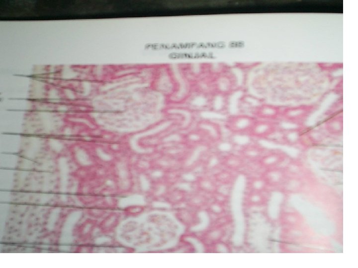
Epitel selapis/simplek a. Epitel selapis gepeng, terdapat di tunika serosa (usus), kapsula Bowman pars parietalis, loop henle desendens ginjal. Endothelium of blood vessel, mesothelium of body cavity, thin segment of Henle‘s loop in kidney
Epithelia: Simple Squamous • • • Single layer of flattened cells with discshaped nuclei and sparse cytoplasm Functions Ø Diffusion and filtration Ø Provide a slick, friction-reducing lining in lymphatic and cardiovascular systems Present in the kidney glomeruli, lining of heart, blood vessels, lymphatic vessels, and serosae
Simple squamous epithelium C M
Epithelia: Simple Squamous Figure 4. 2 a
Epitel selapis/simplek • Epitel selapis kubis, terdapat di folikel tiroid, epitel pigmen dari retina, kapsula lensa, glandula parotis, dll.
Epithelia: Simple Cuboidal • • • Single layer of cubelike cells with large, spherical central nuclei Function in secretion and absorption Present in kidney tubules, ducts and secretory portions of small glands, and ovary surface
Epithelia: Simple Cuboidal Figure 4. 2 b
Epithelia: Stratified Squamous • • • Thick membrane composed of several layers of cells Function in protection of underlying areas subjected to abrasion Forms the external part of the skin’s epidermis (keratinized cells), and linings of the esophagus, mouth, and vagina (nonkeratinized cells)
Epithelia: Stratified Squamous Figure 4. 2 e
Stratified squamous epithelium keratinized
Stratified squamous epithelium nonkeratinized
Epithelia: Stratified Cuboidal and Columnar • Stratified cuboidal – Quite rare in the body – Found in some sweat and mammary glands – Typically two cell layers thick • Stratified columnar – Limited distribution in the body – Found in the pharynx, male urethra, and lining some glandular ducts – Also occurs at transition areas between two other types of epithelia
Stratified cuboidal epithelium Thin skin, stratified cuboidal epithelium Identify: n A stratified cuboidal epithelium
Epitel bertingkat • Epitel bertingkat silindris bersilia, tdp duktus epididimis testis epiglotis and in the male urethra • Epitel bertingkat silindris bersilia dengan sel goblet, tdp trakea.
Epithelia: Pseudostratified Columnar • Single layer of cells with different heights; some do not reach the free surface • Nuclei are seen at different layers • Function in secretion and propulsion of mucus • Present in the male sperm-carrying ducts (nonciliated) and trachea (ciliated)
Duktus epididimis testis
Pseudostratified epithlium
Pseudostratified columnar epithelium Trachea, pseudostratified columnar epithelium, low & med. mag. Identif The pseudostratified ciliated epithelium
Pseudostratified epithlium
Epitel transisional lapisan paling superfisial td dari sel yang besar dan cembung pada perrmukaan bebasnya yg disebut sel payung. terdapat pada vesika urinaria.
Epithelia: Transitional • • • Several cell layers, basal cells are cuboidal, surface cells are dome shaped Stretches to permit the distension of the urinary bladder Lines the urinary bladder, ureters, and part of the urethra
Bangunan penghubung Membran epitelial • Membrana basalis/ basement membrane – Yaitu struktus ekstra sel yang terlihat pada dasar sel dari membran epitel atau kelenjar, terletak diantara epitel dan jaringan ikat longgar. • Junctional complex, (perlekatan lateral antar sel) td: – – – Zonula occludens/tigh junction Gap jaunctional Zonula adherens/intermediate junction Macula adherens/desmosome hemidesmosome
Basement Membrane • Region located between the epithelial cell and the underlying connective tissue
• Zonula occludens/tigh junction Perlekatan dua membran sel pada tepi bebasnya berada di duatidak membran selinterseluler pada tepi bebasnya berada di basis. Perlekatan mikrovili dan ada zat diantaranya. ujung selbasis sangat mikrovili kuat berlekatan dan tidak ada membentuk zat interseluler pemadatan diantaranya. ujung membran sel sangat kuat sel berlekatan disebut membentuk terminal barpemadatan membran sel disebut terminal bar
Anchoring, Intermediate, or Attachment junctions Zonula Adherens
Gap jaunction Perlekatan pada jungtional compleks di mana strukur hampir mirip dengan zonula occludens, tetapi ruang pemisah sangat sempit.
Zonula adherens/intermediate junction • Dibawah Zonula occludens/tigh junction, di mana membran sel yang berdekatan tidak saling bersentuhan. tetapi ada zat interseluler di dalam nya. Macula adherens/desmosome Mrp perlekatan paling basal, membran sel tidak saling bersinggungan. zat interseluler lebih padat.
Hemidesmosome • Desmosome yang tidak melekat pada permukaan lateral sel tetapi pada permukaan basal sel (membrana basalis)
• • Perlindungan/ proteksi: epidermis kulit Penyerapan/ absorbsi: epitel di gaster, usus, nefron Pengeluaran zat/sekresi: kelenjar Pernafasan: Epitel di paru Reproduksi: testis/ovarium Pelicin: epitel di gaster, usus Penerima rangsang: alat sensoris, pengecap, FUNGSI MEMBRAN EPITELIAL