Starter Structure of the thorax Put them in


 Trachea")


 Liver (Sugar levels adjusted) Examples of specialised exchange surfaces")


 Bronchiole and (b) trachea in transverse section")


")

(i)State one function for each of the following components of the bronchus")
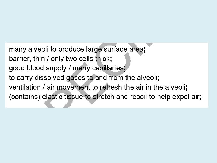
 Gaseous exchange occurs across the walls of the alveoli. Explain why")
 One feature of the disease emphysema is that the alveoli lose")


 Exhaling (Expiration) Volume of thorax Diaphragm muscle Diaphragm Relaxes and resumes to")
 Exhaling (Expiration) Volume of thorax Increases Decreases Diaphragm muscle Contracts Relaxes Diaphragm")
")


- Slides: 42
Starter- Structure of the thorax Put them in the correct order Nose Terminal bronchioles Secondary bronchi Alveoli, alveolar ducts and alveolar air sacs. Right and left primary bronchi Tertiary bronchi Trachea
The structure of the thorax Nose Trachea / Windpipe, Right and left primary bronchi Secondary bronchi, Tertiary bronchi Terminal bronchioles Alveoli, alveolar ducts and alveolar air sacs.
AS Biology- Module 2 The mammalian lung This lesson you should be able to… Grade C- Describe the features of the mammalian lung that adapt it to efficient gas exchange. Grade B – Outline the mechanism of ventilation. Grade A/A* - Describe and explain the distribution and functions of the different tissues found in the lungs.
O 2 from the atmospheric air entering the alveoli enters the blood by diffusion across the alveolar wall down its concentration gradient Total barrier to diffusion is less than 1 um CO 2 from the blood enters the alveoli by diffusion across the alveolar wall down its concentration gradient
Oxygenated blood to heart via pulmonary vein Low CO 2 concentration Surfactant produced by alveoli – reduces cohesive surface tension forces between H 2 O molecules in film of H 2 O ST forces may cause collapse of alveoli during exhalation – prevented by surfactant Diffusion gradient maintained by rhythmic ventilation (replaces air in alveoli) and a rich blood supply - high [O 2] on supply side (alveoli); low [O 2] on demand side (capillaries) – vice versa for CO 2 Squamous (flattened) epithelium O 2 CO 2 Deoxygenated blood from heart via pulmonary artery High CO 2 concentration Capillaries Close contact with alveolar wall 1 cell thick – short diffusion distance Narrow – space enough for passage of single RBC – allows close contact with capillary wall Large surface area – numerous and tubular Alveoli – spherical and numerous (600 million); large surface area (c. 70 m 2) Film of H 2 O – dissolves gases - evaporates Alveolus wall Capillary wall Plasma – carries 85% of CO 2 as hydrogen carbonate ions, 5% dissolved in plasma, and 10% in combination with haemoglobin Each 1 cell thick RBC’s – contain haemoglobin - carries O 2 as oxyhaemoglobin
The structure of the thorax 1. 6. 3. 7. 8. 4. 5. 9. 10.
The structure of the thorax C-shaped cartilage Trachea Right lung Left lung Intercostal muscle Rib Bronchus Bronchiolus Diaphragm
Adaptations of Respiratory System for Gas Exchange • Cleans, warms, and moistens the air entering the respiratory tract during inhalation – achieved by hairs, mucus, and blood capillaries (warm blood) in the nasal cavity and cilia in the trachea • A large number of spherical alveoli (600 million) 100 -300 um across – giving a large surface area of approx. 80 -100 m 2 for rapid gas exchange between the blood and atmosphere; alveolar wall is one cell thick (squamous epithelium) – short diffusion path between alveoli and blood in the pulmonary capillaries • Numerous and tubular capillaries in close contact with alveoli - wall also made of squamous epithelium and one cell thick; total diffusion distance is less than 1 um • Capillaries are narrow – red blood cells are squeezed against the capillary wall – making contact with the wall to reduce diffusion distance. The blood cells are also slowed down – increasing the time for exchange
Adaptations of Respiratory System for Gas Exchange • Constant ventilation - replacing the air in the alveoli to maintain a concentration gradient between the alveolar air and the blood • Alveoli are surrounded by a large number of capillaries – a rich blood supply, replaces the blood constantly, to maintain a concentration gradient between the blood and alveolar air. • Haemoglobin (Hb) in red blood cells carries oxygen and has a high affinity for oxygen – helps in maintaining a steep concentration gradient for oxygen • Moist exchange surface – allows gases to dissolve • Water in the alveoli contains a surfactant (phospholipid) – reduces surface tension – prevents collapse of alveoli • Alveoli contain phagocytic cells - for defence
Respiratory Tree Direction of air flow during inhalation Atmospheric air Larynx (“voice box”) Trachea Bronchus (2) Bronchioles Direction of air flow during exhalation Alveoli (“air sacs”) Site of gas exchange Gases exchanged by diffusion down their concentration gradients O 2 from alveoli to blood in alveolar capillaries CO 2 from blood into alveoli (for excretion) Capillaries (blood) Red blood cells transport absorbed O 2 from lungs to body tissues CO 2 transported from tissues to lungs for excretion 10
GAS EXCHANGE . . . describe with the aid of diagrams and photographs, the distribution of cartilage, ciliated epithelium, goblet cells, smooth muscle and elastic fibres in the trachea, bronchioles and alveoli of the mammalian gaseous exchange system Rings of cartilage in the walls of the trachea and bronchi provide support It is strong but flexible It stops the trachea and bronchi collapsing when pressure drops
Structural components of the respiratory system Trachea Smooth muscle Elastic fibres C-shaped rings of cartilage Fill in the functions
Smooth muscle and elastic fibres in the trachea Smooth muscle is found in the walls of the trachea, bronchi and bronchioles Smooth muscle relaxes during exercise, widening the lumen Results in less resistance to airflow Elastic fibres stretch on inspiration and recoil to help push air out when exhaling
Describe and explain the distribution and functions of the different tissues found in the lungs. Structure Cartilage Function/Characteristics
Structural components of the respiratory system Fill in function Inside surface of trachea – epithelial lining Goblet (mucus) cells Ciliated epithelium Loose tissue
Ciliated epithelium Simple columnar epithelial cells Fine hair-like outgrowths Rapid, rhythmic, wavelike beatings Movement of mucus Usually found in the air passages like the nose, uterus and fallopian tubes
Ciliated epithelium Cilia beat the mucus Helps to prevent lung infections
Goblet cells Specialised as gland cells Synthesising and secreting mucus
Goblet cells Secrete mucus Mucus traps microorganisms and dust particles in inhaled air
Small intestine (absorption of nutrients) Liver (Sugar levels adjusted) Examples of specialised exchange surfaces Root hair of plants (water and minerals absorbed) Hyphae of fungi (nutrients absorbed)
TASK -Look at the diagrams below. Draw a table to describe the function and characteristics for each part. (a) Bronchiole and (b) trachea in transverse section
Cross section of bronchiole Alveolus wall § Thin - single cell thick § Squamous epithelium § Reduces diffusion distance Bronchiole wall § Ciliated epithelium (cilia move mucus upwards) § Goblet cells (secrete mucus) Blood capillary § Close to alveoli § Thin - single cell thick § Squamous epithelium § Reduces diffusion distance Pulmonary vein § Carries oxygenated blood to heart
(a) Bronchiole and (b) trachea in transverse section
Structure Function/Characteristics Cartilage Smooth muscle Elastic fibres Goblet cells and glandular tissue Ciliated epithelium Bronchioles .
Exam Question 4. The diagram below shows the detailed structure of a small part of the mammalian lung. (i) State the name of the structure shown between lines D and E. . . . . . . . [1]
Exam Question List three features of the structure which you have identified in (i) which make it suitable for gas exchange. 1. . . . . . . . . . . . . . . 2. . . . . . . . . . . . . . . 3. . . . . . . . . . . . . . . [3] [Total 4 marks]
Exam Question 5. The different parts of the gaseous exchange system, such as the bronchi, show structural adaptations to their functions. The diagram below shows a section through the wall of a bronchus as seen with a light microscope.
Exam Question (a)(i)State one function for each of the following components of the bronchus wall. goblet cell. . . . . . . . . . . . . cartilage. . . . . . . . . . . . . [2] (ii)State two ways in which the structure of the wall of the bronchus would be different in a long-term smoker. 1. . . . . . . . . . . . . . 2. . . . . . . . . . . . . . [2]
Exam Question (b) Gaseous exchange occurs across the walls of the alveoli. Explain why the walls of the alveoli contain elastic fibres. . . . . . . . . . . . . . . . . . . . . . . . . . . . . . . [2]
Exam Question (c) One feature of the disease emphysema is that the alveoli lose their elasticity. Explain the effects of this loss of elasticity on the gaseous exchange system of a person with emphysema. . . . . . . . . . . . . . . . . . . . . . . . . . . . . . . . . . . . . . . . . . . . . . . . . . . . . . . . . . . . . [4] [Total 10 marks]
The lungs and associated structures © Pearson Education Ltd 2008 This document may have been altered from the original
The lungs and associated structures 2. 1. 3. 4. 5. 6. 7. 9. 8. 10. © Pearson Education Ltd 2008 This document may have been altered from the original
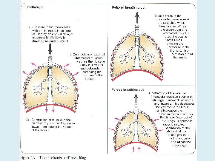
Ventilation The ribcage, intercostal muscles and diaphragm all work together to move air into and out of the lungs, where gas exchange occurs across the thin (single-celled) walls of the alveoli Ventilation is a physical process, relying on the principle of Boyle’s Law – which state “Pressure is inversely proportional to volume” The mechanism can be illustrated using a bell jar model of the respiratory system – however, the model does not illustrate involvement of the rib cage and the intercostal muscles in ventilation Breathing out (expiration / exhalation) Internal intecostals contract in forced expiration Breathing in (inspiration / inhalation)
INSPIRATION EXPIRATION Atmospheric pressure = 760 mm. Hg Diaphragm & external intercostals contract Rib cage raised (upwards and outwards) Diaphragm lowered (becomes flatter) Volume of chest cavity increases Pressure in chest cavity drops to below atmospheric pressure to 758 mm. Hg Diaphragm & external intercostals relax Rib cage lowered Diaphragm raised (dome shape) due to push from abdominal organs Volume of chest cavity decreases Pressure in chest cavity increases to above atmospheric pressure to 763 mm. Hg Air moves into lungs from atmosphere Active process Air forced out of lungs into atmosphere Aided by elastic recoil and abdominal organs Passive process
Inhaling (Inspiration) Exhaling (Expiration) Volume of thorax Diaphragm muscle Diaphragm Relaxes and resumes to dome shape External intercostal muscles Rib cage Pressure in chest cavity Movement of air Decreases below atmospheric pressure
Inhaling (Inspiration) Exhaling (Expiration) Volume of thorax Increases Decreases Diaphragm muscle Contracts Relaxes Diaphragm Flattens and pushes digestive organs down Relaxes and resumes to dome shape External intercostal muscles Contracts/expands Relaxes Rib cage Upward and outward Inward and downward Pressure in chest cavity Decreases below atmospheric pressure Increases below atmospheric pressure Movement of air Into the lungs down pressure gradient Air forced out of lungs
Exam question (5 marks)
Exam Questions - OCR Name five tissues, cells or cell structures found in the mammalian gas exchange system and explain the function of each [5 marks]
Name five tissues, cells or cell structures found in the mammalian gas exchange system and explain the function of each [5 marks] -Goblet cells secrete mucus, the mucus traps bacteria and dust particles -Some cells are ciliated, cilia move mucus towards the throat to be removed -Elastic fibres stretch on inspiration and recoil at expiration -Smooth muscle relaxes to make air passages wider when exercising -Cartilage provides support and keeps trachea open