Shock William Lawson MD Division of Allergy Pulmonary


- Slides: 41
Shock William Lawson, MD Division of Allergy, Pulmonary, and Critical Care Medicine
Outline for Today • • • Definition Ramifications Physiologic determinants Classification Approach to the patient with shock Treatment strategies
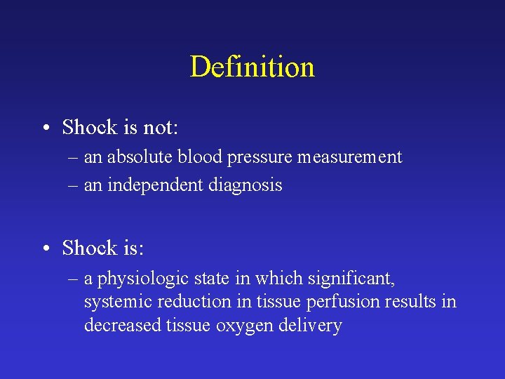
Definition • Shock is not: – an absolute blood pressure measurement – an independent diagnosis • Shock is: – a physiologic state in which significant, systemic reduction in tissue perfusion results in decreased tissue oxygen delivery
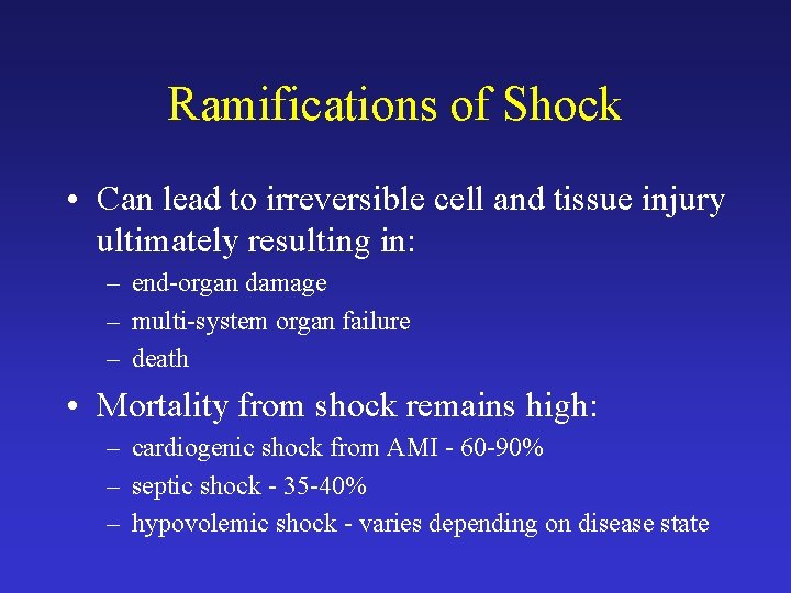
Ramifications of Shock • Can lead to irreversible cell and tissue injury ultimately resulting in: – end-organ damage – multi-system organ failure – death • Mortality from shock remains high: – cardiogenic shock from AMI - 60 -90% – septic shock - 35 -40% – hypovolemic shock - varies depending on disease state
Physiologic Determinants P = CO * SVR MAP – CVP = CO * SVR P = change in pressure MAP = mean arterial pressure CVP = central venous pressure CO = cardiac output SVR = systemic vascular resistance
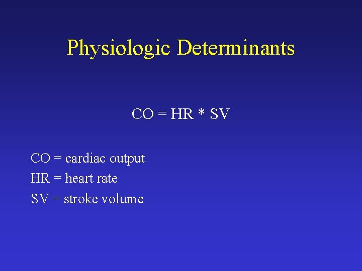
Physiologic Determinants CO = HR * SV CO = cardiac output HR = heart rate SV = stroke volume
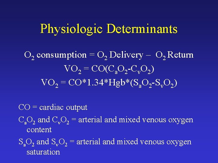
Physiologic Determinants O 2 consumption = O 2 Delivery – O 2 Return VO 2 = CO(Ca. O 2 -Cv. O 2) VO 2 = CO*1. 34*Hgb*(Sa. O 2 -Sv. O 2) CO = cardiac output Ca. O 2 and Cv. O 2 = arterial and mixed venous oxygen content Sa. O 2 and Sv. O 2 = arterial and mixed venous oxygen saturation
Classification of Shock • Hypovolemic • Cardiogenic • Distributive (vasodilatory)
Hypovolemic Shock • Results from decreased preload • Preload is one of the determinants of stroke volume • Thus, when preload drops, cardiac output drops
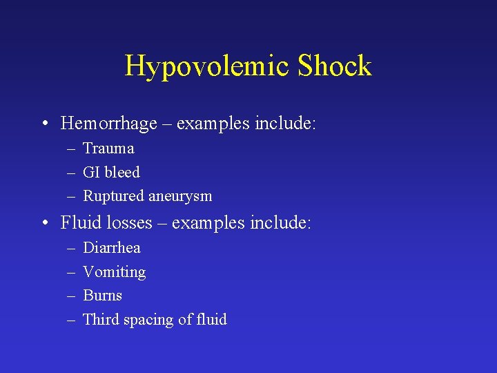
Hypovolemic Shock • Hemorrhage – examples include: – Trauma – GI bleed – Ruptured aneurysm • Fluid losses – examples include: – – Diarrhea Vomiting Burns Third spacing of fluid
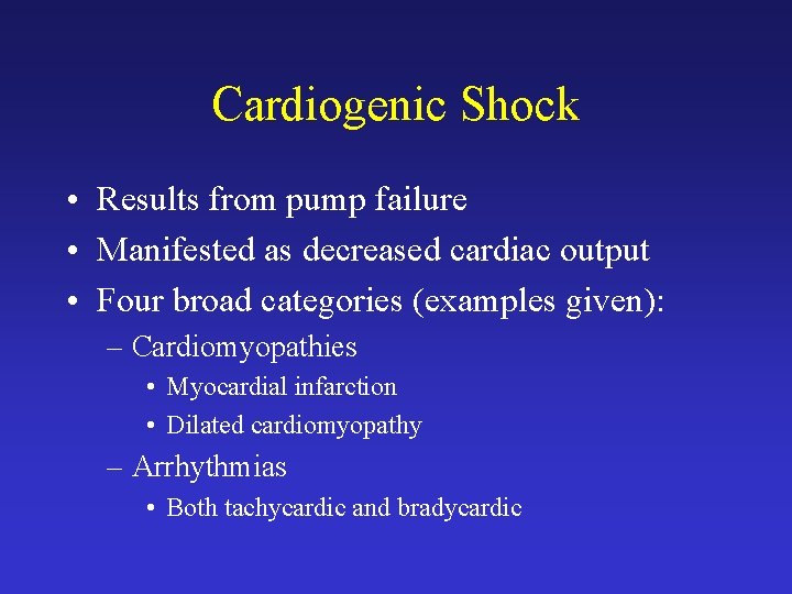
Cardiogenic Shock • Results from pump failure • Manifested as decreased cardiac output • Four broad categories (examples given): – Cardiomyopathies • Myocardial infarction • Dilated cardiomyopathy – Arrhythmias • Both tachycardic and bradycardic
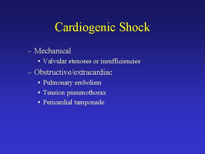
Cardiogenic Shock – Mechanical • Valvular stenoses or insufficiencies – Obstructive/extracardiac • Pulmonary embolism • Tension pneumothorax • Pericardial tamponade
Distributive Shock • Also referred to as vasodilatory shock • Results from a severe decrease in SVR • Examples include: – septic shock – systemic inflammatory response syndrome (SIRS) – anaphylaxis – neurogenic shock – Addisonian crisis
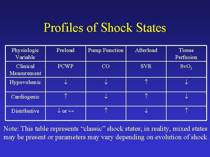
Profiles of Shock States Physiologic Variable Preload Pump Function Afterload Tissue Perfusion Clinical Measurement PCWP CO SVR Sv. O 2 Hypovolemic Cardiogenic Distributive or ↔ Note: This table represents “classic” shock states; in reality, mixed states may be present or parameters may vary depending on evolution of shock.
Common Features of Shock • • • Hypotension Cool, clammy skin Oliguria Altered mental status Metabolic acidosis
Evaluation of the Patient in Shock • Primary – History – Physical Examination – Laboratory – Radiographic • Secondary – pulmonary artery catheterization – echocardiography
Pulmonary Artery Catheterization • Allows measurement of: – vascular pressures including PCWP – CO by thermodilution – calculation of SVR and PVR – mixed venous oxygen saturation (Sv. O 2) • Most cases of shock can be managed without a PA catheter • Debate ongoing on utility and safety of PA catheter
Management of the Patient in Shock • Shock is an emergency state: – initial focus is on ABC’s of resuscitation • airway, breathing, circulation • Adequate venous access – central venous access not always required, but often necessary • Optimization of volume status • Identification of cause of shock and directed therapy
Management - Hypovolemic Shock • Volume replacement key for all causes of hypovolemic shock – nonhemorrhagic: crystalloid fluid replacement – hemorrhagic: crystalloid fluid replacement and blood product replacement • Directed therapy to correct the cause of hypovolemic shock
Management - Cardiogenic Shock • General points for cardiogenic shock – PA catheter commonly used – vasopressor support (specific discussion later) often needed – may need intra-aortic balloon pump – hypotension may or may not be present – identification of type of cardiogenic shock critical for optimal management
Management - Cardiogenic Shock • Myocardial infarction – most common cause of cardiogenic shock – directed therapy for MI • aspirin, heparin, glycoprotein IIb/IIIa inhibitors, revascularization • Pulmonary embolism – most common cause of obstructive shock – avenues available to rapidly resolve clot burden • thrombolysis, interventional radiology directed clot extraction, surgical embolectomy
Management - Distributive Shock • Identification of cause and directed therapy – e. g. Addisionian crisis - steroid replacement • Septic shock – most common form of distributive (vasodilatory) shock – early and aggressive fluid replacement is important and often underutilized – vasopressor support often required
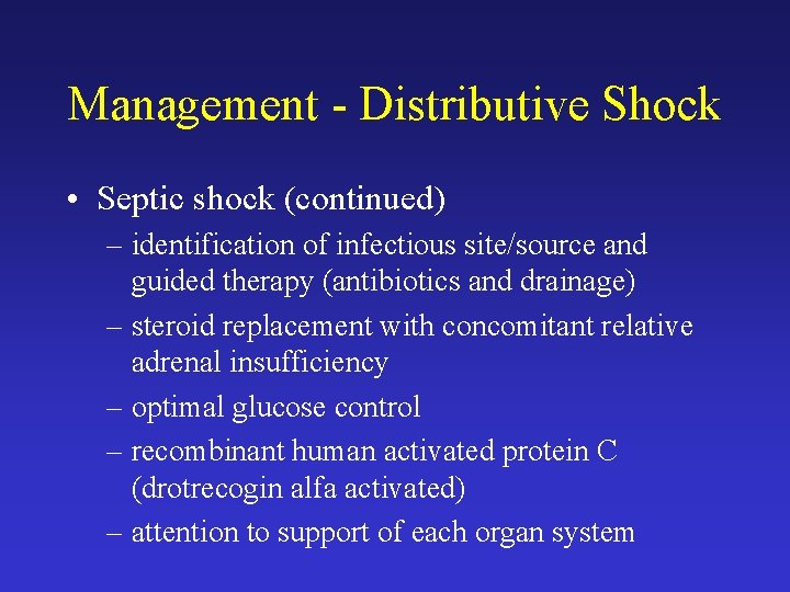
Management - Distributive Shock • Septic shock (continued) – identification of infectious site/source and guided therapy (antibiotics and drainage) – steroid replacement with concomitant relative adrenal insufficiency – optimal glucose control – recombinant human activated protein C (drotrecogin alfa activated) – attention to support of each organ system
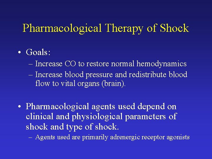
Pharmacological Therapy of Shock • Goals: – Increase CO to restore normal hemodynamics – Increase blood pressure and redistribute blood flow to vital organs (brain). • Pharmacological agents used depend on clinical and physiological parameters of shock and type of shock. – Agents used are primarily adrenergic receptor agonists
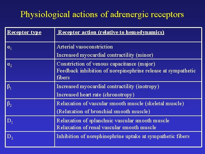
Physiological actions of adrenergic receptors Receptor type Receptor action (relative to hemodynamics) α 1 Arterial vasoconstriction Increased myocardial contractility (minor) α 2 Constriction of venous capacitance (major) Feedback inhibition of norepinephrine release at sympathetic fibers β 1 Increased myocardial contractility (inotropy) Increased heart rate (chronotropy) β 2 Relaxation of vascular smooth muscle (skeletal muscle) (Relaxation of bronchial smooth muscle) D 1 Relaxation of splanchnic vascular smooth muscle Relaxation of renal vascular smooth muscle D 2 Inhibition of norephinephrine uptake at sympathetic fibers
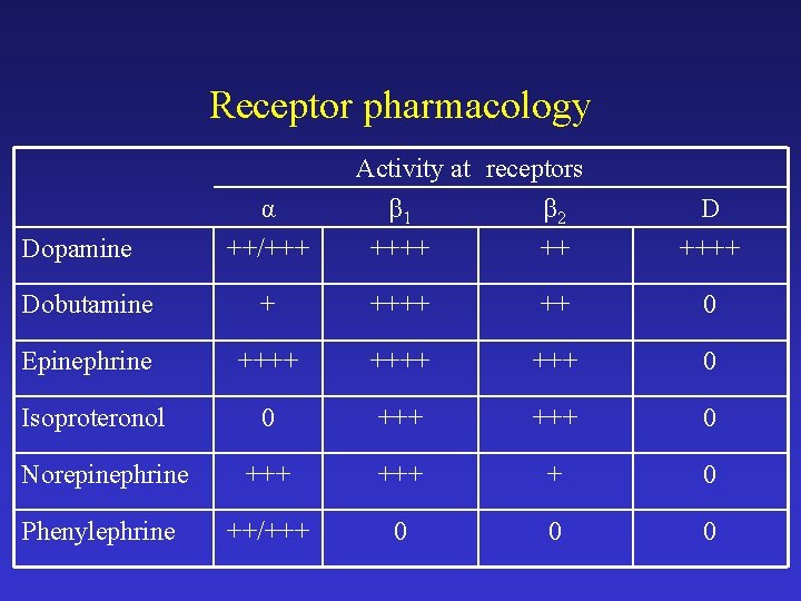
Receptor pharmacology Dopamine α ++/+++ Activity at receptors β 1 β 2 ++++ ++ D ++++ Dobutamine + ++++ ++ 0 Epinephrine ++++ +++ 0 Isoproteronol 0 +++ 0 Norepinephrine +++ + 0 Phenylephrine ++/+++ 0 0 0
Dopamine • Pharmacodynamics – Low and moderate doses – primarily binds dopaminergic and β 1 receptors – Higher doses – α 1 receptors stimulated • Clinical effects – Increase heart rate (chronotropy) and contractility (inotropy) – Increase SVR by arteriolar vasoconstriction (higher doses).
Dopamine • Clinical application – Most commonly used vasopressor • common starting dose – 5 μg/kg/min – Cardiogenic or distributive shock (shock of any etiology) • Adverse effects – – Tachyarrhythmias, excessive tachycardia Precipitate myocardial ischemia Excessive vasoconstriction (digital ischemia) Nausea and vomiting (central effects)
Dobutamine • Pharmocodynamics – Primarily stimulates β 1 receptors – Lesser binding to β 2 and α receptors • Clinical effects – Increase CO (primarily inotropic effect) – SVR without significant change or a decline – Starting dose – 2 -5 μg/kg/min
Dobutamine • Clinical application – Cardiogenic shock and congestive heart failure • Common starting dose – 2 -5 μg/kg/min – Sometimes used in distributive shock (when CO is normal or low which can occur in septic shock) • Adverse effects – Tachycardia and tachyarrhythmias – Myocardial ischemia
Epinephrine • Pharmacodynamics – Extremely potent, high affinity binding of all α and β receptors. • Clinical effects – Increase CO – Increase SVR by vasoconstriction • High doses cause prominent vasoconstriction
Epinephrine • Clinical applications – Refractory shock of all types – starting at 0. 2 μg/kg/min – Cardiopulmonary resuscitation – 1 mg IV push – Anaphylaxis – 0. 2 -0. 5 ml of 1: 1000 dilution subcutaneously • Adverse effects – – Excessive vasoconstriction Tachycardia and tachyarrhythmias Myocardial ischemia Hyperglycemia
Isoproteronol • Pharmacodynamics – β 1 and β 2 receptor agonist – No α effects • Clinical effects – Increased CO – Decreased SVR – Variable effect on blood pressure
Isoproteronol • Clinical application – Not useful for shock because of prominent β 2 effect and lack of α – may decrease MAP – Rare uses to increase heart rate (as in Torsade de Pointes)
Norepinephrine • Pharmacodynamics – β 1, α 2 receptor agonist – Low doses (< 0. 03 μg/kg/min) have prominent β 1 effect – Higher doses (> 0. 06 μg/kg/min) stimulate α receptors • Clinical effects – Increase CO – Increase SVR
Norepinephrine • Clinical applications – Distributive shock – Profound, refractory shock of any type • Adverse effects – Excessive vasoconstriction – Arrhythmia – Myocardial ischemia
Phenylephrine • Pharmacodynamics – α 1 and α 2 receptor agonist – Lacks β effect • Clinical effects – Increased SVR – Starting dose - 30 μg/min
Phenylephrine • Clinical application – Distributive shock (especially if very high heart rate or history of tachyarrhythmia) – Less commonly used than norepinephrine • Adverse effects – Excessive vasoconstriction – Reflex bradycardia
Nonadrenergic Vasopressors and Inotropes • Vasopressin – antidiuretic hormone (ADH) analoge – can be used as a second line pressor agent – recent studies suggest a relative deficiency of ADH in sepsis – often added at a fixed rate of 0. 04 units/min
Nonadrenergic Vasopressors and Inotropes • Phosphodiesterase inhibitors – amrinone and milrinone – have inotropic and vasodilatory actions – effects are similar to dobutamine – used in cardiac failure
Summary • Treat shock as an emergency • Remember the physiology that governs shock - your goal in treatment is to intervene appropriately to help correct the variable that is driving the shock state.