Pulmonary Embolism Prepared by Dr Sanaa Saber Mohamed

Pulmonary Embolism Prepared by Dr/ Sanaa Saber Mohamed Critical care nursing department
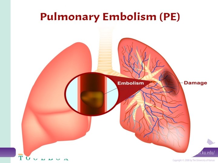

Introduction • Pulmonary embolism occurs when a thrombus, which has broken loose and migrated to the pulmonary arteries, obstructs part of the pulmonary vascular tree. A pulmonary embolism usually results from a deep vein thrombosis (DVT) that has formed in the lower extremities.

Definition: • A pulmonary embolism is a blockage in one of the blood vessels in the lungs. It happens when part, or all, of a blood clot blocks the blood supply to the lungs, • Pulmonary embolism is a clot (thrombotic emboli) or fat, tumors, amniotic fluid, air and foreign bodies (non thrombotic emboli) lodges in the pulmonary arterial (PA) system disrupting the blood flow to a region of the lungs.
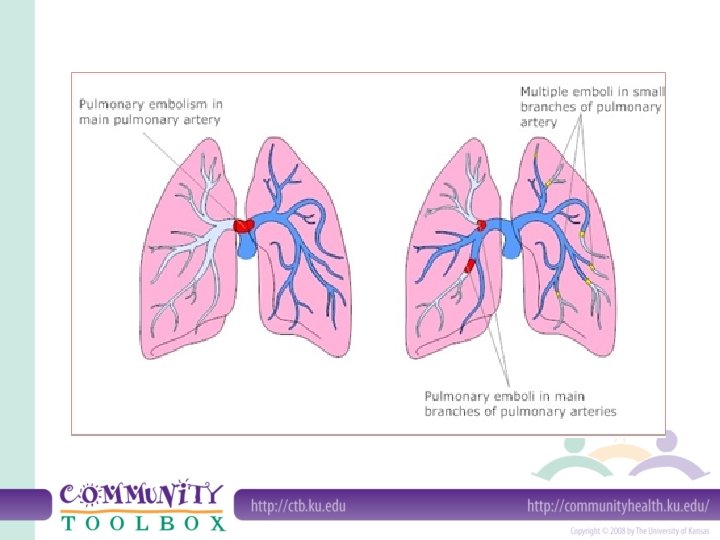

Types of embolism according to cause • Pulmonary embolism: usually formed in the leg (known as a deep vein thrombosis or DVT), lodges in one of the arteries of the lungs. • Brain embolism: If a blood clot travels to the brain, this causes an ischemic stroke or TIA (transient ischemic attack). • Retinal embolism: Small clots can block the smaller blood vessels feeding the retina at the back of the eye. The result is usually sudden blindness in one eye. • Septic embolism: caused by infection in the body reach the bloodstream and block blood vessels.

• Amniotic embolism: . In pregnancy, the Amniotic fluid can embolize and reach the mother's lungs, causing pulmonary amniotic embolism. • Air embolism: Scuba divers who rise to the surface too rapidly can generate air embolism, bubbles in the blood that can block arterial blood flow. • Fat embolism: If fat or bone marrow particles are introduced into the blood circulation, they may block blood vessels the way a blood clot or air bubble can.

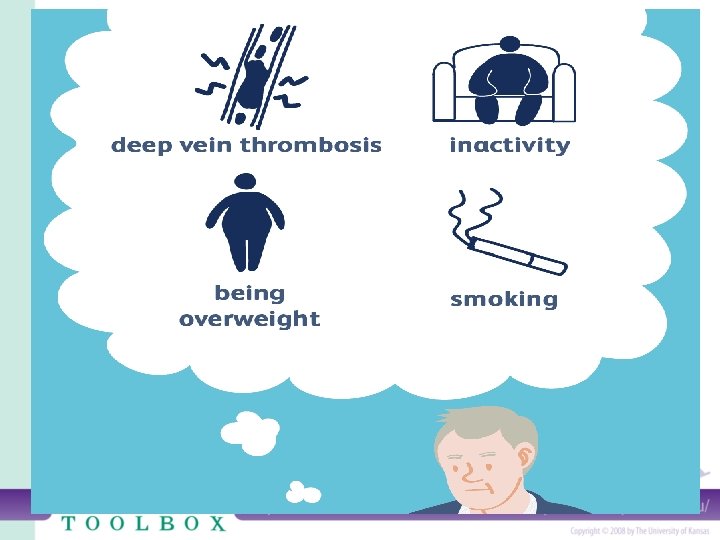
Risk factors: – Cardiovascular disease, specifically heart failure. – Cancer: especially pancreatic, ovarian and lung cancers. – Surgery: for this reason, medication to prevent clots may be given before and after major surgery such as joint replacement. – Bed rest: when the lower extremities are horizontal for long periods, the flow of venous blood slows and blood can pool in the legs. – Smoking: tobacco leads to blood clot formation. – Obesity : excess weight increases the risk of blood clots – Sedentary lifestyle – Supplemental estrogen. – Pregnancy: the weight of the baby pressing on veins in the pelvis can slow blood return from the legs.
_ Small to Moderate Embolus ü Chest pain -")
Clinical manifestation of pulmonary embolism a)_ Small to Moderate Embolus ü Chest pain - sharp, stabbing pain that might become worse when breathing ü Increased or irregular heartbeat ü Dyspnea ü Diaphoresis - Wheezing ü Dizziness ü Difficulty breath - this could occur suddenly, or slowly develop over time ü Rapid breathing ü Cough - normally a dry cough but sometimes blood, or mucus containing blood may be coughed up.
- Massive embolus (above manifestation plus the following) § Cyanosis § Restlessness § Anxiety")
b)- Massive embolus (above manifestation plus the following) § Cyanosis § Restlessness § Anxiety § Confusion § Cool, clammy skin § Decreased urinary output
_ Signs of Pulmonary Embolism in Intensive Care Patients o Worsening hypoxemia or hypocapnia")
c)_ Signs of Pulmonary Embolism in Intensive Care Patients o Worsening hypoxemia or hypocapnia in a patient on spontaneous ventilation o Worsening hypoxemia and hypercapnia in a sedate patient on controlled mechanical ventilation o Worsening dyspnea, hypoxemia, and a reduction in Pa. CO 2 in a patient with chronic lung disease and known carbon dioxide retention o Unexplained fever o Sudden elevation in pulmonary artery pressure or central venous pressure in a hemodynamically monitored patient.

Diagnostic tests of pulmonary embolism Ø Physical exam. Ø History for the previous risk factors Ø ECG. Ø Arterial blood gases Ø Chest X-ray. Ø Pulmonary angiography. Ø Doppler ultrasound (to rule out DVT).

Medical treatment of pulmonary embolism § Anticoagulant therapy with low-dose or adjusted-dose heparin, LMW heparin. § Thrombolytic therapy as streptokinase. § Elastic stoking or intermittent pneumatic leg compression devices. § Surgical intervention (pulmonary embolectomy).

Collaborative management • Administer oxygen therapy, Intubate patient, Initiate mechanical ventilation • Administer medication As bronchodilators, intropic agents , Sedatives, and analgesics • Administer fluids • Position patient to optimize ventilation/perfusion matching • Maintaining surveillance for complications such as ( Bleeding , ARDS)

Nursing care plan • Impaired gas exchanged related to decrease pulmonary perfusion • Acute pain related to transmission and perception of cutaneous, visceral, muscular, or ischemic impulses • Impaired gas exchange related to ventilation /perfusion mismatching • Anxiety related to threat biological, psychologic, and /or social integrity

Patient education for preventing recurrent embolism § § § § § Apply elastic stoking. Elevation of the legs Active/passive range of motion (ROM) exercises Adequate hydration Avoid crossing, dangling of the legs. Avoid sitting or lying for prolonged periods. Avoid wearing of constricting clothes. Do not left the IV lines for prolonged periods. Do not take aspirin or any drugs without doctor's order. When traveling, change your position frequent early

Complications of pulmonary embolism Ø Sudden cardiac death Ø Obstructive shock Ø Pulseless electrical activity Ø Atrial or ventricular arrhythmias Ø Secondary pulmonary arterial hypertension Ø Cor pulmonale Ø Severe hypoxemia Ø Right-to-left intracranial shunt Ø Lung infarction Ø Pleural effusion

- Slides: 19