Regulation of Respiration Prof dr Zoran Vali Department


 2) 3) several groups of neurons in the medulla oblongata")
 2) 3) control of inspiration & respiratory rhythm")

 it begins")
 2) control of the rate of increase of the ramp signal control of")

 2) 3) 4) RVLM; nucleus ambiguus & retroambiguus inactive")


 & aortic bodies (vagus) – dorsal")


 2) q chemical changes –")

")


 2) it takes few seconds")


- Slides: 61
Regulation of Respiration Prof. dr. Zoran Valić Department of Physiology University of Split School of Medicine
q q q nervous system normally adjusts the rate of alveolar ventilation almost exactly to the demands of the body oxygen pressure (PO 2) and carbon dioxide pressure (PCO 2) in the arterial blood are hardly altered heavy exercise!
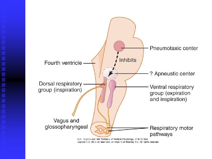
Respiratory Center q 1) 2) 3) several groups of neurons in the medulla oblongata and pons of the brain stem dorsal respiratory group (inspiration) ventral respiratory group (mainly expiration) pneumotaxic center (dorsally in the pons, controls rate and depth of breathing)
Dorsal Respiratory Group q q 1) 2) 3) control of inspiration & respiratory rhythm nucleus of the tractus solitarius and reticular substance (vagus and glossopharyngeus) peripheral chemoreceptors baroreceptors in the lungs
Rhythmical Inspiratory Discharges q q q transection experiment ( repetitive bursts of inspiratory neuronal action potentials) cause is unknown – prof. Đogaš! neural networks, neurons from adjacent areas of the medulla
Inspiratory "Ramp" Signal q q q transmitted to the inspiratory muscles (diaphragm) it begins weakly and increases steadily in a ramp manner for about 2 s, ceases abruptly for approximately the next 3 s “ramp signal” – causes a steady increase in the volume of the lungs during inspiration, rather than inspiratory gasps
1) 2) control of the rate of increase of the ramp signal control of the limiting point at which the ramp suddenly ceases – shortening of inspiration (shortening of expiration – increasing frequency of respiration)
Pneumotaxic Center q q q located dorsally in the nucleus parabrachialis of the upper pons control the "switch-off" point of the inspiratory ramp (0, 5 -5 s) limit inspiration, increasing the rate of breathing (from 3 -5 to 30 -40 breaths per minute)
Ventral Respiratory Group q 1) 2) 3) 4) RVLM; nucleus ambiguus & retroambiguus inactive during normal quiet respiration do not appear to participate in the basic rhythmical oscillation increased respiratory drive both inspiration and expiration (abdominal muscles)
The Hering-Breuer Inflation Reflex q q q sensory nerve signals from the lungs stretch receptors (muscular portions of the walls of the bronchi and bronchioles – vagus) "switches off" the inspiratory ramp – increases the rate of respiration
q q in humans not activated until the tidal volume increases to more than three times normal (VT > 1. 5 L per breath) protective mechanism for preventing excess lung inflation
Chemical Control of Respiration q q q ultimate goal of respiration is to maintain proper concentrations of O 2, CO 2 & H+ in the tissues CO 2 or H+ act directly on the respiratory center O 2 does not have a significant direct effect – acts almost entirely on peripheral chemoreceptors (carotid and aortic bodies)
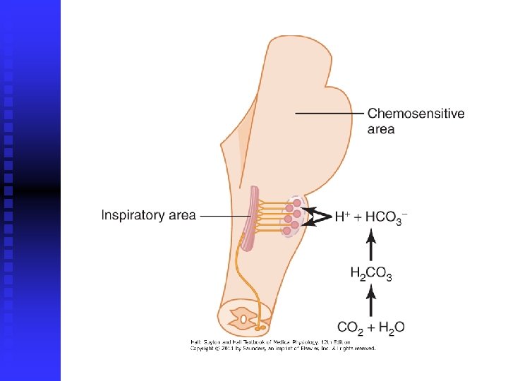
Direct Chemical Control q q q additional neuronal area – chemosensitive area located bilaterally, lying only 0. 2 millimeter beneath the ventral surface of the medulla highly sensitive to changes in either blood PCO 2 or H+ concentration
Influence of q q + H may be the only important direct stimulus for chemosensitive neurons however, H+ do not easily cross the bloodbrain barrier – less effect than PCO 2
Influence of PCO 2 q q q little direct effect in stimulating the neurons in the chemosensitive area potent indirect effect – action through H+ ions blood-brain barrier almost do not exist for CO 2
Decreased Stimulatory Effect of CO 2 q q CO 2 excites respiratory center greatly the first few hours after the PCO 2 first increases, but then it gradually declines over the next 1 to 2 days to 1/5 the initial effect action of the kidneys (increased levels of HCO 3 -) – in blood, cerebrospinal fluid and intercellular fluid
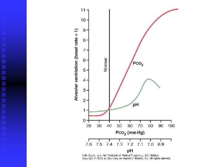
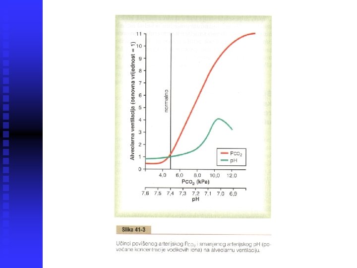
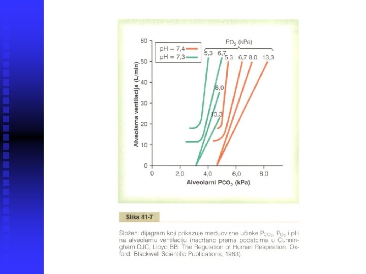
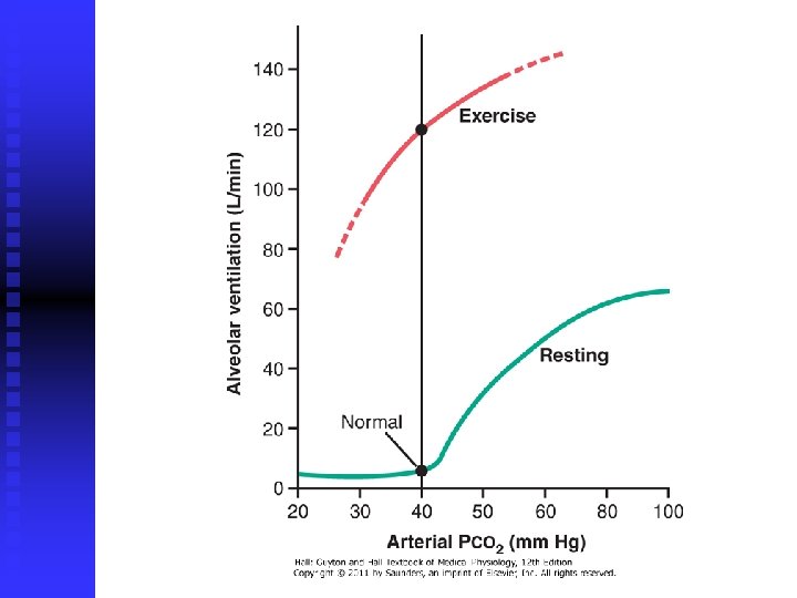
Quantitative Effects of PCO 2 and H+ on Alveolar Ventilation q q normal range of PCO 2 in blood is between 35 and 75 mm Hg (4. 5 – 10. 0 k. Pa) normal range of p. H: 7. 3 – 7. 5
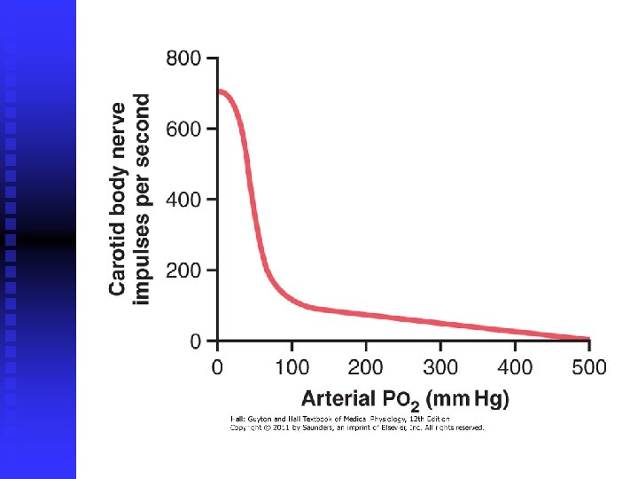
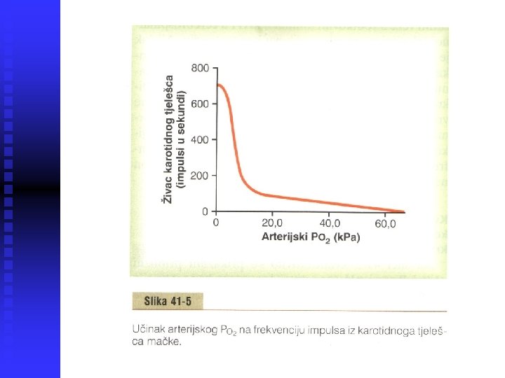
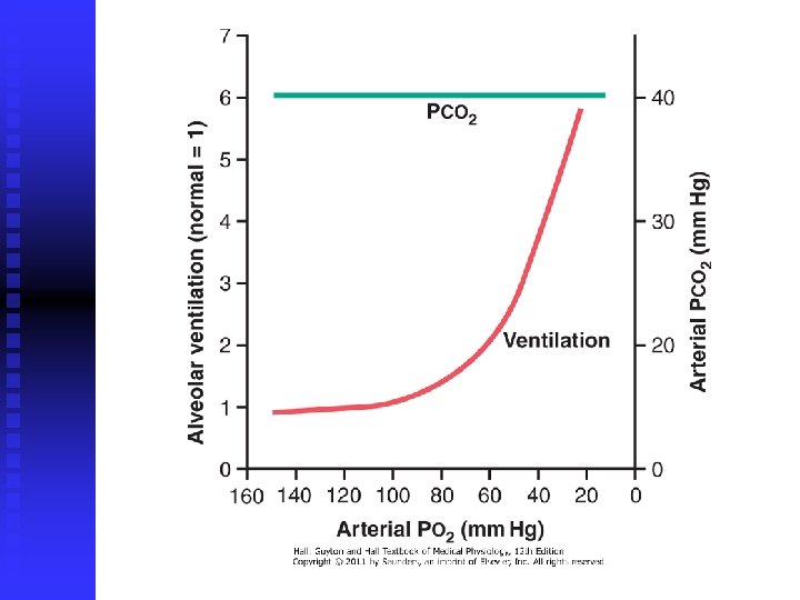
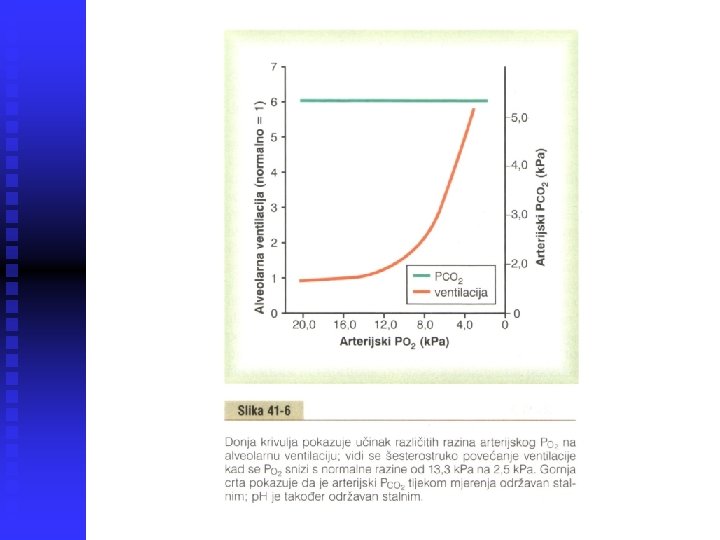
Direct Effect of O 2 q q q virtually no direct effect on the respiratory center very effective hemoglobin-oxygen buffer system (from 60 to 1000 mm. Hg) in special conditions with lack of oxygen – peripheral chemoreceptors (PO 2 below 70 mm. Hg)
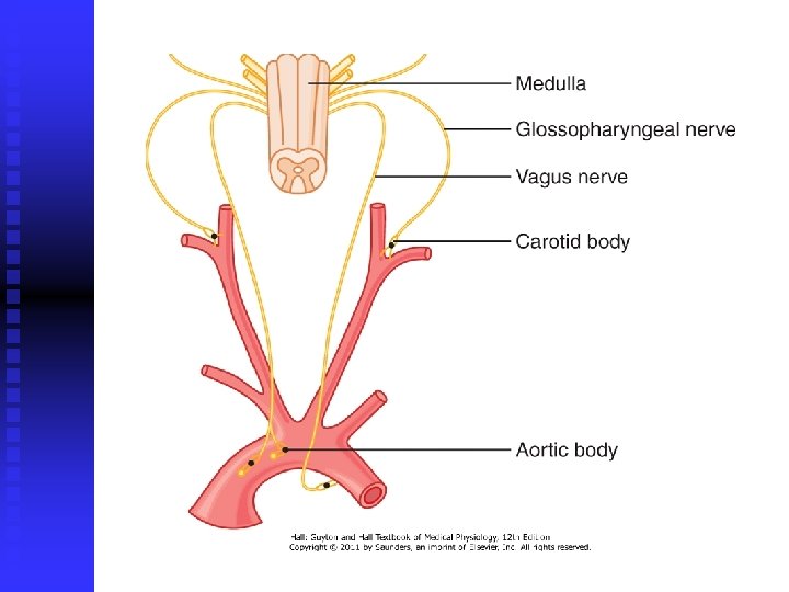
Peripheral Chemoreceptor System – Role of Oxygen q q special nervous chemical receptors – chemoreceptors several areas outside the brain changes in O 2, CO 2 and H+ transmit nervous signals to the respiratory center
q q carotid bodies (Hering's nerves, glossopharyngeal nerves) & aortic bodies (vagus) – dorsal medullary respiratory area minute artery, blood flow 20 times the weight of the bodies themselves (percentage of O 2 removed from the flowing blood is virtually zero)
Influence of CO 2 & H+ on Chemoreceptors q q q also excite the chemoreceptors direct effects of both these factors in the respiratory center itself are much more powerful (7 x) 5 x as rapidly as central stimulation – onset of exercise!
Basic Mechanism of Stimulation of the Chemoreceptors by Oxygen q q q the exact means are still unknown! glomus cells (glandular-like cells) – synapse directly or indirectly with the nerve endings opposing opinions (nerve endings might function as the chemoreceptors)
Phenomenon of Acclimatization q q within 2 to 3 days, the respiratory center loses about 4/5 of its sensitivity to changes in PCO 2 and H+ instead of 70 percent increase (acute), alveolar ventilation increases 400 to 500 percent after 2 to 3 days of low oxygen
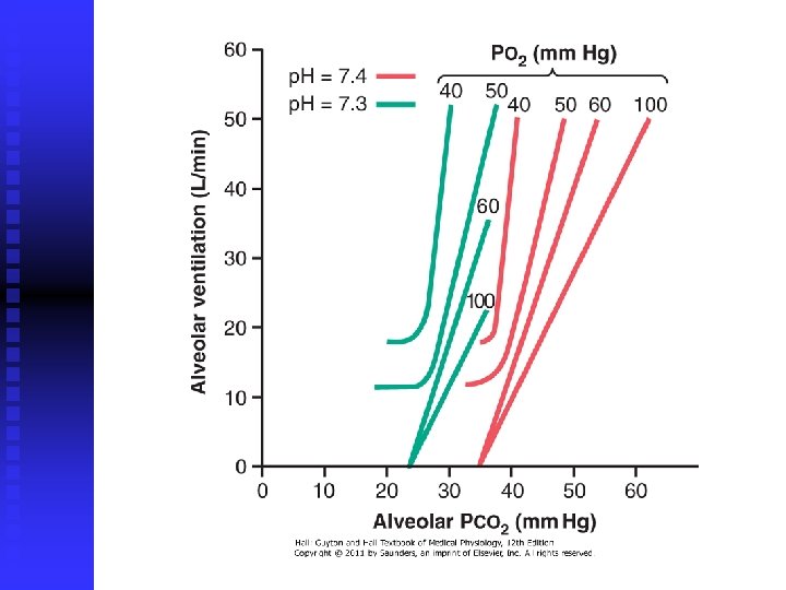
Composite Effects of PCO 2, p. H & PO 2 on Alveolar Ventilation
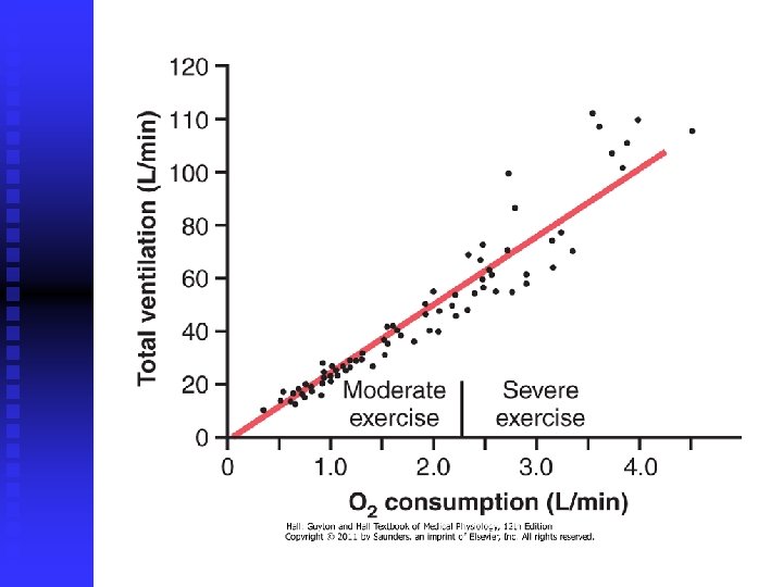
Regulation of Respiration During Exercise q q q O 2 consumption and CO 2 formation can increase as much as 20 -fold alveolar ventilation ordinarily increases almost exactly in step with the increased level of oxygen metabolism arterial PO 2, PCO 2, and p. H remain almost exactly normal
What causes the increased ventilation during exercise? q 1) 2) q chemical changes – NO collateral impulses into the brain stem to excite the respiratory center (analogous to the increase in arterial pressure) excitation of proprioreceptors in joints and muscle – excitation of respiratory center (passive movements & severed nerves) hypoxic muscles, variations in PO 2, PCO 2
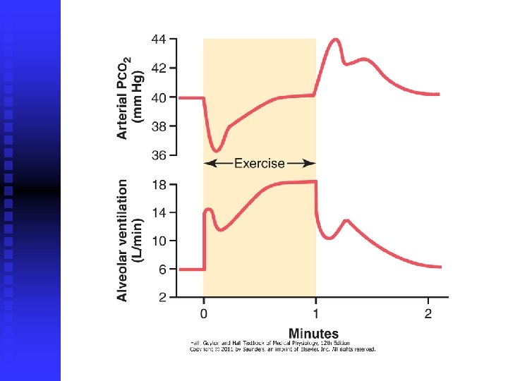
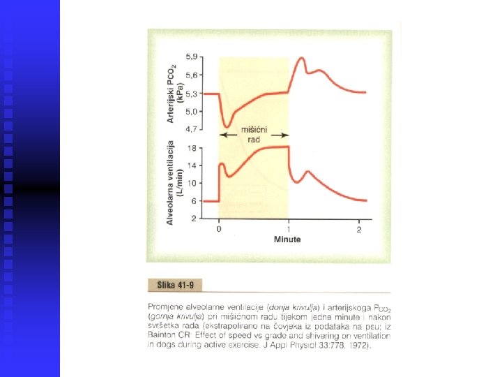
Interrelation Between Chemical and Nervous Factors q q q occasionally the nervous respiratory control signals are either too strong or too weak chemical factors play a significant role in the final adjustment of respiration increase in alveolar ventilation – decreases arterial PCO 2 below normal at the onset of exercise
- neurogeni pomak ventilacijske krivulje - zadržavanje oblika krivulje (funkcije)
q q brain's ability to shift the ventilatory response curve during exercise is at least partly a learned response with repeated periods of exercise, the brain becomes progressively more able to provide the proper signals
q even the cerebral cortex is involved in this learning because experiments that block only the cortex (anesthesia) also block the learned response
Voluntary Control of Respiration q q one can hyperventilate or hypoventilate to such an extent that serious derangements in PCO 2, p. H, and PO 2 can occur in the blood directly from the cortex or other higher centers
Effect of Irritant Receptors in the Airways q q epithelium of the trachea, bronchi, and bronchioles is supplied with sensory nerve endings called pulmonary irritant receptors cause coughing and sneezing
Function of Lung "J Receptors" q q q few sensory nerve endings in the alveolar walls in juxtaposition to the pulmonary capillaries stimulated when the pulmonary capillaries become engorged with blood or when pulmonary edema occurs (heart failure) their excitation may give the person a feeling of dyspnea
Effect of Brain Edema q q q depressed or even inactivated respiratory center by acute brain edema resulting from brain concussion therapy: intravenous injection of hypertonic solutions such as highly concentrated mannitol solution
Anesthesia q q overdosage with anesthetics or narcotics – respiratory arrest sodium pentobarbital, halothane & morphine
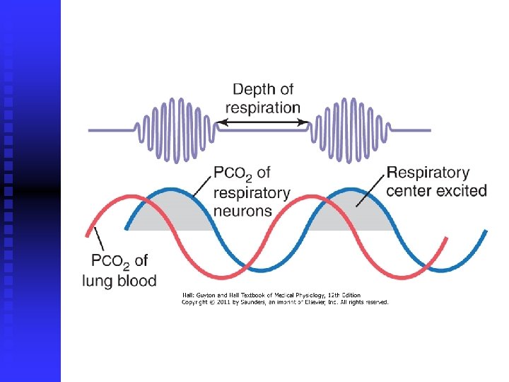
Periodic Breathing q q q occurs in a number of disease conditions person breathes deeply for a short interval and then breathes slightly or not at all for an additional interval most common – Cheyne-Stokes breathing (slowly waxing and waning respiration occurring about every 40 to 60 seconds)
Basic Mechanism of Cheyne. Stokes Breathing q q 1) 2) it takes few seconds before the pulmonary blood can be transported to the brain under normal conditions, this mechanism is highly "damped" long delay occurs for transport of blood from the lungs to the brain (severe cardiac failure) increased negative feedback gain (brain damage – a prelude to death)
Sleep Apnea q q q apnea – absence of spontaneous breathing occasional apneas occur during normal sleep in persons with sleep apnea – frequency and duration are greatly increased occurring 300 to 500 times each night, lasting for 10 seconds or longer obstruction of the upper airways or impaired CNS respiratory drive
Obstructive Sleep Apnea q q caused by blockage of the upper airway muscles of the pharynx normally keep passage open during sleep, these muscles usually relax the airway passage remains open enough to permit adequate airflow
q q some individuals have an especially narrow passage, and relaxation of these muscles during sleep causes the pharynx to completely close so that air cannot flow into the lungs loud snoring and labored breathing occur apnea PO 2, PCO 2 great stimulation of respiration fragmented, restless sleep excessive daytime drowsiness & increased sympathetic activity
in older, obese persons nasal obstruction, a very large tongue, enlarged tonsils, or shapes of the palate therapy: q q q 1) 2) surgical removal of excess fat tissue at the back of the throat (uvulopalatopharyngoplasty), enlarged tonsils or adenoids, or creation of opening in the trachea (tracheostomy) to bypass the obstructed airway during sleep nasal ventilation with continuous positive airway pressure (CPAP)
"Central" Sleep Apnea central nervous system drive to the ventilatory muscles transiently ceases causes: q q q damage to the central respiratory centers abnormalities of the respiratory neuromuscular apparatus may have decreased ventilation when they are awake, although they are fully capable of normal voluntary breathing caused by stroke