RECURRENCE AFTER TRANSPLANT SOME HAPPY ENDINGS AND SOME


 • USG KUB s/o B/L small, shrunken kidneys ( RK 7. 3")
 • Worked up for renal transplant; his father underwent pre-transplant work up")
 • Feb 2018 - c/o on & off fever, decreased appetite and")

 • Blood C/S – no growth • Urine C/S – no growth")


 with 18% glomerular crescents. Diagnosis: Ig.")


 for 3 days, f/b oral")
 • A 14 y/ male/ normal birth and")
 • USG KUB S/O bilateral small, shrunken kidneys ( RK 5. 8")
 • His grandmother underwent pre-transplant work up and was found to be")
 • USG of Transplanted Kidney size : 9. 8 x 5. 4")

 • ? Acute rejection")


- Slides: 45
RECURRENCE AFTER TRANSPLANT: SOME HAPPY ENDINGS AND SOME NOT-SO-HAPPY ONES ! Dr. Adel Moideen IInd Year Resident Department of Nephrology Dr. D Y Patil Medical College Pune
CASE - 1 HISTORY • 24 y/ Male/ Nov 2014 - C/o giddiness, nausea and vomiting, DOE, pedal edema and decreased U. O • On presentation, Ø BP 160/100 mm of Hg Ø Urea-80 mg/dl, S. Creat 4. 0 mg/dl; Ø Urine R/M- Protein-+++, RBC-2 -3/ hpf, Pus cells-0 -1/hpf, UPCR-2. 5; Ø viral markers and autoimmune workup - negative; Ø Opthalmology & ENT evaluation-normal.
HISTORY (Contd…) • USG KUB s/o B/L small, shrunken kidneys ( RK 7. 3 x 3. 5 cm & LK 8. 0 x 3. 7 cm) with loss of CMD. • Diagnosed with CKD stage V/ ? CGN; maintained on conservative management. • Lost for F/U; Jan 2015 - dyspnea, hypervolemia, pedal edema and HTN • Urea-164 mg/dl and S. Creat -9 mg/dl • Initiated on hemodialysis; thrice weekly maintenance hemodialysis/ Right IJV tunneled perm-cath as access.
HISTORY (Contd…) • Worked up for renal transplant; his father underwent pre-transplant work up and was found to be fit as donor. • Underwent live related renal transplant on 08/06/2015 (3/6 mismatch) • No induction (financial reasons); On Triple immunosuppresion therapy : Tacrolimus, Mycophenolate sodium, Prednisolone. • Discharged on POD-10 with baseline S. Creat of 1. 1 mg/dl. • Asymptomatic for next 2 years with stable S. Creat and Tac levels; but lost for F/U again for the next 7 months.
HISTORY (Contd…) • Feb 2018 - c/o on & off fever, decreased appetite and generalised weakness since 15 days (unreliable drug compliance). • O/E - Conscious, Oriented, afebrile • PR- 84/min, B. P. - 140/80 mm Hg, RR-20/min • No Pallor, edema, icterus • Transplanted kidney was palpable with scar mark seen in right iliac fossa. • Systemic examination- unremarkable
INVESTIGATIONS • Hb-12. 5 gm%, TLC-10000 cells/cumm, Platelet-2. 3 lakhs/cumm • Blood Urea-89 mg/dl, S. Creat- 5. 8 mg/dl, S. Na-142 mmol/l, S. K-4. 2 mmol/l • S. Uric acid – 7. 8 mg/dl, S. PO 4– 5. 7 mg/dl, S. Ca- 7. 6 mg/dl • Urine R/M- Protein-+++, RBC-6 -8/ hpf, Pus cells-2 -3/hpf; • UPCR- 0. 8
INVESTIGATIONS (Contd…) • Blood C/S – no growth • Urine C/S – no growth • CMV load – Normal • B K Virus load – Normal • Tac level-3. 4 ng/ml • USG of Transplanted Kidney size : 11. 4 x 4. 3 cm, normal echogenicity & CMD maintained and normal graft vessel doppler.
DIFFERENTIAL DIAGNOSIS • ? Chronic allograft nephropathy • ? Recurrence of primary disease • ? CNI toxicity
• Patient underwent graft kidney biopsy in March 2018 • Light microscopy: Ø 11 glomeruliv 2 glomeruli - globally sclerosed v 2 glomeruli - Fibrous crescents v 1 glomerulus - segmental sclerosis vremaining glomerular tuft - mild mesangial matrix expansion with mesangial hypercellularity. Ø Diffuse acute tubular injury, moderate interstitial inflammation and interstitial edema.
Ø Few interstitial blood vessels show moderate medial thickening. Ø No glomerulitis or chronic glomerulopathy/ No necrotizing lesion seen/ No peritubular capillaritis. Ø No evidence of BKV nephropathy or CNI toxixity .
Immunofluorescence: ØIg. G: Negative ØIg. A: 3+ granular in mesangium ØIg. M: Negative ØC 3: Negative ØC 1 q: Negative ØKappa: 2+ granular in mesangium ØLambda: 2+ granular in mesangium ØC 4 d: Negative in peritubular capillaries (C 4 d 0) Immunohistochemistry: ØSV 40 -Negative
Overall features are of Ig. A nephropathy (denovo/recurrent) with 18% glomerular crescents. Diagnosis: Ig. A nephropathy (denovo/recurrent) with 18% glomerular crescents.
H & E Stain Mesangial proliferation and focal matrix expansion can be seen. Cellular crescent with partial collapse of glomerular tuft.
Silver Stain Mild to moderate mesangial hypercellularity can be seen.
MT Stain Mesangial proliferation and matrix expansion.
TREATMENT • IV Pulse therapy (methylpred 1 gm ) for 3 days, f/b oral pred 40 mg/day • Triple immunosuppressants continued. • Underwent 5 cycles of Plasmapheresis and received 3 doses of IV CYC 500 mg each. • Was on regular F/U and asymptomatic with S. Creat maintained (btw 2 -3 mg/dl x 5 months). • Again lost for F/U for 3 months and this time when he presented, he had been already initiated on maintenance HD outside since the last 1 month
CASE - 2 • HISTORY (Contd…) • A 14 y/ male/ normal birth and development history/ April 2018 - c/o nausea and vomiting, DOE, decreased appetite & generalised weakness, facial puffiness & pedal edema and decreased U. O • On presentation, Ø BP -144/96 mm of Hg. Ø Urea-128 mg/dl, S. Creat-7. 2 mg/dl Ø Urine R/M- Protein-+++, RBC-0 -1/ hpf, Pus cells-0 -1/hpf, UPCR-4. 8 Ø viral markers and autoimmune workup – negative Ø Opthalmology & ENT evaluation-normal.
HISTORY (Contd…) • USG KUB S/O bilateral small, shrunken kidneys ( RK 5. 8 x 2. 5 cm & LK 5. 5 x 3. 0 cm) with loss of CMD. • He was initiated on Hemodialysis; thrice weekly maintenance HD with Right IJV tunnelled permcath as access. • Diagnosed with CKD stage Vd/ ? CGN- ? FSGS • He tolerated HD well with interdialytic weight gain of 0. 5 -1 kg.
HISTORY (Contd…) • His grandmother underwent pre-transplant work up and was found to be fit as donor. • Underwent live related renal transplant on 27/08/2018 (6/6 mismatch) • ATG induction (TOTAL-150 mg) ; On Triple immunosuppresion therapy : Tacrolimus, Mycophenolate sodium, Prednisolone. • Developed fever with increasing facial puffiness, B/L lower limb swelling, scrotal swelling and decreasing U. O since POD 3 (Serum creatinine-1. 1 mg/dl).
HISTORY (Contd…) • USG of Transplanted Kidney size : 9. 8 x 5. 4 cm, normal echogenecity & CMD maintained with normal graft vessel and lower limb doppler. • O/E - Conscious, Oriented, afebrile • PR- 114/min, B. P. - 134/90 mm Hg, RR-24/min, • No Pallor, icterus. B/L lower limb and scrotal edema+ • Transplanted kidney was palpable with scar mark seen in right iliac fossa. • Systemic examination- unremarkable
INVESTIGATIONS • Hb-6. 9 gm%, TLC-6400 cells/cumm, Platelet-1. 8 lakhs/cumm • Blood Urea-25 mg/dl, S. Creat- 0. 7 (POD-2) 1. 1 mg/dl (POD-3), S. Na-134 mmol/l, S. K-3. 6 mmol/l • S. Alb-2. 48 g/dl, S. Uric acid – 6. 6 mg/dl, S. PO 4– 4. 3 mg/dl, S. Ca- 7. 6 mg/dl (corrected-8. 7) • Urine R/M- Protein-++++, RBC-1 -2/ hpf, Pus cells-1 -2/hpf; UPCR- 10. 6 • Blood C/S – no growth; Urine C/S – no growth • S. TAC level-8. 3 ng/ml
DIFFERENTIAL DIAGNOSIS • ? Recurrence of primary disease (? FSGS) • ? Acute rejection
TREATMENT • It was decided to perform a graft biopsy - refused by the relatives. • Given IV Pulse therapy (methylpred 500 mg) for 3 days, f/b oral pred. • Triple immunosuppressants continued. • ACEi added on. • Underwent 5 sessions of plasmapheresis on alternate days with FFP as replacement • Edema began to resolve and repeat urine protein by dipstick was 1+ with UPCR-1. 3.
• Discharged on post op day 14 with a baseline S. Creat of 0. 84 mg/dl and UPCR of 0. 8. • At present, he is on regular F/U and asymptomatic with S. Creat of 0. 7 mg/dl and UPCR of 0. 2 with adequate Wt and Ht gain (6 cm in 8 months).
RECURRENCE AFTER TRANSPLANT
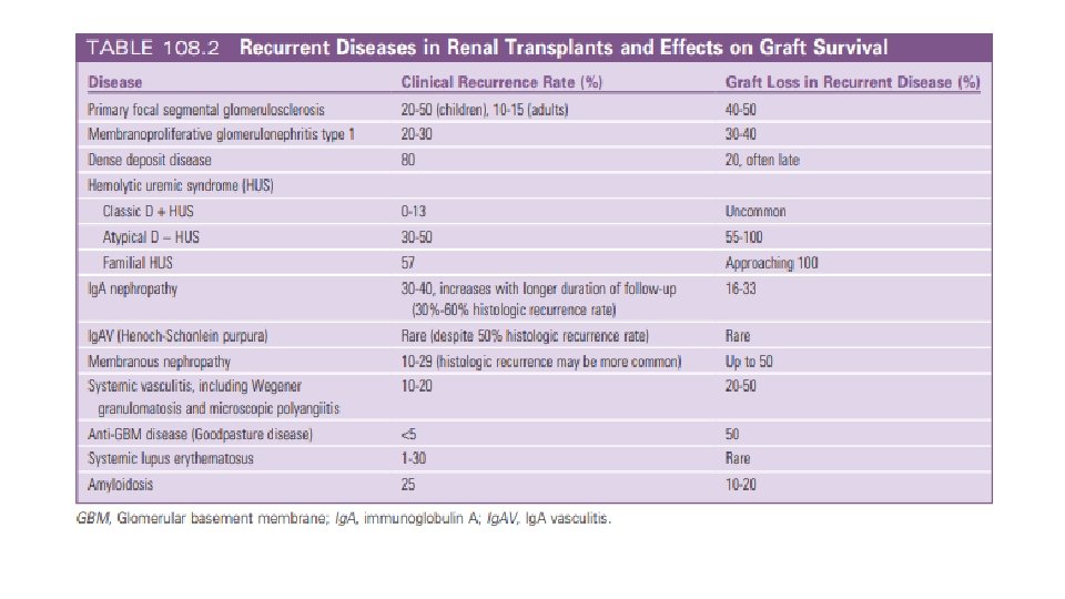
• GN and DM – 2 leading causes of ESRD • As per US Renal Allograft Disease Registry data, prevalence of GN recurrence : -2. 8% at 2 yrs -9. 8% at 5 yrs -18. 5% at 8 yrs of F/U after Tx • As per ANZDATA, biopsy proven recurrence causing graft loss was: -0. 5% within 1 yr -3. 7% within 5 yrs -8, 4% within 10 yrs after Tx
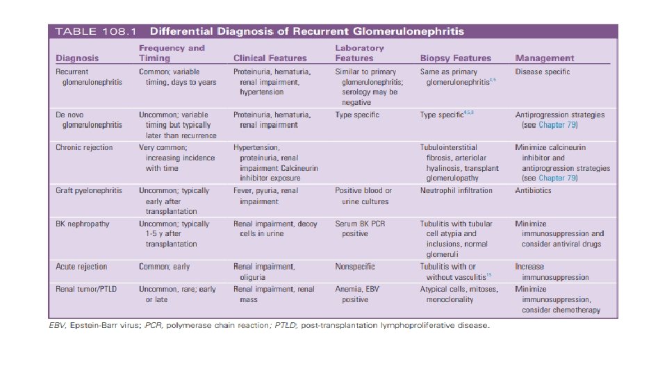
• Recurrent disease: Requires histologic demonstration of the same disease involving both the native & transplanted kidneys. • Incidence underestimated – histologic Dx of primary kidney ds not always obtained and most Tx biopsies are done without keeping recurrent ds in mind. • Recurrent disease – 3 rd most common cause of graft failure beyond 1 st yr after Tx.
Ig. A NEPHROPATHY • M. C form of GN l/t ESRD. • Histologic recurrence – frequent and can occur +/- symptoms; Recurrence - difficult to predict. • Early stages of Ig. AN recurrence - no proteinuria, hematuria, or graft dysfunction. • Published recurrence (8% to 53%)- variation d/t study differences in biopsy indication (protocol or clinical) and length of post-transplant follow-up.
• Risk factorsØ younger age at transplantation Ø rapid progression of the native Ig. AN Ø degree of proteinuria Ø donor factors (presence of Ig. A deposits in donor kidney, HLA matching) • More recurrence in living donor grafts than deceased donors - not confirmed. • 2 Australian registry studies - those with 0 mismatch kidneys have higher rates of recurrence than those with >1 HLA mismatches • ABO-incompatible transplants - lower rates of recurrence (? differences in immunosuppression regimens).
• Graft loss within the first 3 years after transplantationØ Uncommon Ø can occur when Ig. AN in the native kidney was rapidly progressive or after previous graft loss resulting from recurrence. • Long-term graft survival is less for pts with recurrent Ig. AN after a decade. • Other causes of Ig. A deposition: liver disease, celiac disease.
• Choice of immunosuppression after transplantation is controversial. • One large USRDS registry analysis suggested - individual drug choices did not affect the risk for graft loss attributed to recurrence. • In contrast, a retrospective analysis of ANZDATA - continuation of corticosteroids was strongly a/w protection from graft loss caused by recurrence of Ig. AN, although not from other types of GN. • Observational data - induction with ATG may afford relative protection
• Treatment of recurrent Ig. AN and Ig. AV has not been systematically evaluated. • No known preventive measure. • Addition of steroid maintenance in steroid free pts with recurrence > unproven but reasonable. • Non-specific, useful measures – tight BP control, RAS blockade, and avoidance of nephrotoxins • A change to MMF, the use of fish oil, antiplatelet agents, and tonsillectomy – questionable benefit.
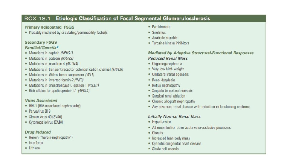
Focal Segmental Glomerulosclerosis • 1500 FSGS pts undergo Tx every year with a 30 -80% risk of recurrence in the new graft. • FSGS accounts for 11% of pediatric kidney transplants.
• Low risk for recurrence – Ø FSGS secondary to vascular disease or reflux nephropathy Ø those with familial or sporadic forms a/w mutation of slit-diaphragm proteins such as podocin (rare cases of post-transplant nephrotic syndrome d/t dev of antibodies against the “neoantigen” within the donor kidney reported).
• High risk - pts with Ø primary FSGS Ø malignant FSGS (heavy proteinuria and renal failure within 3 years of onset) Ø age < 15 yrs Ø mesangial hypercellularity on biopsy Ø with recurrence in a previous graft.
Mophologic variants of FSGS: 1. 2. 3. 4. 5. FSGS NOS FSGS, perihilar FSGS, cellular FSGS, collapsing FSGS, tip
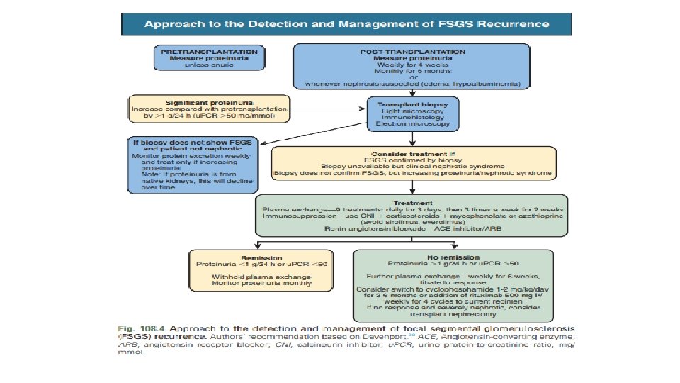
• Rate of recurrence >75% in subsequent grafts when 1 st graft was lost d/t recurrence. • Recurrence – Ø occurs typically within the 1 st month post-transplantation Ø manifests initially with heavy proteinuria, f/b HTN and graft dysfunction. Ø more susceptible to acute rejection and AKI, as well as graft loss. Ø a/w early graft loss in up to 50% of patients Ø Plasmapheresis delays graft loss in most patients and decreases the incidence of overall graft failure.
• Primary FSGS - caused by a circulating factor that targets podocytes (specific factor– unknown). • su. PAR and the costimulatory protein B 7 -1 (CD 80) – ? role in recurrence. • Plasma exchange / immunoadsorption with either a protein A or anti-Ig. G column - effective in pts with recurrent FSGS - removes the circulating permeability factor (disease remission reported in most pts receiving Rx within 2 weeks of recurrence). • So, a course of plasma exchange is warranted in all patients without contraindications.
• Immunosuppression should include a CNI, and addition of an ACEi should be considered. • Pts with incomplete response / relapse -> require repeated / long-term plasma exchange or concurrent treatment with secondary agents such as Ritux or CYC. • For such pts - a course of rituximab 500 mg weekly for 4 weeks + existing immunosuppression and weekly plasma exchange. • Pre-transplantation plasma exchange – controversial role in preventing recurrence. • MCD – rare cause of ESRD; Recurrence after transplantation reported; difficult to be sure that the underlying disease was not FSGS.
Reference No: of Patients Era / Controls No: of Plasmapheresis sessions Result Gonzales et al. 2011 34 pediatric pts 1996 -2007 No controls 1 -10 x 1. 5 exchanges with 5% albumin replacement Not useful Gohh et al. 2005 9 adults and 1 child 1999 -2003 (only high risk pts No controls enrolled; prior recurrence or rapid progression to FSGS) 8 x 1. 0 exchanges with 5% albumin replacement 30% recurrence which they concluded was lower than the reported 52% Ohta et al. 2001 20 japanese children 1 -3 x 1. 5 exchanges with 5% albumin replacement 67% recurred without PP vs 33% with PP 1984 -1991: controls 1991 -1997: cases
THANK YOU !