Polyomaviridae Molecular Virology Introduction Group Ids DNA virus


 • persist as latent infections without causing disease •")

 ( REFERENCE STRAIN ) • Common Type BK")


-linked sialic acids • BFPy.")
")


 •")


.")


 polyomaviruses, KI (Karolinska Institute) and WU (Washington")


- Slides: 42
Polyomaviridae Molecular Virology
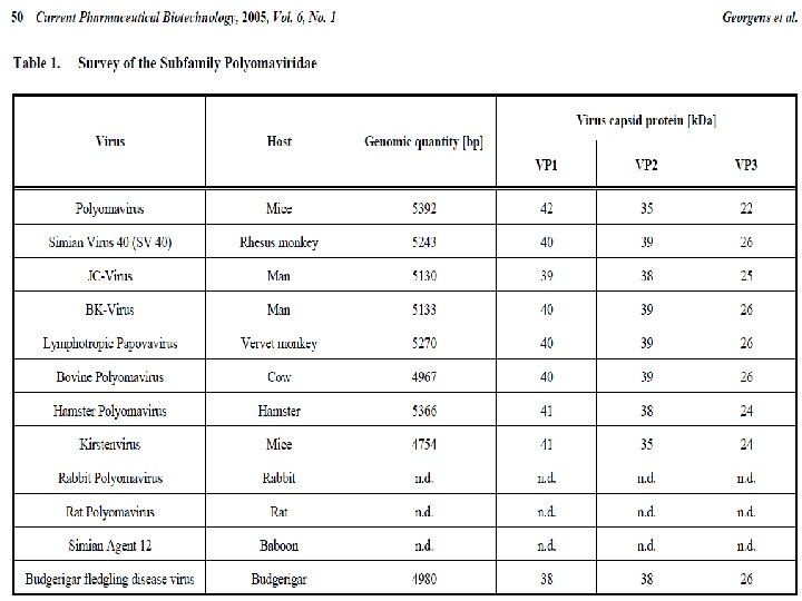
Introduction • Group I:ds DNA virus • Family: Polyomaviridae • Genus: Polyomavirus – Diameter: 40 -50 nm(small) – Cubic symmetry (icosahedral) – no envelope – Genome size: 5 kbs – eg. SV 40, may cause tumors
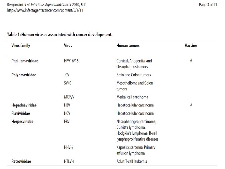
Introduction • potentially oncogenic (tumor-causing) • persist as latent infections without causing disease • They may produce tumors in a host of a different species, or a host with an ineffective immune system • The name polyoma refers to the viruses' ability to produce multiple (poly-) tumors (oma)
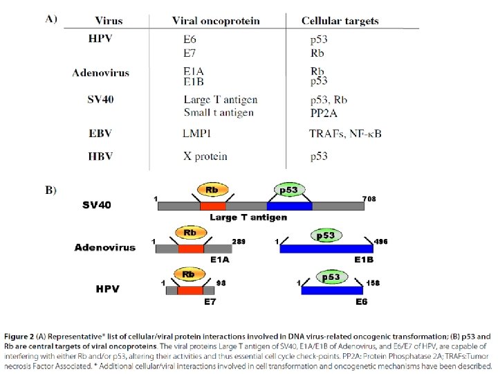
Introduction • Polyomaviruses have been extensively studied as tumor viruses in humans and animals, leading to fundamental insights into carcinogenesis, DNA replication and protein processing. • The tumor suppressor molecule p 53 was discovered, for example, as a cellular protein bound by the major oncoprotein (cancer-causing protein) • T antigen made by Simian vacuolating virus 40 (SV 40).
Taxonomy • Simian virus 40 (SV-40) ( REFERENCE STRAIN ) • Common Type BK polyomavirus (BKPy. V) JC polyomavirus (JCPy. V) Bovine polyomavirus (BPy. V) Murine polyomavirus (MPy. V)
Taxonomy • Murine polyomviruswas the first polyomavirus discovered by Ludwik Gross in 1953. • Mammals and birds GROSS L (June 1953). Society for Experimental Biology and Medicine 83 (2): 414– 21.
Classification
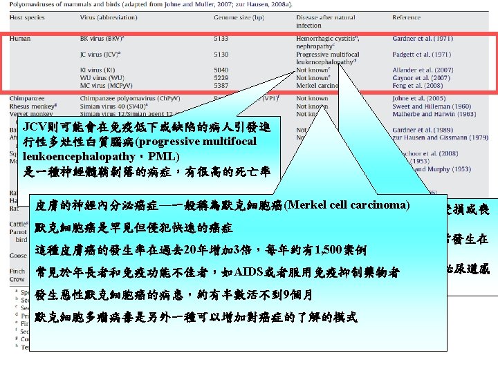
Classification Human Polyomaviruses JCV and BKV first isolated in 1971 (JCV-brain tissue of patient with progressive multifocal leukoencephalopathy (PML) BKV from urine of renal transplant patient Epidemiology: BKV first acquired by age 3 -4 JCV by age 10 -14 60 -80% of adults are seropositive for both in pre-HIV era PML was seen in older adults with hematologic malignancies now seen in ~5% of HIV/AIDS patients
Classification • Polyomaviruses are divided into three major genetically-related groups Pérez-Losada M, Christensen RG, Mc. Clellan DA, et al. (June 2006). "Comparing phylogenetic codivergence between polyomaviruses and their hosts". Journal of Virology 80 (12): 5663– 9.
Cell Receptor • SV-40: Gangliosides JCPy. V: terminal alpha(2, 6)-linked sialic acids • BFPy. V and MPy. V: alpha(2, 3)-linked sialic acids Polyomavirus的receptor為sialic acid醣類而非proteins Polyomavirus需要蛋白質coreceptor才能順利感染 coreceptor包括MHC-1 (SV 40)、α 4β 1 integrin (m. Py. V)或血清素receptor (JCV) polyomavirus感染細胞的方式: 連結帶有sialic acid的coreceptor (或未知的真正receptor? )之後 virus被帶引到coreceptor而結合 這樣的結合強化了細胞的內吞作用而進入到細胞質內,反向地經過內質網而進入到 細胞核
CELL RECEPTOR (S)
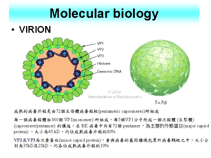
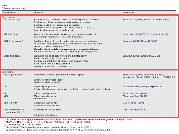
Genome • Circular ds. DNA about 5 kb in size • associated with cellular histones in a chromatin-like complex • genomes divided into two regions, early and late • Early proteins LT, MT and s. T antigens expressed from differentially spliced m. RNAs • Late, capsid proteins VP 1, VP 2 and VP 3 (viral capsid) • BKV and JCV : 75% (nucleotide sequence Homology) • 70% with SV 40
Replication of DNA Viruses 1
Virus replication 1. Cellular receptors for polyomaviruses are sialic acid residues of gangliosides. The attachment is mediated by viral protein 1 (VP 1) via the sialic acid attachment region. 2. Virus is endocytosed into vesicles in the host cell 3. Virion transits through endoplasmic reticulum where host protein disulfide isomerases rearrange its capsid structure 3. 1 Export of misfolded virion to the cytoplasm 3. 2 Loss of VP 1 in the low-calcium conditions of the cytosol 4 - 5. Import of genomic DNA into host nucleus. Then by an unknown mechanism the virus is exported to the nucleus
Virus replication 6 -9. Transcription of early genes (LT and s. T genes) • Early gene expression is responsible for the synthesis of non-structural proteins. • The role of the non-structural proteins is to regulate the cellular mechanisms and gene expression. • Close to the N terminal end of polyomavirus genome are enhancer elements which induce activation and transcription of a molecule known as the T-antigen. • Early m. RNA’s, encoding T-antigen are produced by host RNA polymerase II. • T-antigen autoregulates early m. RNA’s, subsequently leading to elevated levels of T-antigen. • At high concentrations of LT-antigen, triggering the late phase of viral infection to begin. 6. & 10. Replication of the DNA genome in the nucleus
Virus replication 10. Genome replication acts to separate the early and late phase gene expression. • The duplicated viral genome is synthesised and processed as if it were cellular DNA • As the daughter viral DNA are synthesised they associate with cellular nucleosomes to form structures that are often referred to as "minichromosomes“ • In this manner the DNA is packaged more efficiently.
Virus replication 10 -14. Transcription of late genes encoding for structural proteins (VP 1, VP 2 and VP 3) • Late gene expression synthesises the structural proteins, responsible for the viral particle composition. • This occurs during and after genome replication. • late gene expression generates an array of proteins as a result of alternative splicing.
Virus replication 15. Assembly of new virions in the nucleus • Within each viral protein are 'nuclear localization signals' which cause the viral proteins to amass in the nucleus. • Assembly of new virus particles consequently occurs within the nucleus of the host cell
Virus replication 16. Virions are released by lysis of the cell. • Release of newly synthesized polyomavirus particles exit the infected cell by one of two mechanisms. • Firstly and less commonly, they are transported in cytoplasmic vacuoles to the plasma membrane, where budding occurs. • More frequently, they are released when the cell lyses due to the cytotoxicity of virus particles present in the infected cell.
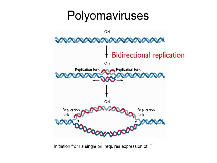
The large T-Antigen • The large T-antigen plays a key role : 1. in regulating the viral life cycle by binding to the viral origin of DNA replication where it promotes DNA synthesis. 2. replicate the host cell needs to be in s-phase for this to begin 3. modulates cellular signaling pathways • This is achieved by a two prong attack of inhibiting tumor suppressing genes p 53 and members of the retinoblastoma (p. RB) family • Stimulating cell growth pathways by binding cellular DNA, ATPasehelicase, DNA polymerase α association, and binding of transcription preinitiation complex factors. • This abnormal stimulation of the cell cycle is a powerful force for oncogenic transformation.
• T-antigen also binds and inactivates tumor suppressor proteins (p 53, p 105). • This causes the cells to leave G 1 phase and enter into S phase, which promotes DNA replication.
The large T-Antigen
SV 40 effects in different cellular environments Infection of permissive cells results in cell death and virion production. SV 40 infection of rodent cells induces S-phase but does not result in cell death or virus production. Integration of viral DNA occurs in a very low percentage of nonpermissive cells, which then become stably transformed
The Middle T-Antigen • The Polyoma Middle T-Antigen is used in animal like the where it is coupled to the MMTV promoter. • There it functions as an oncogene, while the tissue where the tumor develops is determined by the MMTV promoter.
The small T-Antigen • The small T-antigen protein is also able to activate several cellular pathways which stimulate cell proliferation. • Such as the mitogen-activated protein kinase (MAPK) pathway, and the stressactivated protein kinase (SAPK) pathway.
Polyomavirus Infection 1. Capable of causing tumors in animals and humans 2. Can cause other diseases 3. BK and JC viruses are endemic worldwide 4. Infection outcome depends on the individual’s immune system • • Normal immune systems tend to prevent latent infections Compromised immune systems allow latent infections to become established in the kidneys
Polyomavirus Infection • All the polyomaviruses are highly common childhood and young adult infections. • Most of these infections appear to cause little or no symptoms. • These viruses are probably life-long persistent among almost all adults. • Diseases caused by human polyomavirus infections are most common among persons who become immunosuppressed by AIDS, old age or after transplantation and include Merkel cell carcinoma, PML and.
Polyomavirus Infection Reactivation events are different between the BK and JC viruses • BK virus – Potentially severe urinary tract infections can develop • JC virus – Can cause progressive multifocal leukoencephalopathy » Viruses infect and kill the white matter of the CNS » Paralysis and death eventually result
PML focal neurological defects due to demyelination parieto-occipital brain involvement associated with muscle weakness, gait disturbance, speech defect, or visual blind spot H&E staining of PML brain biopsy; black cerebellum involvement associated with broadlegged gait and imbalance Diagnosis: CT or MRI lumbar puncture for JCV PCR Treatment: none, except for highly active antiretroviral therapy( HAART) in AIDS patients arrows point out some of the reactive astrocytes; blue arrow points toward an oligodendrocyte nucleus which has been markedly enlarged with viral particles, giving it a magenta color
BK nephropathy: no evidence that BKV causes disease in the immune competent population possible route of infection through contaminated food or water or respiratory spread remains latent in lymphocytes, urogenital tract, and brain but may be reactivated if the host becomes immunocompromised. viral inclusions and cellular changes in BK nephropathy are seen in tubular epithelium of a renal transplant case especially associated with disease in renal transplant patients; thought to cause graft failure in 2 -5% of this population treatment is to decrease immunosuppression as much as possible without causing rejection Cidofovir appears to reduce BKV-associated nephropathy
Polyomavirus Infection • Two recently discovered (2007/2010) polyomaviruses, KI (Karolinska Institute) and WU (Washington University) viruses, are closely related to each other and have been isolated from respiratory secretions and skin secretions. • In January 2008, a new virus, Merkel cell polyoma virus was described and shown to cause most Merkel skin cancer.
Polyomavirus Infection • In March, 2011, a ninth polyoma virus HPy. V 9, related to a monkey lymphotropic virus (LPV) was cultured from the blood of immunosuppressed patients.
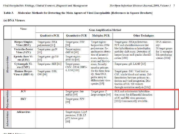
Diagnosis