Osteoarthritis RIZWAN MANSOOR M D CONSULTANT RHEUMATOLOGIST OA


- Slides: 27

Osteoarthritis RIZWAN MANSOOR M. D. CONSULTANT RHEUMATOLOGIST

OA - Definition Heterogeneous group of conditions resulting in common histopathologic and radiologic changes involving Entire joint organ, including: the articular cartilage the subchondral bone and the synovium.

Epidemiology Internationally, osteoarthritis is the most common articular disease. Estimates of its frequency vary across different populations. 80 -90% of individuals older than 65 years have evidence of radiographic osteoarthritis. the prevalence of osteoarthritis is higher among women than among men. Interethnic differences in the prevalence of osteoarthritis have been noted.

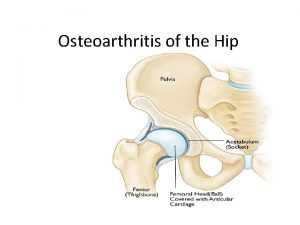
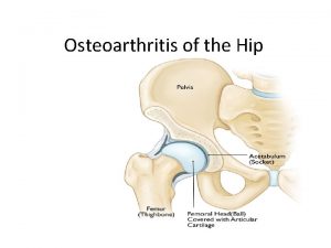
Involved joints Weight-bearing joints, including: the knees the hips cervical and lumbosacral spine feet. Non weight bearing joints: the(DIP), the(PIP), and the(CMC) joints.

Synovial Joints Articular cartilage Subchondral bone Synovial membrane Synovial fluid Joint capsule

Synovial joint anatomy

The normal articular surface of synovial joints articular cartilage ( chondrocytes) surrounded by extracellular matrix includes proteoglycans and collagen. The cartilage facilitates joint function and protects the underlying subchondral bone by distributing large loads, maintaining low contact stresses, and reducing friction at the joint.

Synovial cartilage

Synovial Fluid Synovial fluid is formed by (synoviocytes). Synovial cells also manufacture hyaluronic acid (HA, also known as hyaluronate), a glycosaminoglycan that is the major noncellular component of synovial fluid. Synovial fluid supplies nutrients to the avascular articular cartilage; it also provides the viscosity needed to absorb shock from slow movements provides elasticity required to absorb shock from rapid movements.

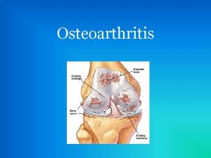
Pathogenesis Swelling of the cartilage usually occurs the level of proteoglycans eventually drops very low, the cartilage softens and lose elasticity and compromising joint surface integrity. Flaking and fibrillations (vertical clefts) develop along on the surface of an osteoarthritic joint. Over time, the loss of cartilage results in loss of joint space. a greater loss of joint space occurs at those areas experiencing the highest loads.

Cartilage changes

Clinical and Radiological

Bone changes Bone denuded of its protective cartilage continues to articulate with the opposing surface. Eventually, the increasing stresses exceed the biomechanical yield strength of the bone. The subchondral bone responds with vascular invasion and increased cellularity, becoming thickened and dense (a process known as eburnation) at areas of pressure.

Bone changes subchondral bone undergo cystic degeneration. Osteoarthritic cysts are also referred to as subchondral cysts, pseudocysts, or geodes and may range from 2 to 20 mm in diameter. Osteoarthritic cysts in the acetabulum are termed Egger cysts.

Joint changes vascularization of subchondral marrow, osseous metaplasia of synovial connective tissue, and ossifying cartilaginous protrusions lead to irregular outgrowth of new bone (osteophytes). Fragmentation of these osteophytes or of the articular cartilage itself results in the presence of intra-articular loose bodies (joint mice).

Joint changes

Etiology Risk factors- Age, obesity, trauma, genetics, hypogonadism, muscle weakness, repetitive use, Infection, crystal deposition, acromegaly, previous inflammatory arthritis (burnt-out rheumatoid arthritis) Heritable metabolic causes (alkaptonuria, hemochromatosis, Wilson disease)Hemoglobinopathies (sickle cell disease and thalassemia)Neuropathic disorders leading to a Charcot joint (syringomyelia, tabes dorsalis, and diabetes)Underlying morphologic risk factors (congenital hip dislocation and slipped femoral capital epiphysis) Disorders of bone (Paget disease and avascular necrosis)Previous surgical procedures (meniscectomy)Diabetes mellitus [44]

Radiographic changes

Radiological changes

Osteoarthritis Progression Stage 1 - breakdown of the cartilage matrix occurs. Stage 2 - involves the fibrillation and erosion of the cartilage surface Stage 3 - a chronic inflammatory response in the synovium. Further Progression - the above events alter the joint architecture, compensatory bone overgrowth occurs. joint architecture is changed mechanical and inflammatory stress occurs on the articular surfaces, the disease progresses unchecked.

PGOA

Erosive OA

Chonromalcia Patellae

Differential Diagnosis Crystalline arthropathies (ie, gout and pseudogout) Inflammatory arthritis (eg, rheumatoid arthritis) Seronegative spondyloarthropathies (eg, psoriatic arthritis and reactive arthritis) Septic arthritis or postinfectious arthropathy Fibromyalgia Tendonitis

Work Up Laboratory Plain Radiography CT scan, MRI scan, ultrasonography Bone scintigraphy Arthrocentesis

Treatment Non pharmacologic. Life style modification, physical and rehab therapy Pharmacotherapy Arthroscopy Osteotomy Arthroplasty Fusion and joint Lavage Stem cell therapy

References 1. 1. Hunter, W. Of the structure and diseases of articulating cartilages. Phil. Trans. Royal Soc. 470, 514– 521 (1743). 2. 2. National Collaborating Centre for Chronic Conditions (UK). 3. National Clinical Guideline for Care and Management in Adults(Royal College of Physicians of London, 2008). 3. 3. Felson, D. T. An update on the pathogenesis and epidemiology of osteoarthritis. Radiol. Clin. North Am. 42, 1– 9 (2004). 4. 4. WHO. The World Health Report 2002: reducing risks, promoting healthy life (WHO, 2002). 5. 5. Centers for Disease Control and Prevention (CDC). Prevalence of doctor-diagnosed arthritis and arthritis-attributable activity limitation—United States, 2010– 2012. MMWR Morb. Mortal. Wkly Rep. 62, 869– 873 (2013).
 Dr eugene lim rheumatologist
 Rheumatologist carefirst bcbs
 Rheumatologist vsp
 Dr forghani rheumatologist
 Dr susan wallis rheumatologist
 Bait rizwan
 Dr raja rizwan
 Saddle pontic
 Raja 105
 Anita roddick leadership style
 Mansoor sufi
 Dr shahid mansoor
 Mansoor hotel hargeisa
 Syed mansoor sarwar
 Jungho ahn
 Syed mansoor sarwar
 Mansoor and gore
 Arshad mansoor
 Zubair
 Joanna ji
 Mansoor amiji
 Osteoarthritis
 Osteoarthritis
 Difference between osteoarthritis and rheumatoid arthritis
 Osteoarthritis
 Kellgren and lawrence classification system
 Generalised osteoarthritis
 What is esr
