LEPROSY Dr Amrita Upadhyaya Assistant Professor Dermatology 29


, dark red or")


 -Single or 2 or 3 -Erythematous or coppery -Dry surface,")


 • Several number, bilateral but asymmetrical distribution")

")


, RR in a case")


")


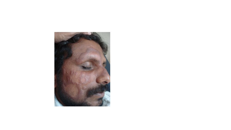
. The facial paralysis in leprosy")


")
")
")

 on palms and soles")


 Pole Tuberculoid Lepromatous Skin Localized lesion, hypopigmented, raised erythematous margins Symmetric")


- Slides: 69
LEPROSY Dr Amrita Upadhyaya Assistant Professor Dermatology 29 th sep 2017
Cardinal signs of Leprosy • 1. Anaesthetic/hypoaeshetic skin lesion or lesions • 2. Enlarged peripheral nerves with impairment of sensations in the area supplied • 3. Acid-fast bacilli in the slit skin smear • Any one of these signs is sufficient for diagnosis of leprosy. Two of them are clinical. Therefore clinical skill of the health care worker is important for diagnosis of leprosy.
Exclusion criteria for diagnosis of leprosy • • • White (de-pigmented), dark red or black in colour Scaly lesion (regressed type 1 lepra reaction lesion may show scaling) Present since birth Seasonal or appears and disappears suddenly Hurts (discomfort may be felt in leprosy reaction )/ itches Presence of sweating
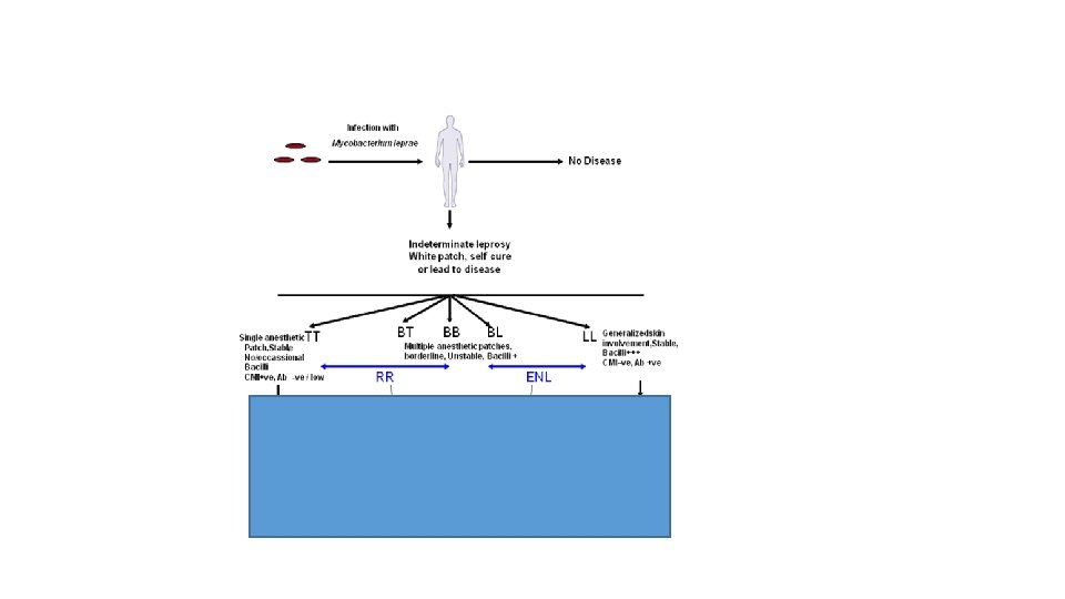
Current WHO classification. MB; Multi bacillary. PB; Pauci bacillary. Criteria MB > 6 skin lesions, or Positive bacterial index PB ≦ 5 skin lesions and Negative bacterial index
Leprosy Spectrum CMI+Ab-ve CMI–ve, Ab+
• Ill-defined patch of indeterminate leprosy
RIDLEY-JOPLING CLASSIFICATION
RIDLEY JOPLING CLASSIFICATION
Tuberculoid leprosy (plaque type) -Single or 2 or 3 -Erythematous or coppery -Dry surface, hairless -Raised well defined edge with sharp outer margin and sloping inside & tendency of central flattening -Sensation(touch, temp. pain) : absent -Feeding nerve to the patch or solitary peripheral nerve may be thickened -AFB: negative Lepromin: +++
TT • WELL DEFINED RAISED OUTER BORDER OF A TT LESION. • ENLARGED GREATER AURICULAR NERVE • INVOLVEMENT OF EAR
BT • Large patches of BT leprosy • SATELLITE LESIONS
BB Leprosy (macular, plaque, inverted saucer shaped) • Several number, bilateral but asymmetrical distribution • Variable size, sloping outer edge & central punched out area • Sensation: slightly diminished • Slightly shiny • Asymmetrical many nerve thickening • AFB: moderate 2+to 3+ • Lepromin: negative
Inverted saucer-shaped lesion of BB
LL (Fine to course infiltration)
• BILATERAL LOSS OF EYEBROWS , MOUSTACHE & BEARD
• glove & stocking distribution of neuropathy.
Pure neuritic leprosy
HISTORY TAKING • • Primary goal of history taking is to establish the diagnosis by excluding other conditions simulating leprosy. History should be taken according to the following steps, mostly like any other medical history; • Personal identification and demographic data. • Presenting complaints. • History of present illness. • Past history. • Family history. • Personal history.
Various presenting features of leprosy IN ABSENCE OF REACTION 1. Skin lesions: ; hypopigmented / erythematous, hypoaesthetic patches, skin-coloured erythematous papules, plaques nodules, which may be waxy ± umbilication. 2. Hypoaesthesia along distribution of peripheral nerves / gloves and stockings hypoaesthesia 3. Spontaneous blisters and ulcers on the hands and/ or feet 4. Trophic changes ; dryness, ichthyosis, fissuring / trophic ulcer 5. Diffuse swelling of hands and feet ( early sign of lepromatous leprosy / leprosy reactions) 6. Nasal stuffiness, epistaxis (early sign of lepromatous leprosy) 7. Irregular thickening of ear and nodules on face (lepromatous leprosy) / WH I LE IN REACTION 1. Tingling and numbness of hands and/ or feet 2. Sudden w eakness of hands and/ or feet / inability to close eyelids 3. Sudden redness, swelling and pain of existing lesions and / or appearance of new lesions with or without constitutional symptoms (type 1 reaction) 4. Sudden appearance of crops of evanescent, erythematous, painful nodules on apparently normal looking skin with or without constitutional symptoms (erythema nodosum leprosum / type 2 reaction) 5. Painful swelling on the dorsal aspect of wrists (tenosynovitis) 6. A c u t e s c r o t a l p a i n ( e p i d y m o - o r c h i t i s ) 7. Pain in and around eyes, redness, photophobia or diminished vision (iridocyclitis) IN PRESENCE OF DEFORMITY 1. 2. 3. Inability to use hands for precision works; e. g. , button a shirt, eating rice with hands, typing, etc; Inability to make a power-grip; e. g. , holding a rod, carrying utensils. Inability to wear slipper. (all these are indicative of peripheral anaesthesia and poor functioning of small muscles of hands and feet).
Looking for other organ involvement in leprosy Table 10. 2: Looking for other organ involvement in l ORGAN Eye SPECIFIC QUESTIONS TO BE ASKED Difficulty in eye closure? (Lagophthalmos) Red eye with pain, watering and sticky discharge? (Corneal ulceration) Red eye with pain, photophobia and diminished vision? (Iridocyclitis) Localized redness, severe radiating pain to temporal region, normal/ slightly reduced vision? (Scleritis) Localized redness, mild pain, normal vision? (Episcleritis) Unable to perceive smell of food and scented materials? (Anosmia) Nose is blocked with occasional bleeding? Change of voice, chronic cough, and occasional breathlessness? (Laryngeal involvement) Any episode of acute respiratory distress? (Laryngeal oedema) Upper respiratory tract Cardiovascular system* Adrenal glands** Male reproductive system Features of adrenal insufficiency? e. g. , hypotension, asthenia, prostration Palpitation? Dyspnoea on exertion? Swelling of feet? Normal / diminished libido? (Testicular atrophy) Enlarging male breasts? ( Testicular atrophy) If married, whether having children? (Sterility) *Cardiovascular involvement in leprosy is very rare and rarely gives rise to symptoms. Involvement of peripheral blood vessels, though frequent, is rarely symptomatic. ** Adrenal suppression is very rare in leprosy. However, it may occur rarely during an episode of T 2 R. .
• RR in a case of BT leprosy, (left), RR in a case of BB leprosy (Right), note the shiny erythematous and oedematous plaques.
• BT lesion with type 1 reaction
• ENL in a case of LL. Note the shiny erythematous papules and nodules (usually tender) over face and thighs. The lesions are evanescent and usually associated with systemic features like fever, malaise, joint pain etc.
• NODULES OF ENL
• Spontaneous blister on finger
• Callosity over lateral malleolus (pressure point)
• Trophic ulcer
• partial or ulnar clawhand
• Bilateral complete claw hands with guttering of dorsal • interosseous spaces and callosities on interphalangeal joints.
• A case of Bell’s palsy (right side). The facial paralysis in leprosy involves upper part of face (due to selective involvement of zygomatic branch of facial nerve by a leprosy lesion in the vicinity). In Bell’s palsy the involvement of facial nerve is higher up, due to oedematous compression in facial canal, affecting all branches, leading to paralysis of both upper & lower parts of face.
• Secondary ichthyosis in lepromatous leprosy with brownish discoloration of the scales due to clofazimine.
• LAGOPHTHALMOS
Lagophthalmos- inability to close the eyes , is in direct proportion to the extent of damage to facial nerve If the eyes cannot be closed regularly by normal blinking, or if the eyes are not closed while sleeping , then the eyes are at risk of exposure keratitis
CLINICAL EXAMINATION • Clinical examination involves the following steps: • 1. General physical examination • 2. Cutaneous and mucosal examination • 3. Ocular examination • 4 Palpation of peripheral nerves and testing for sensory impairment. • 5. Examination of musculoskeletal system • 6. Examination of external genitalia • 7. Other systemic examination, wherever indicated.
Precautions for examination of skin lesion • • • Examine patient under good light (preferably natural light) Provide privacy to the patient Examine the whole skin from head to toe as much as possible. Always use the same order of examination, so that you do not forget to examine any part of the body. Ensure presence of an assistant of the same sex as that of the patient to assist you. Especially, if the patient is of the opposite sex.
• A quick general cutaneous survey should be done followed by examination of individual skin lesions. • Prior explanation to the patient along with gaining his / her confidence is of immense importance in achieving patient cooperation.
• DIFFUSE COARSE INFILTRATION OF THE SKIN- LL
• MORE SEVERE INFILTRATION WITH NODULAR LESIONS (LEONINE FACIES)
LL (Leonine face)
Lepromatous nodule on tongue and palate (same pt. )
• Oedema of hands and feet in a patient with untreated LL
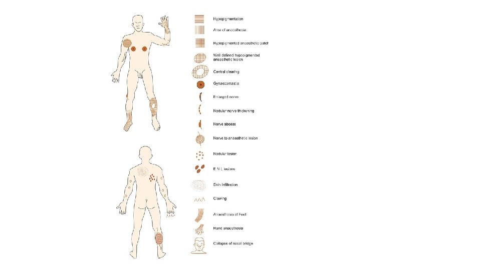
• Recommended sensory testing sites (WHO) on palms and soles
OCULAR EXAMINATION • Tested by- fine wisp of cotton - lightly blowing a puff of air into each cornea (Hutchison’s 21 ed) - aesthesiometer (fine filament –calibrated) - observing infrequent blinking (because the stimulus for eye closure is decreased)
• TESTING CORNEAL SENSATION
PALPATION OF PERIPHERAL NERVES • Clinician should be well aware of the specific sites / bony land-marks along which the peripheral nerves commonly involved in leprosy are to be palpated, as well as the area of distribution of sensory nerves. • Nerve palpation should be gentle (using pulps of fingers rather than tips) to avoid causing pain to an inflamed nerve (neuritis).
Nerve Site / bony landmark Patient position Method Supraorbital notch at the junction of medial 1/3 rd and lateral 2/3 rd of supraorbital ridge Sitting / standing with Palpate with both thumbs on both sides (Fig 38) Supratrochlear Medial to the supraorbital nerve Same as above Infra orbital foramen, just below the medial part of inferior Same as above orbital margin Same as above Branches of facial nerve: Zygomaic & temporal. Zygomatic arch Same as above Head & Neck head kept straight. Infraorbital Great auricular Lateral side of neck, crosses sternomastoid muscle, from lateral side to infra-auricular area. Sitting / standing with head turned completely to opposite side Easily visible, crossing sternomastoid obliquely. May be palpated with 2 fingers Clavicular Shaft of clavicle Sitting / standing straight Fingers rolled along the shafts of both clavicles Sitting / standing with elbow flexed at 90°. Spiral groove on humerus, posterior to the deltoid insertion. Examiner’s right hand holds patient’s right hand in shaking-hand manner (& vice versa). Examiner’s left fingers roll the nerve in the radial groove. Examiner’s left little finger locate the nerve in the groove; other fingers palpate the nerve upwards along medial aspect of arm (Fig 39). Ulnar groove on medial epicondyle of humerus, medial to the point of elbow. Same as above. (3 sets) Upper extremity Radial Ulnar (Both the nerves may be palpated simultaneously for better comparison) Lateral border of radius, just proximal to the wrist; thereafter along the proximal part of extensor pollicis longus tendon, which stands out prominently on ulnar side of the anatomical snuff box Same as above. Proximal to the flexor aspect of wrist joint (proximal to flexor retinaculum), between the tendons of palmaris longus and flexor carpi radialis Sitting / standing with elbow flexed at 90°, & wrist in supination. Examiner’s left hand stabilizes patient’s right hand vice versa Examiner’s right fingers palpate the nerve deep between the tendons. Patient is asked to extend the thumb to visualize the anatomical snuff box. Examiner’s left fingers roll the nerve against radius (Fig 40). It can be further traced and rolled from side to side on extensor pollicis longus tendon &dorsum of hand. Radial cutaneous Median
• Palpation of supraorbital nerve with; fingers are used to stabilize the head.
• Palpation of radial cutaneous nerve.
• Palpation of ulnar nerve
• Palpation of lateral popliteal nerve.
• Palpation of sural nerve.
• Palpation of posterior tibial nerve.
• Sensory distribution of feet by various nerves
• Sensory distribution of HANDS by various nerves
• Terminal branches of facial nerve
MUSKULOSKELETAL SYSTEM EXAMINATION
• Z THUMB DEFORMITY
• Ochsner’s clasping test with pointing index.
• Pen test.
• Book test with positive Froment’s sign (right side
• Positive Wartberg’s sign: earliest evidence of ulnar nerve palsy
Examination of External Genitalia • genitalia of both male and female patients should be inspected for presence of skin lesions • in male patients, testicles should be palpated gently to see for: • • Size and consistency: • Whether the testicles are firm, resilient and of appropriate size or small and soft (sequel of advanced disease and / or repeated type 2 reaction). • • Testicular sensation: • If absent, indicates fibrosis resulting from repeated episodes of epididymo-orchitis (type 2 reaction). • • Tenderness: • Acute testicular pain indicates epididymo-orchitis, a part of type 2 reaction and this may be the presenting feature of type 2 reaction.
OTHER SYSTEM EXAMINATION • Upper respiratory tract is the most common site of involvement. Though several other organs are involved in lepromatous leprosy and also during type 2 reaction, clinical manifestations of such involvement are unusual • A hoarse voice, dry, hacking cough and occasional breathlessness are indicative of laryngeal involvement and or uvular dysfunction
LEPROSY (QUICK FACTS) Pole Tuberculoid Lepromatous Skin Localized lesion, hypopigmented, raised erythematous margins Symmetric diffuse lesions, papules/macules/nodules, massive tissue destruction Nerves Epitheloid granulomas Poorly organised granulomas, vacuolated MΦ, few T cells M. Leprae Classification WHO RIDLEY-JOPLING Immunology Adaptive Rare Abundant Paucibacillary TT Multibacillary BT Th 1, cell mediated, IL-2, IFN-γ Borderline BL LL TH 2, Humoral mediated, IL-4, IL-10 Innate ? ?
Thank you!