Mohs micrographic surgery SWAG network service Update May




- Slides: 26
Mohs micrographic surgery SWAG network service Update May 2016 Dr Adam Bray Consultant Dermatologist Dermatological & Mohs surgeon Bristol
Bristol Mohs service • Since Feb 2015 • Approximate waiting list 3 months once seen (less for eyelids) • Second Mohs surgeon Dr Pawel Bogucki • Excellent links with local Plastic surgery, Oculoplastics, and Maxillofacial • Southmead Hospital, North Bristol NHS Trust (NBT) • Any interested clinicians are welcome to arrange a visit for their professional development
Referrals • Please write to me stating: • ‘Referral for Mohs surgery, Dermatology department’: • Southmead Hospital, North Bristol Trust • NBT. outpatientbookings@nhs. net
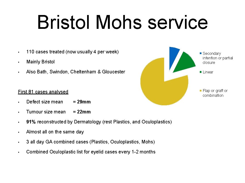
Bristol Mohs service • 110 cases treated (now usually 4 per week) • Mainly Bristol Secondary intention or partial closure • Also Bath, Swindon, Cheltenham & Gloucester Linear Flap or graft or combination First 81 cases analysed • Defect size mean = 29 mm • Tumour size mean = 22 mm • 91% reconstructed by Dermatology (rest Plastics, and Oculoplastics) • Almost all on the same day • 3 all day GA combined cases (Plastics, Oculoplastics, Mohs) • Combined Oculoplastic list for eyelid cases every 1 -2 months
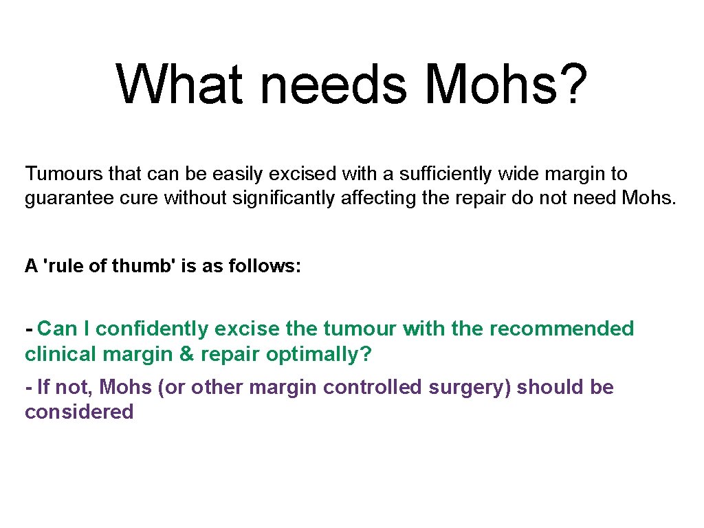
What needs Mohs? Tumours that can be easily excised with a sufficiently wide margin to guarantee cure without significantly affecting the repair do not need Mohs. A 'rule of thumb' is as follows: - Can I confidently excise the tumour with the recommended clinical margin & repair optimally? - If not, Mohs (or other margin controlled surgery) should be considered
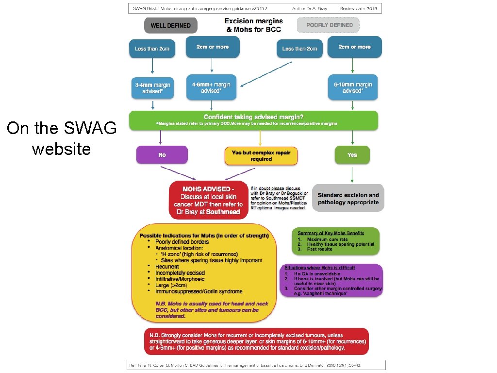
On the SWAG website
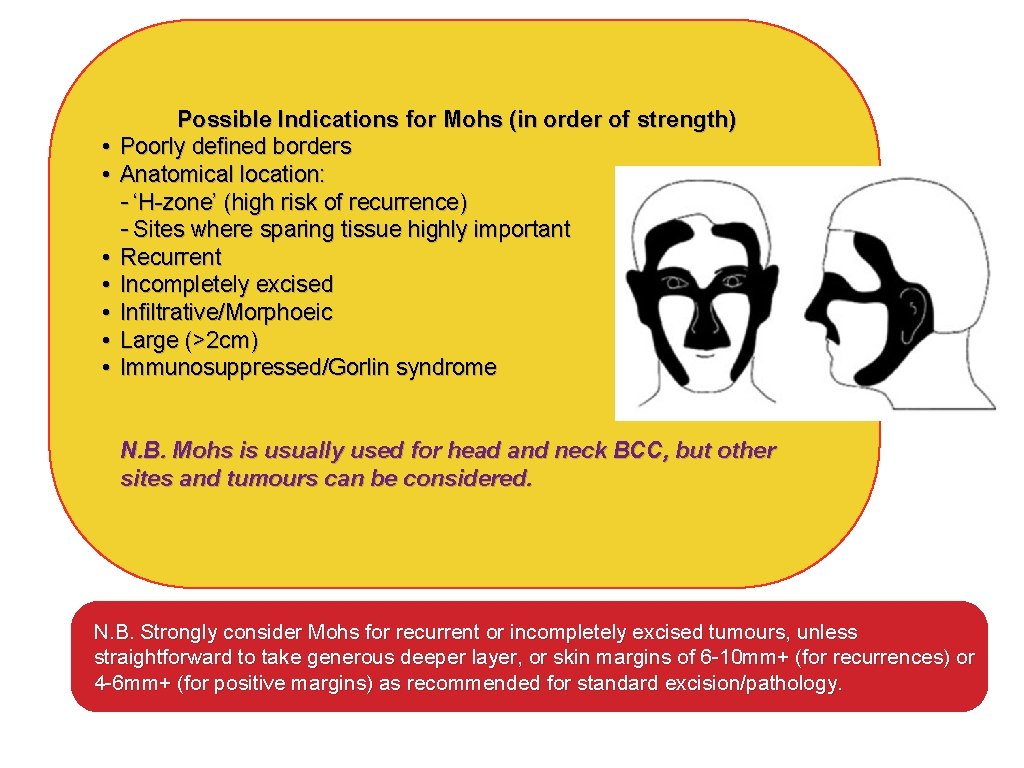
• • Possible Indications for Mohs (in order of strength) Poorly defined borders Anatomical location: - ‘H-zone’ (high risk of recurrence) - Sites where sparing tissue highly important Recurrent Incompletely excised Infiltrative/Morphoeic Large (>2 cm) Immunosuppressed/Gorlin syndrome N. B. Mohs is usually used for head and neck BCC, but other sites and tumours can be considered. N. B. Strongly consider Mohs for recurrent or incompletely excised tumours, unless straightforward to take generous deeper layer, or skin margins of 6 -10 mm+ (for recurrences) or 4 -6 mm+ (for positive margins) as recommended for standard excision/pathology.
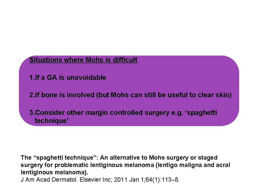
Situations where Mohs is difficult 1. If a GA is unavoidable 2. If bone is involved (but Mohs can still be useful to clear skin) 3. Consider other margin controlled surgery e. g. ‘spaghetti technique’ The “spaghetti technique”: An alternative to Mohs surgery or staged surgery for problematic lentiginous melanoma (lentigo maligna and acral lentiginous melanoma). J Am Acad Dermatol. Elsevier Inc; 2011 Jan 1; 64(1): 113– 8.
Summary of Key Mohs Benefits 1. Maximum cure rate 2. Healthy tissue sparing potential 3. Fast results
What needs Mohs? Consider Mohs over delayed repair Why? The pathology is FAR more accurate Avoid intra-operative frozen section analysis Why? Shown to be VERY inaccurate
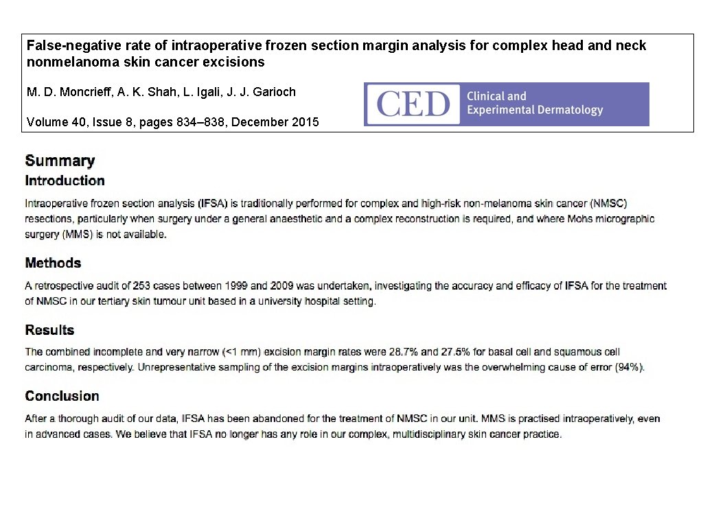
False-negative rate of intraoperative frozen section margin analysis for complex head and neck nonmelanoma skin cancer excisions M. D. Moncrieff, A. K. Shah, L. Igali, J. J. Garioch Volume 40, Issue 8, pages 834– 838, December 2015
Mohs Micrographic Surgery Q. Why are tumours incompletely excised? A. -Because they are difficult to see -Because they grow unpredictably Q. Why do tumours recur? A. Because they were not fully removed 13
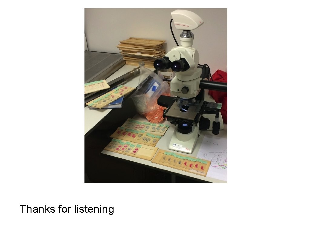
Thanks for listening
Referrals • Please write to me stating: • ‘Referral for Mohs surgery, Dermatology department’: • Southmead Hospital, North Bristol Trust • NBT. outpatientbookings@nhs. net
What needs Mohs? Tumours that can be easily excised with a sufficiently wide margin to guarantee cure without significantly affecting the repair do not need Mohs. A 'rule of thumb' is as follows: - Can I confidently excise the tumour with the recommended clinical margin & repair optimally? - If not, Mohs (or other margin controlled surgery) should be considered
End
The Mohs procedure explained
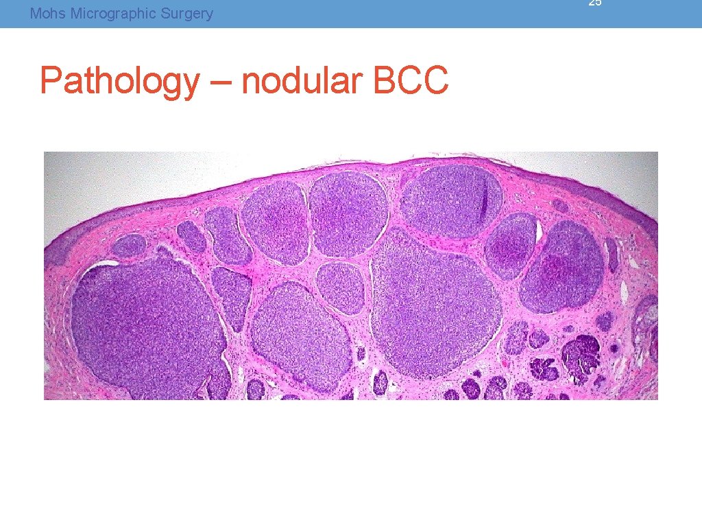
Mohs Micrographic Surgery Pathology – nodular BCC 25
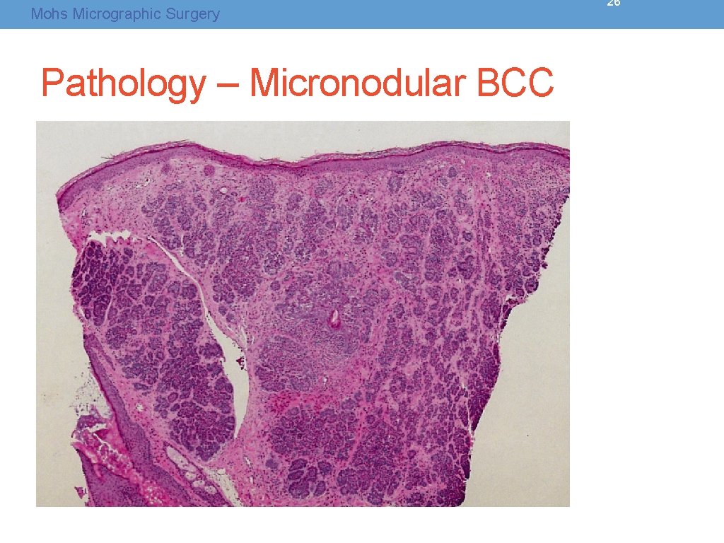
Mohs Micrographic Surgery Pathology – Micronodular BCC 26
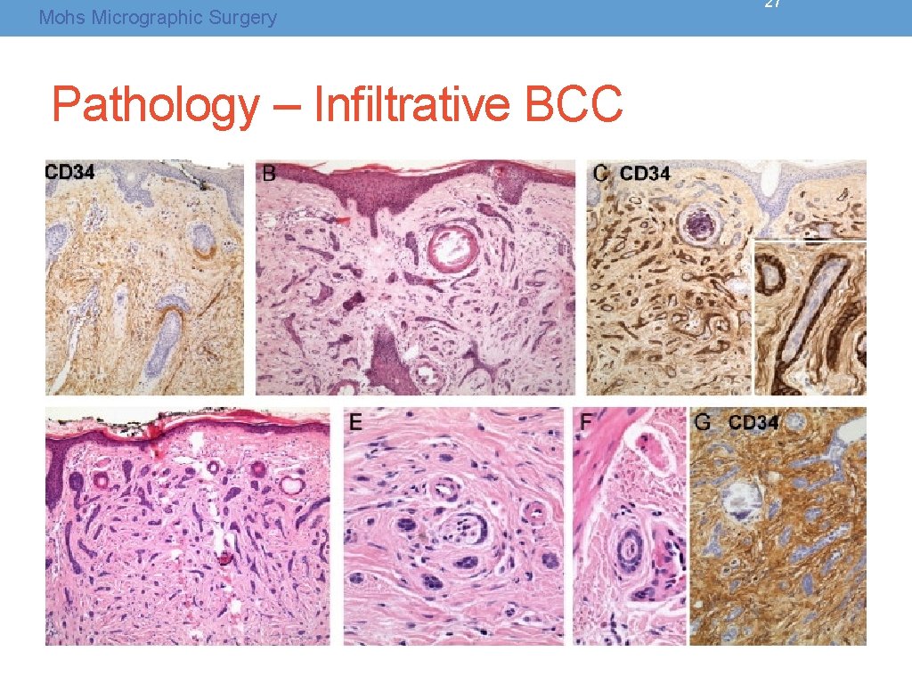
Mohs Micrographic Surgery Pathology – Infiltrative BCC 27
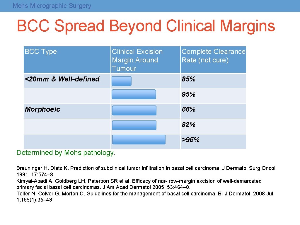
Mohs Micrographic Surgery BCC Spread Beyond Clinical Margins BCC Type <20 mm & Well-defined Morphoeic Clinical Excision Margin Around Tumour 3 mm Complete Clearance Rate (not cure) 4 mm - 5 mm 95% 3 mm 66% 5 mm 82% 13 mm – 15 mm >95% 85% Determined by Mohs pathology. Breuninger H, Dietz K. Prediction of subclinical tumor infiltration in basal cell carcinoma. J Dermatol Surg Oncol 1991; 17: 574– 8. Kimyai-Asadi A, Goldberg LH, Peterson SR et al. Efficacy of nar- row-margin excision of well-demarcated primary facial basal cell carcinomas. J Am Acad Dermatol 2005; 53: 464– 8. Telfer N, Colver G, Morton C. Guidelines for the management of basal cell carcinoma. Br J Dermatol. 2008 Jul. 1; 159(1): 35– 48.
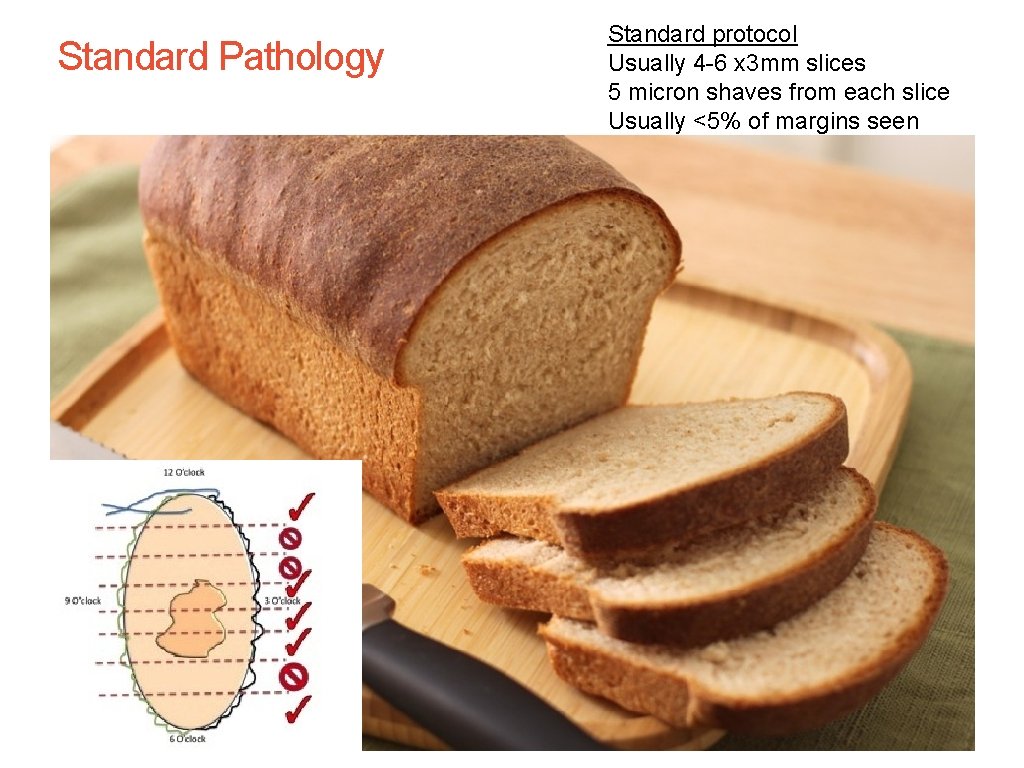
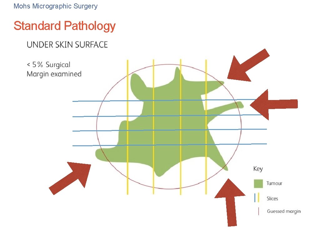
Of 75 Standard Pathology Standard protocol Usually 4 -6 x 3 mm slices 5 micron shaves from each slice Usually <5% of margins seen
Mohs Micrographic Surgery Standard Pathology
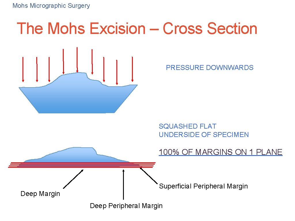
32 Mohs Micrographic Surgery The Mohs Excision – Cross Section PRESSURE DOWNWARDS SQUASHED FLAT UNDERSIDE OF SPECIMEN 100% OF MARGINS ON 1 PLANE Deep Margin Superficial Peripheral Margin Deep Peripheral Margin
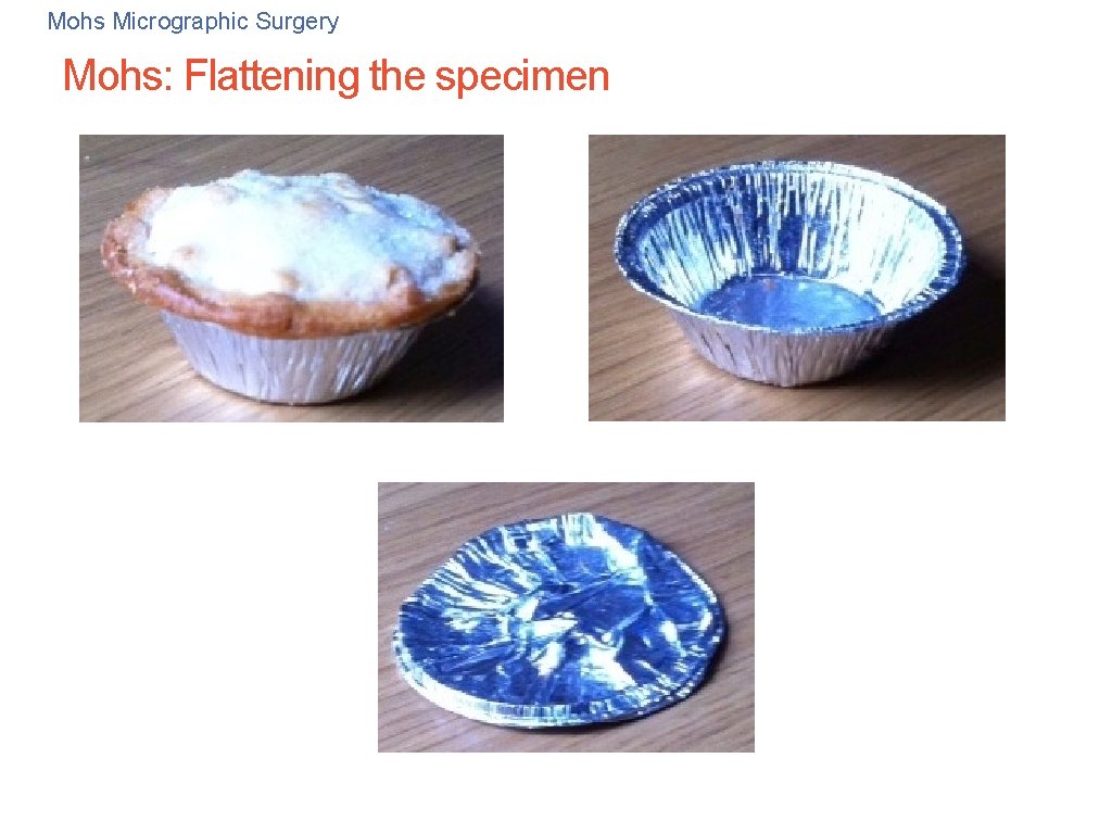
Mohs Micrographic Surgery Mohs: Flattening the specimen 33