Leishmania donnovani Dr Bindhusaran M D Hom Assistant
 Assistant Professor Dept. of Pathology &")
Leishmania donnovani Dr. Bindhusaran M. D. (Hom. ) Assistant Professor Dept. of Pathology & Microbiology

FLAGELLATES • 1. INTESTINAL ORAL AND GENITAL • Infecting the intestinal canal, oral cavity and the genital tract. • They are mostly non-pathogenic. • 2. BLOOD AND TISSUE FLAGELLATE • Infecting the vascular system and various tissues of the body. Include two genera which are pathogenic to man: • (a) Trypanosoma and (b) Leishmania.

Leishmania donnovani The parasite causing visceral leishmaniasis or kalaazar
 Indian Kala-azar •")
CLINICAL CLASSIFICATION OF LEISHMANIASIS I. Visceral leishmaniasis or Kala-azar • (a) Indian Kala-azar • (b) Mediterranean Kala-azar II. Mucocutaneous leishmaniasis

Leishmania donnovani • It is named after the discoverers, Leishman and Donovan, both of whom reported on the organism simultaneously; Leishman from London in May 1903 and Donovan from Madras in July 1903. • Geographical Distribution. Endemic in many places in India, China, Africa, Southern Europe, South America and Russia. • In India, it is specially common in Assam and Bengal along the coasts of the Ganges and the Brahmaputra. It is also endemic in Bihar, Orissa, Madras and the eastern parts of Uttar Pradesh as far as Lucknow.
 the parasite is always intra-cellular, occurring in")
Habitat • Inside the vertebrate host (man) the parasite is always intra-cellular, occurring in the amastigote form. • It is essentially a parasite of the R. E. System

Morphology • 1. Amastigote Stage: Occurs in man. • 2. Promastigote Stage: Occurs in (a) gut of insect (sandfly) and (b) artificial culture
. L. D bodies • The parasite at this stage resides")
AMASTIGOTE STAGE (AFLAGELLAR STAGE). L. D bodies • The parasite at this stage resides in the cells of the reticulo-endothelial system of vertebrate hosts (man, dog and hamster)

The characteristics of the amastigote • Shape and Size. It is a round or oval body measuring 2 to 4 mm. Cell membrane is delicate • Nucleus measures a little less than 1 µm in diameter. It is oval or round and is usually situated in the middle of the cells or along the side of cell-wall. • Kinetoplast lies at right angles to the nucleus. It comprises a DNA-containing body and a mitochondrial structure. • Axoneme (rhizoplast), a delicate filament extending from the kinetoplast to the margin of the body. It represents the root of the flagellum.

Amastigote form in R. E cells
. • cultures and in insect vectors (sandflies).")
PROMASTIGOTE STAGE (FLAGELLAR STAGE). • cultures and in insect vectors (sandflies).
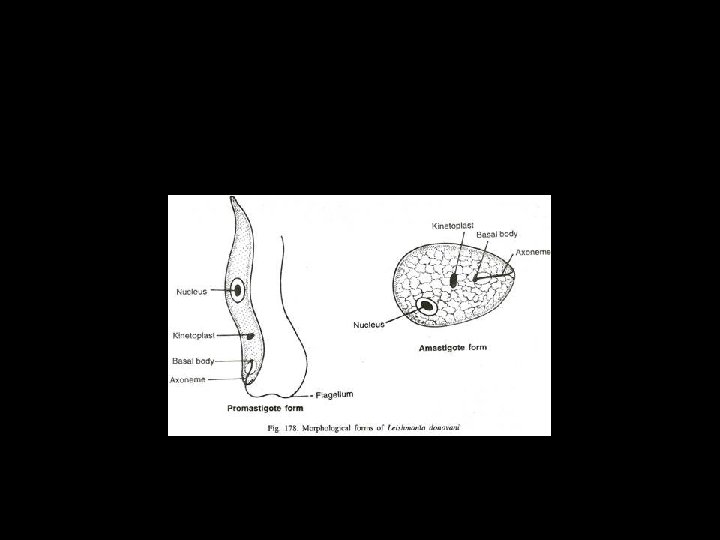

Promastigote form • Shape and Size. The earlier ones are short oval, or pear-shaped bodies • Nucleus is situated centrally. • Kinetoplast lies transversely near the anterior end. • Easinophilic vacuole, a light staining area lying in front of the kinetoplast over which the root of the flagellum runs. • Flagellum may be of the same length as the body or even longer, projecting from the front.
 • L. donovani can be cultured in a medium composed of")
Cultivation( NNN medium) • L. donovani can be cultured in a medium composed of two parts of salt agar and one part of defibrinated rabbit’s blood. • The medium was first introduced by Novy and Mac. Neal, later modified by Nicolle and is commonly referred to as N. N. N. medium. • The material for culture is inoculated into the medium and incubated at 22° to 24°C.

Immunology. • Amastigotes developing from promastigotes excite a cellular reaction comprising histiocytic proliferation followed by invasion of lymphocytes and plasma cells. The former give shelter to Leishmania inside which the parasites multiply. • ( Histiocyte- a stationary phagocytic cell present in connective tissue. )

Life Cycle • The parasite has two stages in its life cycle: • 1. The amastigote form, occurring in man (also in dog in some areas). • 2. The promastigote form, occurring in sandfly.

HUMAN CYCLE • The amastigote form while residing in the cells of the reticulo-endothelial system, multiplies by binary fission. • Multiplication goes on continuously till the cell becomes packed with the parasites. • The host-cell is thereby enlarged and eventually ruptures (as many as 50 to 200 or even more may be found embedded in the cytoplasm of the enlarged host-cell).
 • The parasites liberated as a result of the")
HUMAN CYCLE (cont. . ) • The parasites liberated as a result of the rupture into the circulation are again either taken up by, or invade fresh cells and the cycle is repeated. • In this way, the entire reticulo-endothelial system becomes progressively infected. • In the blood stream, some of the free amastigotes are phagocytosed • A blood-sucking insect draws these free amastigote forms as well as those within the monocytes during its blood-meal.

In sand fly • In certain species of sandfly, these amastigote forms develop into promastigote forms which again multiply by binary fission producing an enormous number of flagellates. • Multiplication proceeds in the mid-gut of the sandfly and the flagellates tend to spread forwards to the anterior part of the alimentary canal (pharynx and buccal cavity). • Metacyclic promastigotes thus formed are introduced into human when this sandfly bite aperson

Sand fly

BLOCKED SAND FLY • The development of flagellates depend upon whether they have fed on suitable fruit or plant juices before their second blood-meal. • The sandfly which subsists on fruit or plant juices after the first blood-meal shows a heavy flagellate infection, its pharyngeal and buccal cavity becoming completely blocked by the flagellates. Bites of such “blocked” sandflies on susceptible persons almost invariably cause infection
congenital infection of a child in utero, (b) transmission by")
Other Methods of Transmission (a)congenital infection of a child in utero, (b) transmission by blood transfusion, (c) transmission by inoculation of cultures of L. donovani, and (d) possible transmission during coitus.

Pathogenicity • Incubation Period. (This is the period between the time of the initial infection and the appearance of clinical manifestation. ) • It generally varies from 3 to 6 months, but it may exceed one and sometimes two years.
 about 1 to 1%")
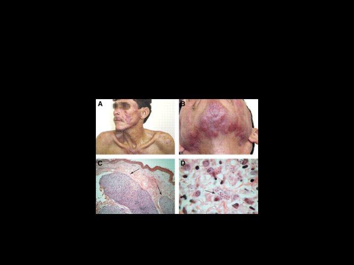
Clinical features • A primary skin lesion (leishmanioma nodule (leishmanioma) about 1 to 1% inch in diameter ) in 1 to 3 weeks’ time • Infection with L. donovani produces the disease kala-azar or visceral leishmaniasis

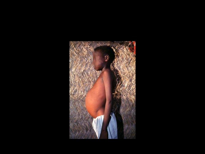
KALA AZAR characterised by • Fever is often an early symptom and it may be continuous or remittent in type, becoming intermittent at a later stage. • Waves of pyrexia may be followed by apyrexial period • Splenic enlargement is one of the most striking features and the organ progressively enlarges. With the progress of the disease, it extends several inches below the costal margin, often filling up the entire abdomen.

General Features. • There is no malaise(a general feeling of discomfort, illness, or unease ) or apathy and the patient may be quite unaware of the high fever. • The patient has a good appetite and a clean moist tongue. • Epistaxis may be a presenting symptom. • In a fully developed case, emaciation and anaemia become noticeable.

• The skin over the entire body is dry, rough and harsh and dark). The hair brittle and falls out. • If left untreated, 75 to 95 per cent of the patients die within a period of 2 years. • Death in kala-azar is always due to amoebic or bacillary dysentery, pneumonia, pulmonary tuberculosis etc.
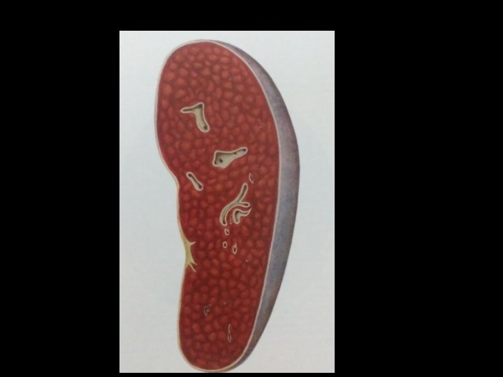
 The organ is enormously enlarged. (ii) The capsule of")
PATHOGENIC LESIONS SPLEEN Macroscopic i) The organ is enormously enlarged. (ii) The capsule of the enlarged Spleen is often thickened due to perisplenitis. • (iii) The organ is soft in consistency • (iv) The cut surface shows marked congestion and has a dull red or chocolate • (v) The substance of the splenic tissue is friable and can easily be broken down by the pressure of the thumb, signifying the absence of any fibrosis. • •
 The vascular spaces are widely dilated • (ii)")
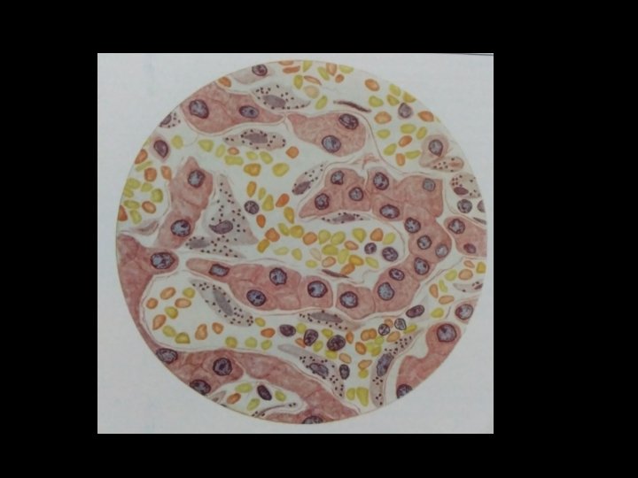
Spleen • Microscopic Appearance • i) The vascular spaces are widely dilated • (ii) The reticular cells of Bilroth cords are markedly increased are packed with amastigote forms of L. donovani • the sinus lining cells (littoral cells) do not contain any parasite.

Spleen Microscopic Appearance

CHANGES IN THE LIVER • The organ is enlarged and congested. The cut surface may show a nutmeg appearance. • Microscopically- The Kupffer’s cells are greatly increased in size and number and their cytoplasms are packed with amastigote forms of L. donovani. • (ii) The sinusoidal capillaries are dilated and engorged with blood.

William Boog Leishman

• a Scottish pathologist and British Army medical officer • Contributions • Leishman’s stain • Leishmania donnovani
- Slides: 37