Tuberculoid and Lepromatous Leprosy Tuberculoid Leprosy Dr Hena

Tuberculoid and Lepromatous Leprosy Tuberculoid Leprosy Dr Hena A. Ansari Deptt. Of Pathology JNMC, AMU

Instructions • Note down the description of the lesion from the slides labelled “Gross” and “Microscopic Appearance” in the same way as you used to wrie in your files. • Draw the diagram as you used to draw from the slide labelled “Diagrammatic representation” • Those who have files can note in them and the others can please note down in a notebook and later transfer to your files. • The rest of the ppt will focus on explanation of the lesion. I have provided an audio narration with the ppt which will play automatically along with slide show as well as with individual slides by clicking on the audio icon in lower right hand corner.

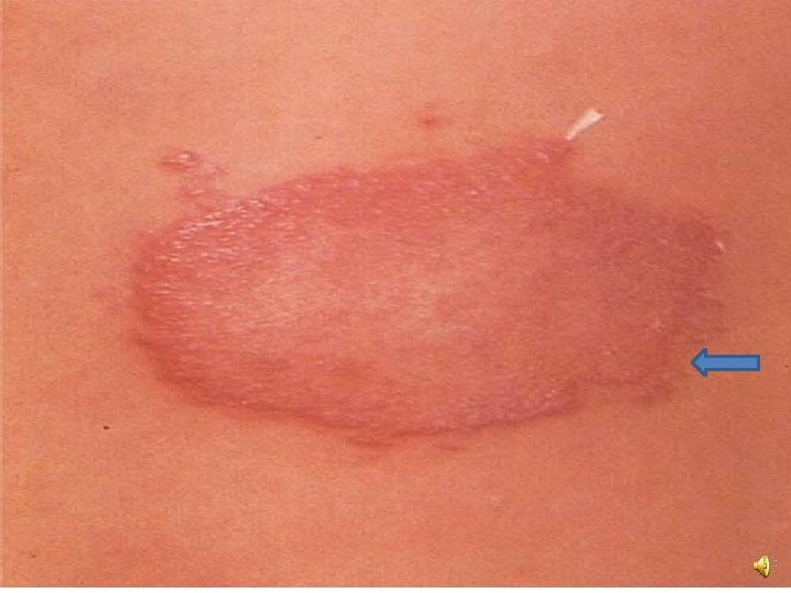
Tuberculoid leprosy--Gross Appearance • Few well defined dry hypopigmented macules or plaques which are usually hypoaesthetic or anaesthetic and anhidrotic with loss of hair. • Distribution is asynmetrical. Neural involvement is usually detected • Commonly face, trunk and extremities are involved

Microscopic Appearance • Hematoxyln and Eosin stained section of skin shows stratified keratinized squamous epithelium and underlying dermis. Epidermis may be atrophic or eroded. • In borderline tuberculoid or polar tuberculoid lesions, well developed granulomatous reaction is seen around the skin appendages(hair follicles, sweat glands) and nerve bundles. • Granulomas consist of lymphocytes , macrophages, epithelioid cells and occasionally Langhan’s giant cells

• There may or may not be necrosis • There is no clear space or Grenz zone between epidermis and dermis • Granulomas become less well developed on moving towards the lepromatous end of the spectrum. • Modified AFB stain—only occasional mycobacteria detected in granuloma or in vicinity of affected nerve bundles (paucibacillary)

Diagrammatic representation - 40 x

Clinical Appearance

Extensive granulomas below epidermis —low power

Granuloma with giant cell and chronic inflammation under epidermis.

Granuloma around nerve and sweat glands Granuloma Nerve

Lymphocytes and epithelioid cells – high power

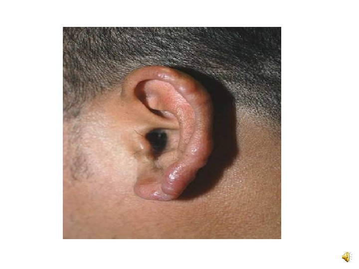
Lepromatous Leprosy –Gross appearance • Macules, plaques, nodules or diffuse skin infiltration may be seen. Lesions are often synmetrically distributed, with poorly defined borders. There is no/little loss of sensation. • Loss of eyebrows and leonine facies may develop • Commonly affected sites include the face, elbows, wrists, ears, etc.

Microscopic Appearance • Hematoxylin and Eosin stained section shows skin with thinned out epidermis with a narrow clear uninvolved subeipderml zone called as Grenz zone. • Dermis is infiltrated by groups/sheets of foamy macrophages called as lepra cells or Virchow cells along with lymphocytes • Granulomas, giant cells or epithelioid cells are not seen
multibacillary form")
• Modified AFB stain ---positive for many lepra bacilli within macrophages (globii)multibacillary form

Diagrammatic representation -40 x

Diffuse reddish patches on back

Subepidermal Grenz zone

Sheets of foamy macrophages in dermis without granuloma formation –low power

Foamy macrophages with clear cytoplasm—high power

Numerous mycobacteria detected on modified ZN stain

Useful links • http: //www. virtualpathology. leeds. ac. uk/ • https: //www. webpathology. com/ • https: //www. pathpedia. com/

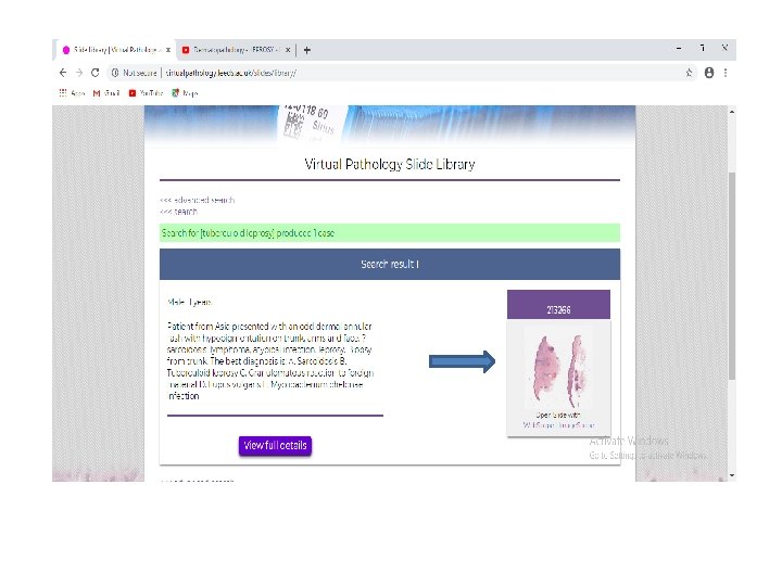
Useful links • You can access whole slide images of these lesions on the Leeds Virtual Pathology site at http: //www. virtualpathology. leeds. ac. uk/ • This is the virtual slide repository of Leeds University, UK. They have an amazing collection of teaching slides which can be viewed on mobile phones and laptops using their online virtual slide viewer.

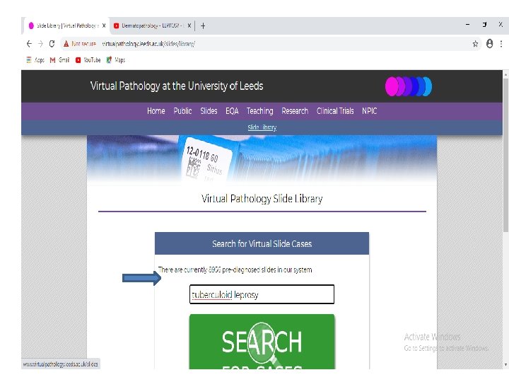
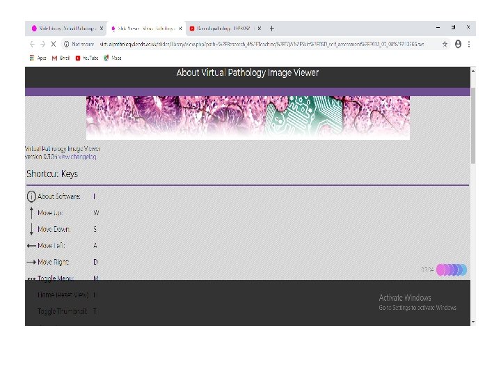
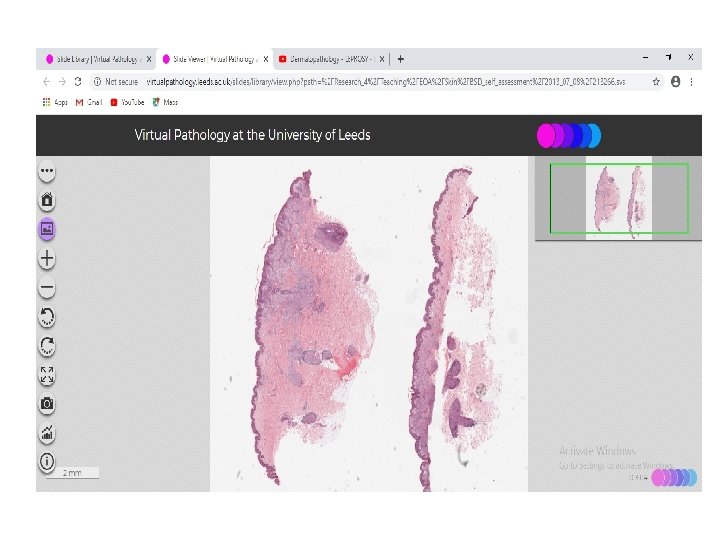
• Go to the website and click on the slide option from the top bar. • Click on the slide viewer icon • Type ‘ tuberculoid leprosy’, or ‘lepromatous leprosy ‘ and then click on the microphotograph image • You can focus on parts of the slide and zoom in or out using the options in the toolbar.

• There are other ways of viewing these slides with more advanced options such as the Aperio Imagescope but this is a software which has to be downloaded on the laptop or desktop and cannot be run on mobile. • The next few slides show stepwise how you can access the slides at leeds, UK. • Happy reading


- Slides: 34