Lecture 9 General Medicine3 rd semester MICROSCOPIC STRUCTURE

 MEDIA ACCESSORY STRUCTURES OF THE EYE OVERVIEW OF DEVELOPMENT")


 - tunica fibrosa - comprises the sclera and the")


 organization in the retina in direction from the")
 is inverse e. g. light must")

 - is a site of leaving axons of ganglion cells")


 Major structural differences between the statokinetic and")
 has 2 main functions: n it serves to hearing n")


 from site to site")


 changes of angular acceleration")


 is very thin and covered from both sides by")
 extends from the insertion of the vestibular")

 consists of supporting cells and hair cells Supporting")


- Slides: 58
Lecture 9 General Medicine_3 rd semester MICROSCOPIC STRUCTURE AND DEVELOPMENT OF THE ORGAN OF VISION MICROSCOPIC STRUCTURE AND DEVELOPMENT OF THE ORGAN OF HEARING
THE EYE AND REFRACTIVE (DIOPTRIC) MEDIA ACCESSORY STRUCTURES OF THE EYE OVERVIEW OF DEVELOPMENT OF THE EYE
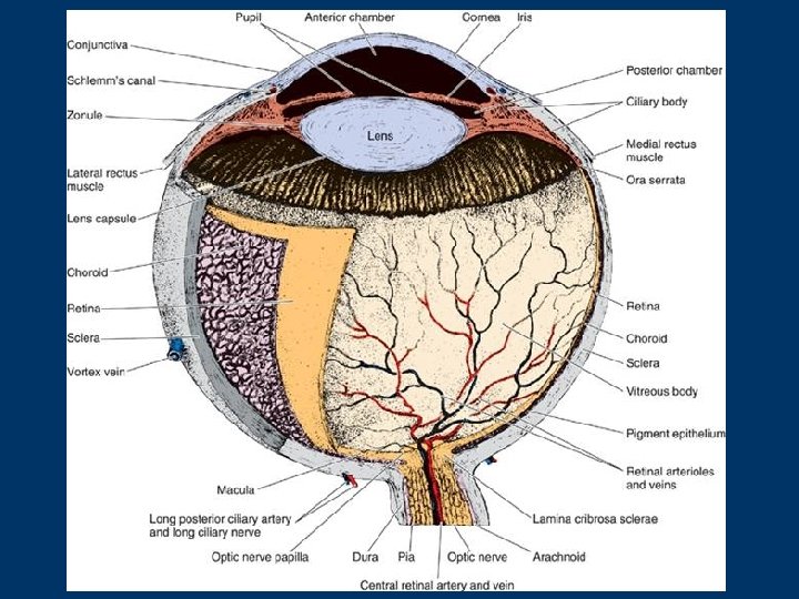
the eye a complex and highly specialized photosensitive organ that permits an accurate analysis of the form, light intensity, and colour reflected from objects it consists of: n n the eyeball (bulbus oculi) the accessory organs of the eye: - conjunctiva, - eyelids, - lacrimal apparatus (lacrimal gland with ducts, 2 lacrimal canaliculi - superior and inferior, lacrimal sac and nasal duct) - and oculomotor muscles on each side THE EYEBALL resembles a sphere, lies in protective bony structure of the skull - the orbit anatomical description of the eyeball uses similar terms as description of the earth globe in geography meridians or meridian planes or sections that transect the poles + the equator or equatorial plane or section oriented upright to the meridian planes and halve them
polus ant. fovea centralis linea visus polus post. axis bulbi ext. n. opticus anterioposterior diameter 24 - 26 mm the eyeball is made up of: - a wall - a content
2 hemispheric segments: the anterior the posterior meridians equator the wall: tunica externa tunica media tunica interna ---------------
n external fibrous coat (layer) - tunica fibrosa - comprises the sclera and the cornea n middle vascular coat (layer) - tunica vasculosa or the uveal tract includes the choroid, ciliary body and iris n internal nervous coat (layer) - tunica nervosa or retina the content of the eyeball are the aqueous humour, lens and vitreous body are known as the refractive (dioptric) media (in broader sense, the cornea is supposed as an integral part of the refractive media) 3 well-defined spaces (compartments) are within the eyeball: n n n the anterior chamber - a space between the cornea and the iris and the anterior face of the lens that is filled with the aqueous humour the posterior chamber - a slit-like shaped space between the iris, ciliary processes, zonular attachment and lens containing also the aqueous humour the vitreous space - a compartment lying behind the lens and zonular attachment and retina; it is occupied by the vitreous body.
Tunica interna oculi, tunica nervosa internal nervous layer - retina the retina lines the eyeball and ends at the margin of the pupil Ø photosensitive portion of the retina - 120– 180 mm Ø not photosensitive portion of the retina - 20 - 25 mm (that is identical with surface epithelium of the ciliary body and the posterior epithelium of the iris) the ora serrata
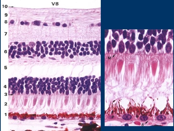
Photosensitive portion of the retina pigment cells, cone and rod cells, bipolar cells, ganglion cells, horizontal cells, amacrine cells, and supporting cells n pigment cells: are columnar in shape with a basal nucleus, basal aspects of cells adhere to Bruch´s membrane, apices have abundant extensions: microvilli and cylindrical sheaths that invest the tips of the rods and cones the cytoplasm contains numerous melanin granules that may migrate from body into microvilli and cylindrical sheaths
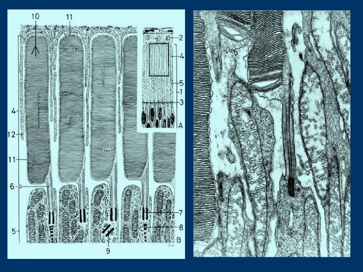
§ Rod and cone cells are thin, elongated and highly specialized nerve cells (50 to 70 x 1. 5 to 3. 0 mm), they form the first neuron of the retina Rod cells consist of: THE PERIPHERAL PART - serves as photoreceptive apparatus; it has two segments - the outer and the inner ones, that are separated each other by a constriction containing an atypical kinocilium the outer segment contains 600 to 1000 flattened membranous disks piled up like a stack of coins. The disks contain visual purple or rhodopsin the inner segment - is rich in glycogen and local accumulation of mitochondria and polyribosomes THE CENTRAL PART - contains nucleus and narrows into the thick process ending with arborizations that contact the dendrites of bipolar cells approx. 120 millions, responsible for night vision The cone cells have similar structure to that of the rod cells but they are shorter and fatter, cells synthesize photopigment iodopsin responsible for visual discrimination and colour vision - total number of cones is about 6 -7 millions
n n n Bipolar cells: have small bodies, one dendrite and one neurit; represent the second neuron of the retina Ganglion cells: show large bodies, numerous dendrites and single axon; cells represent the third neuron of the retina Horizontal cells - association nerve cells that establish contact between different photoreceptors. Cells are located in the internal nuclear layer (near to the outer plexiform layer) Amacrine cells - association nerve cells that establish contact between the ganglion cells. Cells are located in the internal nuclear layer (near to the inner plexiform layer) Supporting cells: are modified glial ones known as Müller cells are columnar in shape and their bodies have many depressions due to neurons Mueller cells occupy practically the entire retina, e. g. its part between the outer and inner limiting membranes (which are formed by their outer or inner cell bases) Nuclei of Müller cells are in the internal nuclear layer
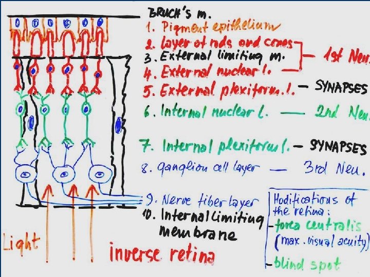
cells described above show layered (laminated) organization in the retina in direction from the Bruch´s membrane to the vitreous space, 10 distinct layers are to distinguish 1. Pigment epithelium (layer) 2. Layer of rods and cones = the peripheral parts of the rod a cone cells 3. External limiting membrane = outer bases of Müller cells THE 1 ST NEURON 4. External nuclear layer= nuclei of rod and cone cells 5. External plexiform layer - contains synapses between the rod (cone) cells and bipolar cells 6. Internal nuclear layer= bipolar cells (it also contains nuclei of Müller cells, bodies of horizontal and amacrine cells) THE 2 ND NEURON 7. Internal plexiform layer - contains synapses between the bipolar and ganglion cells 8. Ganglion cell layer = bodies of multipolar neurons THE 3 RD NEURON 9. Nerve fiber layer = axons of multipolar neurons converging to blind spot 10. Internal limiting membrane = inner bases of Müller cells
Human retina (similar to retina of all vertebrates) is inverse e. g. light must pass through most layers of retina (in all, through 7 layers from 10 th to 3 rd ones) than is absorbed of peripheral parts of rods and cones
Regional differences of the retina the fovea centralis and blind spot show modified microscopic structure n fovea centralis - macula lutea is a depressions lying in the optical axis, in whose centre only cones occur (bipolar and ganglion cells lie at the periphery) - light is directly absorbed with photoreceptors fovea centralis is responsible for maximal visual acuity
blind spot (or papilla) - is a site of leaving axons of ganglion cells no photoreceptors n
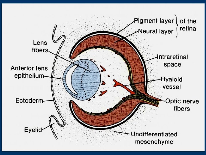
DEVELOPMENT OF THE EYE the eye primordia appear very early (about on the 22 nd day) as optic sulci in the neural folds at the site of forebrain as neural folds fuse, the optic sulci evaginate to form paired hollow diverticula called optic vesicles the optic vesicle then grows laterally on each side and its connection with the forebrain becomes to narrow and forms optic stalk
later both optic vesicles invaginate and become double-walled, cup-like structures - optic cusps they reach up to the surface ectoderm that becomes thickened and form lens placode the central region of each lens placode invaginates and sinks below the surface, forming a lens pit. the edges of the lens pit gradually come together and fuse to form a spherical lens vesicle n Remember: the lens vesicle and optic cusp derive from the ectoderm resp. neuroectoderm and are completely surrounded with head mesenchyme
The retina develops from the double-layered optic cusp the outer layer becomes the pigment epithelium, and the inner layer differentiates into the remaining layers (rod and cone, bipolar, and ganglion cells) intraretinal space, presented initially between the outer and inner layers gradually disappears so that the pigment epithelium and remaining retinal layers fuse the junction of definitive pigment layer with the layer of rods and cones is not so firm as elsewhere so that detachment of retina may occur (after traumatic injury of the eye) the edge of the optic cusp gives rise to the ciliary epithelium and posterior epithelium of the iris is identical with the not photosensitive portion of the retina
The middle and external layers develop from the mesenchyma that envelops the external surface of the optic cusp The lens develops from the lens vesicle the anterior wall of the vesicle gives rise to the anterior epithelium of the lens, the cells of the posterior wall gradually lengthen and form lens fibers the lens capsule is produced by the epithelial cells of both aspects of the lens vesicle nutrition of the lens during development is provided by the hyaloid artery, a branch of the ophthalmic artery rests of the hyaloid artery found in vitreous body are known as hyaloid canal (Cloqueti) The anterior eye chamber originates as cleft-like space that forms between the lens and the surface ectoderm The cornea develops from the surface ectoderm and mesenchyme adhering to it after forming of the anterior eye chamber the stalk of the optic cusp becomes the optic nerve
MICROSCOPIC STRUCTURE OF THE EAR (VESTIBULOCOCHLEAR ORGAN) Major structural differences between the statokinetic and acoustic compartments OVERVIEW OF DEVELOPMENT OF THE VESTIBULOCOCHLEAR ORGAN
the ear (vestibulocochlear organ) has 2 main functions: n it serves to hearing n it serves to maintain equilibrium the ear consists of 3 parts: n the external ear receiver of sound waves - n the middle ear conducts acoustic waves from air to bone and it amplifies them n the inner ear transduction of acoustic waves to specific nerve impulses
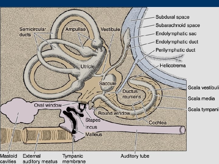
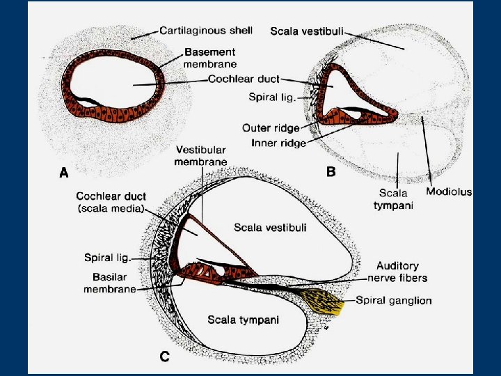
INTERNAL EAR within petrous portion of the temporal bone two labyrinths: Ø the bony (osseous) Ø the membranous the bony labyrinth includes series of spaces: - the vestibule - 3 semicircular canals - cochlea
the membranous labyrinth the utricle and the saccule - are housed within the vestibule Ø the 3 semicircular ducts lie within semicircular canals Ø cochlear duct (scala media) - is housed within the cochlea Ø individual segments are interconnected each other space between the bony and membranous labyrinths = perilymphatic space
the membranous labyrinth is lined with simple epithelium (ectodermic origin) from site to site is differentiated in specialized sensory fields: the maculae (in the utricle and saccule) serve to vestibular function Ø the cristae (in the semicircular ducts) serve vestibular function Ø the organ of Corti (in the cochlear duct) serves auditory function Ø are continuous each other contain endolymph ( fluid with low sodium and high potassium content; the protein concentration is extremely low) a narrow space between the membranous and bony labyrinths is lined with flat cells and is filled with perilymph (has similar ionic composition as the endolymph)
HISTOLOGY OF THE MEMBRANOUS LABYRINTH Saccule and utricle both compartments are bound to the periosteum of the bony labyrinth by thin strands of dense fibrous connective tissue their wall is composed of - a thin sheath of connective tissue - a simple squamous epithelium the epithelium is in one site higher and forms sensory field - the macula (of the saccule/utricle) maculae consist of receptor hair cells supporting cells (and nerve endings) hair cells - on apices have 40 - 80 long rigid stereocilia (highly specialized microvilli) arranged in rows of increasing length and one kinocilium (probably immotile); cells contain numerous mitochondria, a welldeveloped Golgi apparatus, and an abundance of smooth endoplasmic reticulum 2 types of hair cells are distinguished (according to the form of their afferent innervation: type I - have a large, cup-shaped ending surrounding most of the base of the cell, type II- have many small afferent endings Supporting cells - are columnar in shape with microvilli on their apical surfaces, between hair cells
surface of both maculae - covered with a thick gelatinous glycoprotein layer, secreted by supporting cells it contains deposits of calcium carbonate crystals = otoliths or otoconia Function: the maculae respond to linear acceleration (by changes in position of the head, the otoliths displace within the glycoprotein membrane and deformate stereocilia of the hair cells, the deformation results in action potentials that are carried to the CNS
Semicircular ducts are 3 oriented in main planes of body receptor area occupies dilated end of each duct = the ampulla and has an elongated and ridge like form - crista ampullaris structurally, cristae are similar to maculae but glycoprotein layer is substantially thicker and has conical form - a cupula it does not contain otoliths
Function: the cristae respond to angular acceleration (increase or decrease) changes of angular acceleration cause a flow of fluid in the semicircular ducts that induces a movement of the cupula over the crista ampullaris and results in bending of the stereocilia on hair cells is followed by arising of action potentials
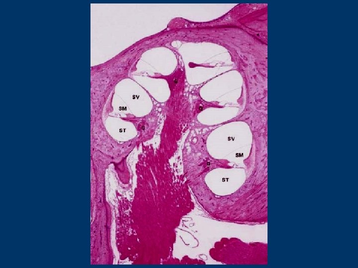
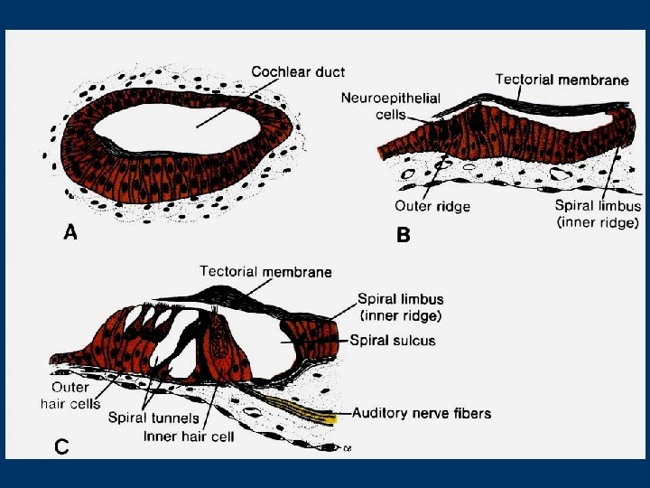
Cochlear duct this compartment of the membranous labyrinth appears to be as diverticulum of the saccule, it is blind and filled with endolymph, about 35 mm long is housed within cochlea it serves as sound receptor The cochlea (saggital section): the cochlear spiral canal - is about 35 mm in total length and makes 2. 5 turns around bony core the modiolus (is penetrated by spaces (chanels) containing blood vessels and cell bodies and processes of acoustic branch of the 8 th cranial nerve - spiral ganglion) the osseous spiral lamina - extends laterally from the modiolus 3 spaces are in the cochlear spiral canal: -- the scala vestibuli - turned to the apical part of the cochlea -- the scala media = cochlear duct -- the scala tympani - turned to the base of the cochlea both scalae communicate each other via opening known as the helicotrema - situated at the apex of the cochlea around the chochlear duct is the perilymph scalae are in realty one long tube, beginning at the oval window and terminating at the round window
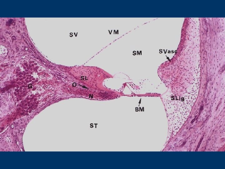
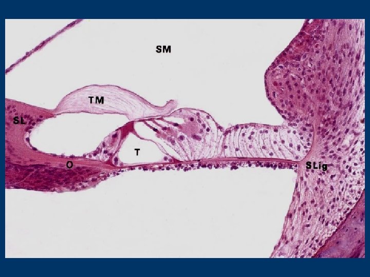
The cochlear duct has triangular profile in sagittal section 3 walls : n a vestibular membrane (Reissner´s membrane) n a lateral wall n a tympanic wall - including the basilar membrane with the organ of Corti peripherally - the osseous spiral lamina centrally
the vestibular membrane (Reissner´s membrane) is very thin and covered from both sides by a simple squamous epithelium extensive tight junction are between cells of both layers
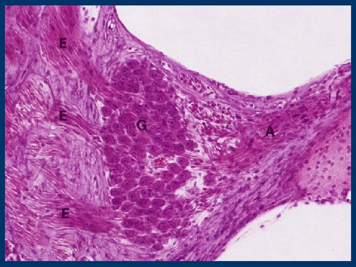
The lateral wall Ductus cochlearis (scala media) extends from the insertion of the vestibular membrane to the spiral ligament its core is a thickened periost covered by highly vascularized stratified epithelium - called as stria vascularis 3 types of cells: marginal, intermediate, and basal marginal cells are responsible for the characteristic ionic composition of endolymph Starting from the prominentia spiralis (thin-walled blood vessel - vas prominens lying here in dense connective tissue), the stria vascularis epithelium is replaced by simple cuboidal epithelium
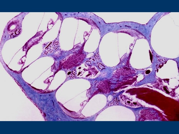
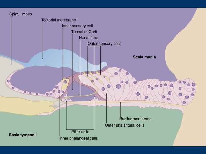
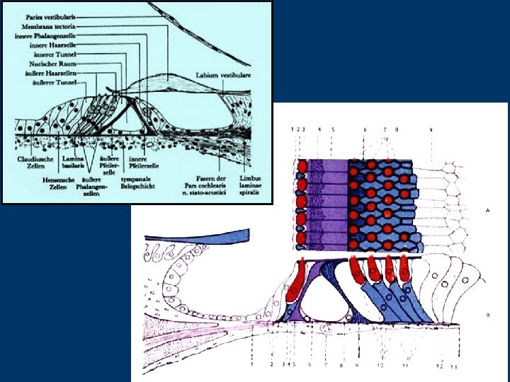
The tympanic wall consists of the basilar membrane and osseous spiral lamina the membrane extends between the crista membranae basilaris and the labium tympanicum of the limbus laminae spiralis is composed of fibrils related to keratin (are produced by cells of the organ of Corti as well as cells lining the scala tympani) the basilar membrane is about 100 mm wide in the basal turn of the cochlea and 500 mm in the apical turn the sound receptor known as organ of Corti rests on the membrane
ORGAN OF CORTI (seu papilla spiralis) consists of supporting cells and hair cells Supporting cells involve several different types lie in rows arranged in direction from sulcus spiralis externus to sulcus spiralis internus: n n n n Claudius´ cells and Boettcher´s cells - columnar in shape and line the external spiral sulcus Hensen's cells - form several rows and are tall and slim outer phalangeal (Deiter's) cells lie in 3 to 5 rows (depending on the position of the turn (apically the number of rows is increasing) outer and inner pillars cells that limit the inner tunnel (of Corti), pillar cells are highly modified epithelial cells whose apices terminate with cuticular plates, their bodies contain numerous tonofilaments the Nuel' s space - between the outer pillar cells and adjacent row of phalangeal cells inner phalangeal cells - lie axially to the inner pillar cells and only in one row border cells - tall and slim cells lowering in height towards the internal spiral sulcus interdental cells - cover the labium vestibulare of the limbus laminae spiralis and produce gelatinous membrana tectoria or tectorial membrane extending above the apices of hair cells.
Hair cells the outer hair cells - lie in 3 -5 rows and are supported by the outer phalangeal cells n the inner hair cell - occur in single row and are supported by the inner phalangeal cells hair cells are columnar, with nuclei located basally, numerous mitochondria and cisternae of smooth endoplasmic reticulum in the cytoplasm n apices of hair cells are provided with stereocilia - W shaped in the outer cells and - linear in the inner ones the height of stereocilia increases from one side of array to the other the tips of the tallest stereocilia of the outer hair cells are embedded in the tectorial membrane
Function of the organ of Corti: because the ossicle form a unit and the base of stapes is inserted in the oval window, vibrations of the tympanic membrane cause the vibrations of perilymph of both scalae (vestibuli and tympani) the pressure changes are transmitted across the vestibular and basilar membrane of the cochlear duct, which result in positional changes of stereocilia against tectorial membrane sequence of mentioned processes induces to arise action potentials within hair cells
OUTLINE OF DEVELOPMENT OF THE EAR THE EXTERNAL EAR n The external acoustic meatus develops from the dorsal end of the 1 st branchial groove; ectodermal cells at the bottom of the groove proliferate and extend inward as a solid epithelial plate - meatal plug; in the fetal period, the central cells of this plug degenerate, forming cavity that becomes the inner part of the external acoustic meatus
n The auricle develops from 6 swellings known as auricle hillocks that surround the margin of the first branchial groove 3 hillocks are on the first branchial (mandibular) arch and 3 on the second (hyoid) branchial arch at the end of the 2 nd month all hillocks fuse to form the definitive pinna The tympanic membrane (TM) derives from the branchial membrane separating the 1 st branchial groove from the 1 st pharyngeal pouch n initially, the membrane is made up of only the ectoderm and endoderm, as development proceeds, mesenchyme grows between both germ layers and is differentiated into the fibrous stratum of the TM, the ectoderm gives rise to the epidermal and the endoderm to the mucous aspect of the definitive TM
THE MIDDLE EAR 2 different embryonic anlages - the 1 st pharyngeal pouch and - cartilages of the 1 st and 2 nd pharyngeal arches n The tympanic cavity - 1 st pharyngeal pouch - its distal expanded end then envelopes the auditory ossicles the proximal unexpanded portion becomes the Eustachian tube
the auditory ossicles n the malleus and incus from dorsal part of Meckel´s cartilage (supporting the first branchial or mandibular arch) n the stapes from dorsal part of Reichert´s cartilage (supporting the second or hyoid branchial arch) during the late fetal period, the tympanic cavity becomes larger and expands into the temporal bone = mastoid antrum
developes from the ectoderm THE INNER EAR first anlage occurs early during the 4 th week as a thickened plate of the ectoderm - otic placode - on each side of the head placode invaginates and sinks below the surface ectoderm into the underlying mesenchyme to form otic pit edges of the pit come together and fuse to form an otic vesicle or otocyst that lies laterally to the rhombencephalon
the otocyst serves a primordium of future membranous labyrinth
two divisions are early recognizable on the otocyst: a dorsal or utricular portion, differentiating into the utricle, semicircular ducts and endolymphatic duct and sac and a ventral or saccular portion that gives rise to the saccule and cochlear duct
Initially, the semicircular ducts form flat-like diverticula growing out from the utricular portion; central parts of them then fuse and disappear the peripheral infused portions of the diverticula become the semicircular ducts from the ventral saccular portion of the otocyst, the coiled cochlear diverticulum grows out starting the 4 th month, differentiation of maculae begin in the utricle and saccule, cristae within semicircular ducts and the organ of Corti within the cochlear duct the mesenchyme around the otic vesicle (later its parts) condenses and differentiates into the bony labyrinth a space separating the membranous labyrinth from the osseous one soon fills the perilymph