IVASIVE DISEASE OF THE VULVA 5 th class


 1 a Confined to vulva and or perineum,")


 ,")


 wide and deep")


- Slides: 33
IVASIVE DISEASE OF THE VULVA 5 th class 2018 -2019 Pro. Dr Esraa Al-maini
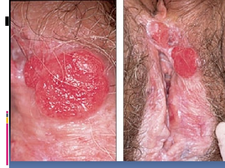
Malignant tumor of the vulva are uncommon, it represent about 4% of female genital malignancies It about three times less common than cervical cancer majority of patients are elderly post menopausal women with mean age at diagnosis is 65 years. 95% of cases are sequmous, and melanoma is the commonest of the reminder (adenocarcinoma , basal cell carcinoma , sarcoma).
Epidemniology: Two different etiological types of vulual cancer: 1 st type seen mainly in younger patient related to HPV and smoking and commonly assosciated with VIN of warty type. 2 nd more common type seen in elderly usually associated with long standing lichen seclerosus is common. Vulual cancer occur in association with lymhpogranuloma venerum and granuloma inguinale. 5% of patients have positive result on serologic testing for syphilis If occur at earlier age and associated with graver prognosis
SPREED Local invasion: into the underlying and surrounding tissues and into the vagina, and the anus vulval cancer spreads predominantly via lymphatic system(lumphatic embolization to regional lymph nodes) The lymph drains from the vulva to the inguinal and femoral gland in the groin and then to the external iliac glands.
Drainage to both groins occurs from: midline structure unilateral structures-( the perineum and the clitoris) some contra lateral spread may take place from other parts of the vulva spread to the controlateral groin occurs in about 25% of those cases with positive groin nodes Lesion less (than 1 mm carry low risk of lymphatic invasion) Hematogenous spread to distant sites like lung, liver, bone , rarely occur in absence of lymphatic spread.
FIGO STAGING OF VULVAL CANCER (1995) 1 a Confined to vulva and or perineum, 2 cm or less maximum diameter, groin nodes not palpable stromal invasion no greater than 1 mm 1 b As for 1 a but stromal invasion more than 1 mm 2 Confined to vulva and or perineum, more than 2 cm maximum diameter , groin nodes not palpable 3 Extends beyond the vulva, vagina , lower urethra or anus or unilateral groin node lymph node metastasis 4 a Involves the mucosa of rectum or bladder upper urethra, or bilateral regional lymph node metastasis and /or pelvic bone 4 b Any distant metastasis including pelvic lymph node
≤ 2 cm
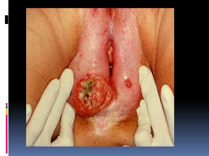
DAIGNOSIS AND ASSESSMENT 71% complain of irritation or pruritus , 57%note a vulval mass or ulcer, it usually not until the mass appears that medical advice is sought. Bleeding 28%and discharge 23%are less common presentation Because of the multicentric nature of female lower gental tract cancer, , the investigation of patient with investigation of a patient with vulvar cancer should include inspection of the cx and cervical cytology. The groin nodes must be palpated carefully and any suspicious nodes sampled by fine-needle aspiration Achest X-ray is always required , but intravenous pyelography or MRI of the pelvis may sometimes be helpful EUA and a full thicknes , generous biopsy are the most important
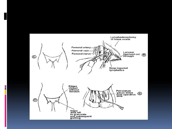
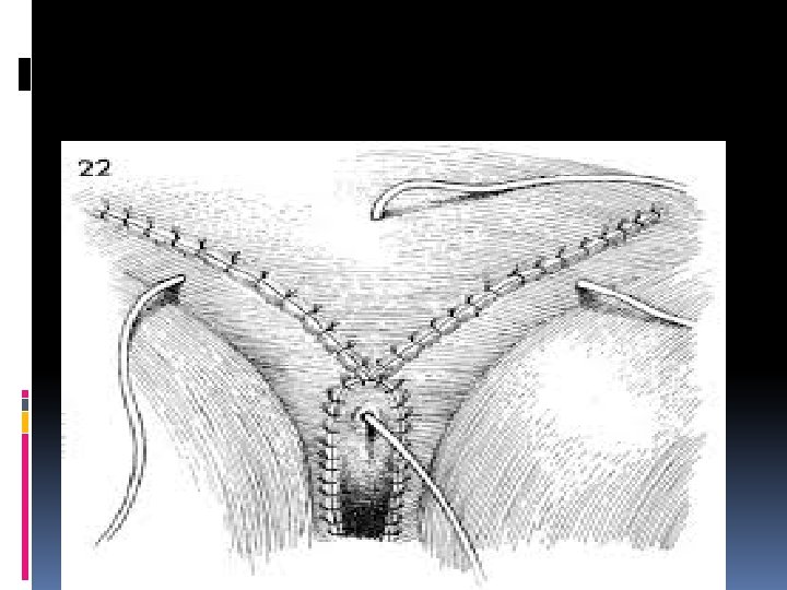
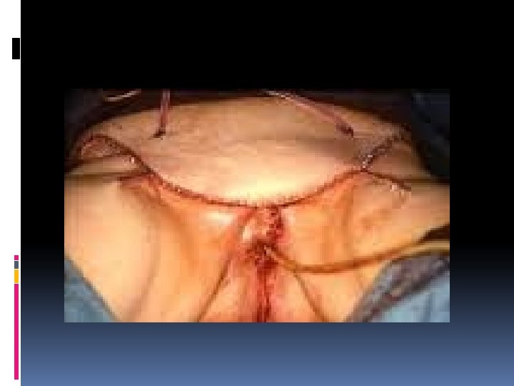
TREATMENT Surgery is main stay of treatment 1 -Radical vulvectomy and bilateral inguinofemoral lymphadenectomy with or without pelvic lymph adnectomy Reduced the mortality from 80% to 40% to control the lymphatic spread, remove large area of normal skin in the groins. The purpose of this operation is to remove the vulva, its adjacent structures, a margin of normal tissue, and the inguinal lymph nodes from the anterior superior iliac spine to the abductor canal in the leg Primary wound closure was rarely achieved.
2 -Modifications of this en-bloc excision were devised to allow primary closure and reduce the considerable morbidity But morbidity still high Impaired psychosexual function was common
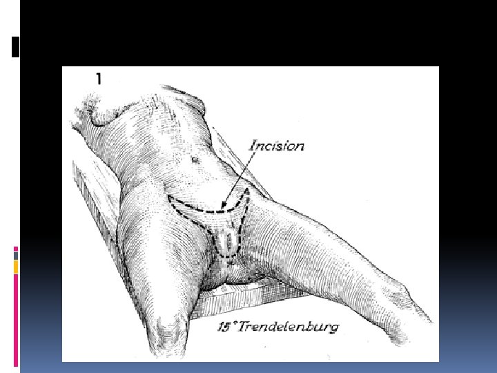
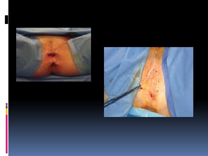
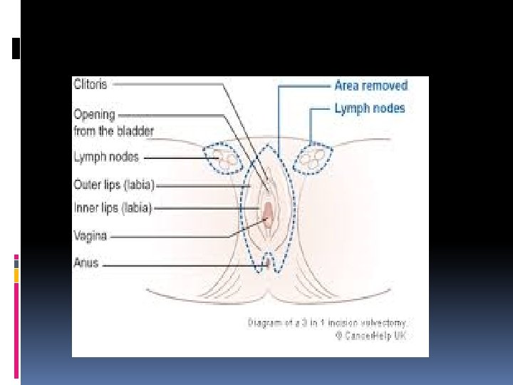
3 -Then replaced by operation using three separate incisions(vulval and groin incision) , this greatly reduced the morbidity of surgery and decrease wound break down (this depended on the principle that lymphatic metastases developed initially by embolization)
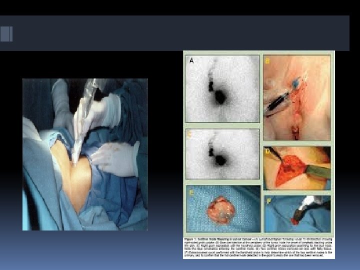
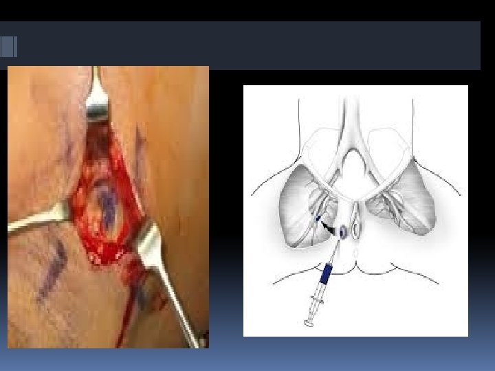
4 -Current research is focusing on identification of a sentinel node or nodes (by injecting blue dye around the primary tumour so lymph node identified and resected. so full groin dissection could be avoided.
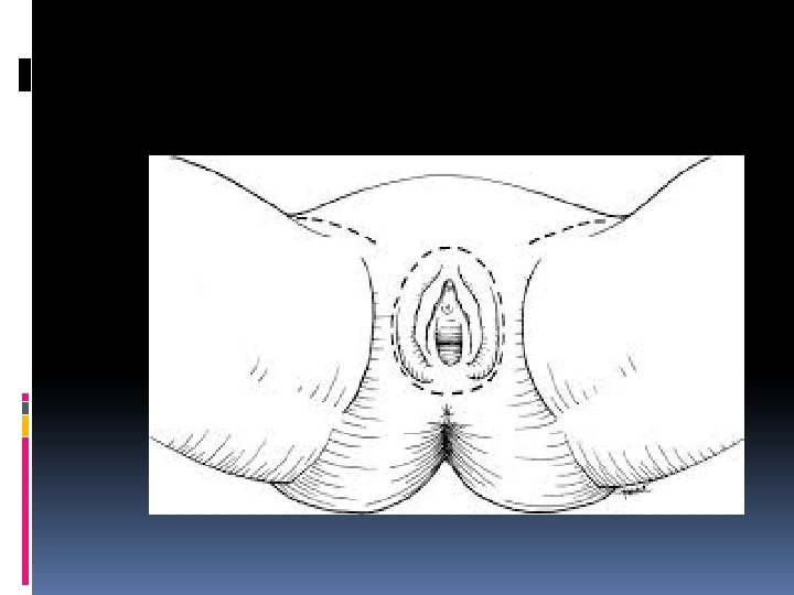
Treatment of early vulval cancer: No t need dissection -Patient with stage Ia do not need groin dissection. wide local excision of tumor with free margin
All patients require at least an ipsilateral inguinal – femoral lymphadenectomy EXCEPT STAGE 1 a - Ipsilateral lesion there is about a 1% risk for involvement of the controlateral nodes if the ipsilateral nodes are negative 1% If -ve
-Patients with stage Ib and IIa(lesion confined to the vulva) wide and deep local excision (Rdical local excision)is effective as radical vulvectomy in preventing local recurrence. Surgical margin should be at least 1 cm free at histopathological examination , with either unilateral or bilateral groin node dissection, if the ipsilateral side +ve then 25%the other side will be involved. 10%chance of local recurrence with either treatment. (local or radical)
Patient with midline lesions invading less than 1 mm , bilateral groin dissection is not necessary Wide local excision, if larger the bilateral groin dissection In patients with midline lesions, less than 1 cm from the midline, an attempt should be made to identify sentinel nodes in each groin. If a sentinel lymph node is not found bilaterally, then a full inguinofemoral d issection is indicated on the side without the sentinel node.
COMPLICATIONS OF SURGERY: 1 -Wound breakdown and infection with triple incision this become minor problem 2 -Osteitis pubis rare need intensive prolonged antibiotic therapy 3 -Thromboembolic disease : reduce by preoperative epidural analgesia to ensure good venous return with subcutaneous heparin begun 12 -24 hours before the operation seems to reduce this risk 4 - 2 nd haemorrhage 5 -Chronic leg oedema may be in 15%
6 -Numbness and par aesthesia over the anterior thigh are common due to the division of small cutaneous branches of the femoral nerve 7 - loss of body image and impaired sex function
Pateints with advance vulval cancer: If proximal urethra , anus , rectovaginal septum involved by the tumor , preoperative radiation or chemoradiaiton should be used to shrink the primary tumor followed by more conservative surgery and avoid the stoma bilateral groin node dissection , or at least removal of any large , positive nodes is usually performed before radiation therapy.
Radiotherapy: 1 - may have a place in reducing the size of a very large lesion prior to surgery. 2 -Radiotherapy used when more than one nodal micrometastaseis (≤ 5 mm in diameter )one or more macrometastases, or evidence of extranodal spread should receive post operative radiation to both groins and to pelvis nodes. 3 -In treatment of tumor involving midline structure clitoris , anus…. .
Prognosis: Patients with positive nodes have a 5 years survival rate of about 50% patients with -ve node 90% 5 years survival rate
THANK YOU