Histology of Nervous System Anatomical Division Central Nervous


 SNS (Sensory) ANS (Sensory) Brain Spinal cord ENS (Sensory)")
 •")
 Nissl bodies (r.")


 • Peripheral nerves contain a considerable amount of connective tissue. The")


: Astrocytes • Star-shaped cells • Form Blood-brain Barrier Forms Perivascular Feet")
: Oligodendrocytes • Most common glial cell type • Each forms Myelin")
: Microglia • Small cells found near blood vessels • Phagocytic Cell")
: Ependymal cells • Form epithelial membrane lining cerebral cavities & central")
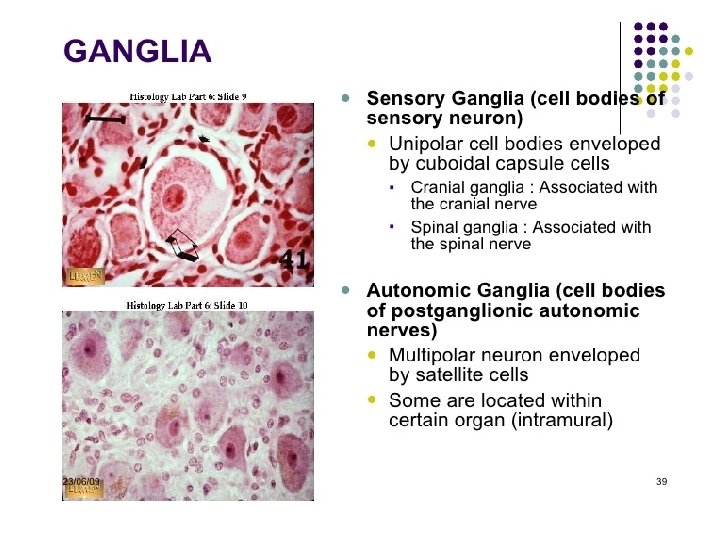
: Satellite Cells • Flat cells surrounding neuronal cell bodies in peripheral")


 Motor Pathways • In Somatic System One Neuron in")


: Schwann Cell • Cells encircling PNS axons • Each cell produces")

")


 2. Saltatory conduction (myelinated fibers)")
 from node")


- Slides: 37
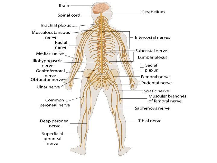
Histology of Nervous System Anatomical Division: Central Nervous System: Brain & Spinal Cord. Peripheral Nervous System: Axons, Spinal Nerves, Cranial Nerves & Ganglia
Nervous Tissue Functions Receive/Analyze/Integrate-External & Internal Stimuli Response in Effector Organs
Nervous Tissue • Controls and integrates all body activities that maintain life • Three basic functions 1. sensing changes with sensory receptors 2. interpreting and remembering those changes 3. reacting to those changes with effectors 5
Organization Sensory Integration SNS (Motor) SNS (Sensory) ANS (Sensory) Brain Spinal cord ENS (Sensory) 6 Motor ANS (Motor)
Gray and White Matter • White matter = myelinated processes (white in color) • Gray matter = nerve cell bodies, dendrites, axon terminals, bundles of unmyelinated axons and neuroglia (gray color) 7
Neurons Functional unit of nervous system • 1. Cell body a) Nissl bodies (r. ER) b) Neurofilaments c) Microtubules d) Lipofuscin pigment clumps 2. Cell processes a) Dendrites b) A single Axons 8
Electron Microscopy One of My Research/Published Light-Stained Purkinje Neurons
Dendrites • Conducts Impulses Towards The Cell Body • Typically short, highly branched & unmyelinated • Surfaces specialized for contact with other neurons • Contains neurofibrils & Nissl bodies (r. ER) 12 impulse
Axons • Conduct impulses Away From Cell Body • Long, thin cylindrical process of cell • Arises at Axon Hillock • Impulses arise from initial segment (Trigger Zone) • Side branches (collaterals) end in fine processes called Axon Terminals • Swollen tips called synaptic end bulbs contain vesicles filled with Neurotransmitters ((Neuromuscular Junction) 13
Axon (Peripheral Nerve) • Peripheral nerves contain a considerable amount of connective tissue. The entire nerve is surrounded by a thick layer of dense connective tissue, the epineurium. Nerve fibres are frequently grouped into distinct bundles, fascicles, within the nerve. The layer of connective tissue surrounding the individual bundles is called perineurium. The perineurium is formed by several layers of flattened cells, which maintain the appropriate microenvironment for the nerve fibres surrounded by them. The space between individual nerve fibres is filled by loose connective tissue, the endoneurium. 14
Axons 15
Structural Classification of Neurons Based on number of processes found on cell body 1. Multipolar = several dendrites & one axon • most common cell type 2. Bipolar Neurons = one main dendrite & one axon • found in retina, inner ear & olfactory 3. Unipolar Neurons = one process only(develops from a bipolar) • are always sensory neurons • 17
Structural Classification of Neurons Based on number of processes found on cell body 1. multipolar = several dendrites & one axon • most common cell type 2. bipolar neurons = one main dendrite & one axon • found in retina, inner ear & olfactory 3. unipolar neurons = one process only(develops from a bipolar) • are always sensory neurons • 18
Structural Classification of Neurons Based on number of processes found on cell body 1. Multipolar Neurons= several dendrites & one axon • most common cell type; Motor Neuron 2. Bipolar Neurons = one main dendrite & one axon • found in retina, inner ear & olfactory (Special Senses) 3. Unipolar Neurons = one process only(develops from a bipolar) • are always Sensory Neurons • 19
Neuroglial Cells Structural Support Cells Half of the volume of the CNS Smaller cells than neurons Cells Can Divide – rapid mitosis in tumor formation (Gliomas) • • • 4 cell types in CNS – astrocytes, oligodendrocytes, microglia & ependymal • 2 cell types in PNS – schwann and satellite cells 20
Neuroglial Cells (CNS): Astrocytes • Star-shaped cells • Form Blood-brain Barrier Forms Perivascular Feet by covering blood capillaries • Metabolize neurotransmitters • Regulate K+ balance • Provide structural support 21
Neuroglial Cells (CNS): Oligodendrocytes • Most common glial cell type • Each forms Myelin Sheath around more than one axons in CNS • Analogous to Schwann cells of PNS 22
Neuroglial Cells (CNS): Microglia • Small cells found near blood vessels • Phagocytic Cell – • Derived macrophages & monocytes 23
Neuroglial Cells (CNS): Ependymal cells • Form epithelial membrane lining cerebral cavities & central canal • Produce Cerebrospinal Fluid (CSF) 24
Neuroglial Cells (PNS): Satellite Cells • Flat cells surrounding neuronal cell bodies in peripheral ganglia • Support Neurons In The PNS Ganglia 25
Spinal Ganglia • Sympathetic Ganglia Cell bodies Lie on posterior Nerve Root of Spinal Cord. • Have Sensory Neurons (Unipolar). • Seen Closely • Multipolar Type: Hence Many Dendrites Hence Widely Apart.
Ganglia • Ganglia Are Masses Of Neuronal Somas, Usually Defined As Being Outside The Central Nervous System. They Seem To Act As Coordinating Way Stations. • Two type Ganglia: • 1. Sensory. 2. Autonomic 27
Anatomy of Autonomic (In. Voluntary) Motor Pathways • In Somatic System One Neuron in CNS connected to effector Organ) • ANS Posses 2 -Neurons between CNS & Effector Organ i. e: • 1. Preganglionic neuron: Lie in CNS, axons Synapse with Multipolar Neuron that is PGN (Acetylcholine) • 2. Postganglionic neuron (PGN): Fibers Connect To Effector Organ (Cardiac, Smooth Muscles & Glands. (Norepinephrine) • Two divisions: • Sympathetic: Prepare Body ((Flight or Fight)) • Parasympathetic: Opposite Function ((Rest or Digest)) 28
29
30
Neuroglial Cells (PNS): Schwann Cell • Cells encircling PNS axons • Each cell produces part of the myelin sheath surrounding an axon in the PNS 32
Myelination • Insulation of axon • Increase speed of nerve impulse
Myelination: PNS • All axons surrounded by a lipid & protein covering (Myelin Sheath) produced by Schwann cells • Neurilemma is fused layers of membranes of Schwann cell – gaps called nodes of Ranvier • Myelinated fibers • Unmyelinated fibers Node of Ranvier
Myelination: CNS • Oligodendrocytes myelinate axons in the CNS • Broad, flat cell processes wrap about CNS axons. One Oligo Can Myelinate Upto 50 Nerves. • Schwan Cell: PNS • Many Schwan Cells Myelinate One Peripheral Nerve • End of one Schwan Segment Node of Ranvier 35
Myelination Process
Electrical Signals in Neurons • • • 37 Neurons are electrically excitable due to the voltage difference across their membrane Communicate with 2 types of electric signals 1. action potentials that can travel long distances 2. graded potentials that are local membrane changes only In living cells, a flow of ions occurs Through Ion Channels In The Cell Membrane
Continuous versus Saltatory 1. Continuous Conduction conduction (unmyelinated fibers) 2. Saltatory conduction (myelinated fibers) 39 Na Na Na A. P.
Saltatory Conduction • Nerve impulse conduction in which the impulse Jumps (Salta) from node to node Na Na Na 40 A. P.
Signal Transmission at Synapses • 41 2 Types of synapses 1. Electrical • ionic current spreads to next cell through gap junctions • faster, two-way transmission & capable of synchronizing groups of neurons 2. Chemical • one-way information transfer from a presynaptic neuron to a postsynaptic neuron – axodendritic -- from axon to dendrite – axosomatic -- from axon to cell body – axoaxonic -- from axon to axon
Synapses