GENERAL MANAGEMENT OF POISONED PATIENTS Dr Nayira A


 Airway Causes of airway obstruction include • Mucosal swelling, Secretions, Posterior")
 Breathing difficulties Due to ventilation or oxygenation failure. that represent major")
 Breathing Causes of oxygenation failure: • Agents which reduce tissue oxygen-utilization")


 Circulation • This should be done to establish that the tissues")
 Depression of the central nervous system • Numerous drugs are capable")
 and Seizures. Generalized seizures secondary to toxins are usually brief")


 atropine. ,")


-Emesis Emetics are substances that cause regurgitation of gastric")
Emesis General conditions in which emesis should not be")
-Gastric lavage • • Gastric lavage is only effective when ingestion of the poison")


 forms a semipermeable membrane")


. It chelates iron to form ferrioxamine. The")


. It is an antagonist")
 and folic acid. Folic")


- Slides: 59
GENERAL MANAGEMENT OF POISONED PATIENTS Dr. Nayira A. Abdel Baky Associate Professor Pharmacology and Toxicology
Remember • All chemicals have potential to be poisons if given in a large enough dose • Poisoning occurs when exposure to a substance adversely affects function of any organ system
Treatment • DECONTAMINATION! ALWAYS be sure that the patient has been decontaminated PRIOR to doing anything to them! • ABC’s… Always stabilize the patient like you would any other patient initially. • Draw labs, obtain your EKG, get x-rays if needed, begin fluids, and if needed. • Decrease absorption or Enhance elimination
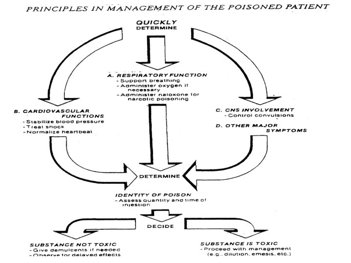
Management Steps 1 -Supportive – Maintain airway clean, and assure respiration. – Correct hypoxia, hypotension, dehydration, hypo- or hyperthermia, and acidosis – Control seizures 2 -Monitor – PR, BP, ECG, Oxygenation 3 -History & Physical examination 4 -General Steps – Absorption – Elimination 5 -Specific antidotes
I-Stabilization of the patient
I-Emergency stabilization *First priorities are ABCDE’s *Vital sign including pulse , oxygen and hypoglycemia must be corrected. *Only in very rare incidences does administration of antidote precede stabilizing ABCDE and vital signs. *Unresponsive pt’s treated empirically with coma cocktail : Oxygen, naloxone, glucose, and thiamine
Evaluation of (A) Airway Causes of airway obstruction include • Mucosal swelling, Secretions, Posterior displacement of the tongue, Foreign bodies and Trauma. Signs and Symptoms • Dyspnea, Dysphoria, Air hunger, Cyanosis, Diaphoresis and Tachypnea Measures to clear the airway obstruction • Clear the airway of secretions with suction and remove any foreign body. • Use of naso-pharyngeal tube or oro-pharyngeal airways in patients to prevent pharyngeal obstruction caused by collapse of the posterior base of the tongue. Intubation in case of comatosed patients without a gag (cough) response to prevent aspiration. • Crico-thyroidotomy or tracheostomy
Evaluation of (B) Breathing difficulties Due to ventilation or oxygenation failure. that represent major cause of morbidity and death. Causes of ventilation failure: • Drug induced respiratory depression, pneumonia, Pulmonary edema, Lung abscess, Pulmonary emboli, Bronchospasm from numerous environmental & occupational sources and Tetanus-induced respiratory failure. • Ventilation is evaluated by measuring the partial pressure of arterial carbon dioxide (Pa. CO 2). It is the ability of the lungs to remove carbon dioxide which is an accurate estimate of alveolar ventilation. Normal Pa. CO 2 levels are 37 -43 mm. Hg. When alveolar ventilation decreases, arterial Pa. CO 2 levels rise. e. g. toxicity with CNS depressants.
Evaluation of (B) Breathing Causes of oxygenation failure: • Agents which reduce tissue oxygen-utilization include cyanide or hydrogen sulphide. • Agents which decrease O 2 -carrying capacity include CO 2 and other agents which cause methemoglobinemia. The level of arterial oxygen is estimated by measuring the arterial oxygen tension (Pa. O 2). Reduced arterial oxygen produces hypoxemia (Pa. O 2 < 55 mm. Hg). Signs of inadequate oxygenation: Cyanosis, Tachypnea, Hypoventilation, Diaphoresis, Suprasternal and or intercostal retractions and altered mental state. Evaluation of inadequate oxygenation • Arterial blood gas measurements. However, A normal Pa. O 2 does not guarantee adequate tissue oxygenation • Chest X-ray • Tidal volume measurement (Tidal volume is the amount of air that is inhaled and exhaled in a single breath).
Treatment 1 -Supplemental Oxygen should be given to all poisoned patients with depressed consciousness or hypoxemia. Supplemental O 2 does not correct hypoventilation. Hypoventilation requires assisted ventilation and supplemental oxygen delivered by nasal catheters and cannulae. Complications of oxygen therapy. • Drying of mucosal membranes. • Trauma associated with tubes, catheters or masks. • Increased formation of free radicals, which interact with cellular enzymes and membranes to produce toxic effects
Cont. ; Treatment 2 -Administer 100% oxygen in carbon monoxide poisoning 3. Give cyanide antidote kit for cyanide poisoning. 4. Remove the patient from the source of exposure to any irritant gas. 5. Administer bronchodilators in case of bronchospasm a. salbutamol inhaler b. If this is not effective, give aminophylline, IV 6 -For patients with bronchospasm and bronchorrhea caused by organophosphate or other anticholinesterase poisoning, give atropine.
Evaluation of (C) Circulation • This should be done to establish that the tissues receive adequate amount of blood. The important sign of inadequate tissue perfusion is shock • Early signs of shock include anxiety and agitation which reflect sympathomimetic excess, Tachycardia, Narrowing of pulse pressure (difference between systolic and diastolic blood pressure), Decreased urinary output (Oliguria), postural hypotension, increased urine specific gravity and decreased urine sodium, Hypotension usually less than 90 mm Hg systolic. Late signs include delirium, stupor and coma. Treatment. • Hypotension is treated with fluids, Position change, Vasopressors (if adequate fluid challenge does not raise the systolic blood pressure above 80 -90 mm. Hg and signs of shock persist after several liters of fluid), a vasopressor should be used as Dopamine and Norepinephrine. Constant cardiac and blood pressure monitoring should be done every 5 minutes
Evaluation of (D) Depression of the central nervous system • Numerous drugs are capable of depressing the CNS. This depression is evaluated by measuring the pupillary size and responsiveness. The maximum miotic response to sympathetic paralysis or parasympathetic excess is 1. 5 -2 mm, whereas, the maximal mydriatic response to sympathetic excess or parasympathetic paralysis is 8 -9 mm. • Good prognostic indications of recovery from hypoxic/ischemic coma include initial presence of the pupillary light reflex, motor responses to pain, and /or spontaneous eye movements. The coma cocktail (GTN) is a group of antidotes be administered to all individuals who present with depressed mental status. It includes: 1. Glucose which is given to guard against hypoglycaemia. 0. 5 -1. 0 g/Kg is given as 50% dextrose in water in adults, 25% dextose in water in children, and 10% dextrose in water in neonates. • 2. Thiamine: 100 mg IM or IV is given as treatment and prevention of Wernicke’s encephalopathy which is characterized by eye movement paralysis, ataxia and mental status changes, occasionally seen in alcoholics and other people with poor nutritional status. 3. Naloxone which is a specific opiate antagonist, is given to comatose patients or those with depressed neurologic signs.
Evaluation of Excitation (E) and Seizures. Generalized seizures secondary to toxins are usually brief and self-limited. Treatments of seizures Diazepam (Valium) or other benzodiazepine as lorazepam (Ativan) Phenytoin (Dilantin) if seizures are persistent Phenobarbital is a third-line drug. Coadministration of phenobarbital and diazepam causes synergistic respiratory depression and should be avoided • General anaesthesia is the last choice in case of persistent status epilepticus. • Intravenous pyridoxine effectively stops seizures duo to isoniazide overdose. • • • Seizures unresponsive to diazepam may result from altered body homeostasis and trauma e. g. acidosis, hypocalcemia, electrolyte imbalance, hypoxia, meningitis and subarachnoid hemorrhage. The presence of seizures may also indicates a serious toxic condition that requires methods to enhance drug elimination as hemodialysis use in seizures induced by theophylline, lithium and salicylate.
The violent patient Toxins and conditions induced violence include -Phencyclidine, alcohol, heroin, amphetamine, and LSD -Head trauma, cerebral edema, stroke, meningoencephalitis, CNS bleeds -Metabolic abnormalities capable of altering consciousness (e. g. hypoglycaemia or hyponatremia). Measures against violence include; -Administration of haloperidol with benzodiazepines. -Stabilization of blood glucose level quickly in any violet patient
II-Diagnosis of poisons
A-History 1 -History is key to the approach of the poisoned patient. 2 -Establish an exposure time , Estimate the dose. 3 -Determine other factors that may be present and affecting your treatment. • What type of exposure was this? • Environmental, industrial, accidental, homicidal, suicidal, or unknown? (Empty bottles , if he was taken from garage…. ? , a factory…etc). • Establish the dose if known. Number of pills in the bottle, time ingested/exposed, or have they vomited since ingestion? • The patient's psychological profile: This is difficult because the poisoned individual may be unconscious, unresponsive or confused. Information usually obtained from relatives or friends. • Identify if the patient performed any self treatment or took any medication?
Cont. ; History 1. Adults • Conscious patients; about 80% of adults who take poisons are conscious on arrival at hospitals and they can give their statements about the nature of the poison and the quantity of the poison taken 2 2. Children. • Diagnosis of poisoning in children is based on • The presence of traces (open container, and /or the hands, face, clothing and surrounding floor soiled with some household product) or disintegrated tablets could point to the accidental poisoning in children. • Presence of abnormal behaviour, convulsions, ataxia or GIT disturbances
B-Physical examination • Physical exam should include all body systems. • Many clues may be obtained from the physical exam. • Some poisons produce clinical characteristics which strongly suggest the type of the poison 1 -Presence of characteristic odour • Breath odour (Ethanol) • Garlic (Arsinic, organophosphate phosphorus) • Biter almonds (cyanide) • Acetone (isopropanol, nail polish remover, salicylate) • Pearlike (chlorahydrate) 2 -Skin manifestation a- Changes in the skin Color - Red in carbon monoxide poisoning. , alcohol, antihistaminics, atropine cyanide - Pale and dry in atropine, amphetamine, narcotics, antihistaminics poisoning. - Excessive sweating (Organophosphorous compounds, nicotine, sympathomimetics) - Cyanosed in cyanide, nitrites, strychnin -Yellow “Jaundice” in acetaminophen, carbon tetrachloride , arsenic, castor bean.
Cont. ; Physical examination b- Needle markers: Indicate addiction by intravenous route e. g. cocaine. c- Alopecia: in Thallium. d-Burns & corrosion: Acids & alkali e-Fingernails may hold many lines in a variety of poisonings. Brittle fingernails in thalidomide ingestion. Lines/Grooves in sodium fluoride. Heavy metals may cause staining or other abnormalities of the nails.
Cont. ; Physical examination 3 -Eye manifestation: a- Eye globe: i- Lacrimation: In organic phosphorous • ii- Nystagmus: In barbiturates, Phenytoin, phencyclidine b- Pupils: i- Miosis: clonidine, carbamates, opiates, organophosphates • ii-Mydriasis: anticholinergics or sympathomimetics , amphetamine, cocaine, LSD, atropine. 4 -Gastro-intestinal manifestations: a- Mouth: i- Salivation: In organophosphorous compounds. ii- Dry mouth: In atropine and other anticholinergic drugs. iii- Corrosion : With corrosives. iv- Gum discoloration: Lead causes blue line. Cadmium causes yellow line. Mercury causes grey line. b- Vomiting: Caffeine, corrosive, heavy metals, phenol, salicylates c- Abdominal colics: Arsenic, heavy metals, organophosphates, mushrooms d- Diarrhea: with arsenic, boric acid, iron, organophosphates, mushrooms. e- Constipation with opium and lead.
Cont. ; Physical examination 5 -Cardio-vascular manifestations: a- Tachycardia: In alcohol (ethanol) atropine. , amphetamine, salicylates b- Bradycardia : In digitalis , narcotics, sedatives opium, & β blockers. c- Rise in blood pressure: Lead, amphetamine, and tricyclic antidepressant. , cocaine, antimuscarinic d- Hypotension: In barbiturates. , Calcium channel blockers, β-blockers, clonidine, sedative hypnotics 6 -Respiratory manifestations: • Acute respiratory failure in barbiturates. • Pulmonary edema due to inhalation of petroleum and arsenic fumes. • Rapid respiration (Salicylate, CO), Slow rate (Alcohol, barbiturates, narcotics) • Paralysis of respiration(Botulism, organophosphates),
C- Analytical toxicological laboratory screening These give the correct diagnosis and could be done on biological fluids. This usually include: • CBC • Serum electrolyte level. • BUN • Blood glucose, prothrombin time. • Urine analysis. • X-ray on the chest. • Spinal tap (meningitis (coma, fever)
General methods • • Since about 15 -20 drugs account for almost 90% of pharmaceutical poisonings, most laboratories limit drug screens to those substances commonly involved in poisonings. Initially the samples are separated into bases and acids. Weak acids (e. g. barbiturates) are unionized in acid p. H and easily extracted from an acidified sample. Weak bases (e. g. opiates) are extracted under alkaline conditions. Simple rapid screening methods (1. 5 -hour turnaround time) can detect 94 -98% of all substances eventually found by chromatographic methods, by combining spot tests, immunoassay, and thin-layer chromatography (TLC). Urine spot tests (i. e. , rudimentary tests generally involving the addition of a reagent to a urine specimen) are available for salicylates, acetaminophen, phenothiazines, glucose, ketone bodies, opiates, barbiturates, benzodiazpines, phencyclidine and tetrahydrocannabinol. Gas liquid chromatography (GLC) generally screens for volatiles (alcohols). Enzyme-mediated immunoassay technique (EMIT) screens for drugs of abuse such as benzodiazepines, opiates, cocaine, phencyclidine, barbiturates. TLC screens both the urine and serum. Positive results are generally confirmed by another method, generally TLC or EMIT.
Types of samples used in laboratories Urine samples are preferred when blood concentrations of known or suspected compounds are too low for detection by conventional methods. Such drugs usually have either rapid elimination or large volumes of distribution. Examples: phenothiazines, barbiturates, benzodiazepines, sedative hypnotics, tricyclic antidepressants and antihistamines. Blood samples. – Blood samples are preferred when detectable levels occur in blood because these concentrations are more representative of the concentration at receptor sites. Not all blood levels correlate with clinical effects – Blood samples are quantitative whereas urine levels are qualitative. – Basic drugs have large volume of distribution and are better detected in urine, whereas acid and neutral drugs are more easily detected in blood screens.
Types of samples used in laboratories Meconium samples. Meconium drug analysis is useful for drug testing in the new-born period and may be highly specific and sensitive. Meconium analysis can be performed with common laboratory techniques for screening purposes and confirmed by GC-MS. Collection of meconium is easy and analysis of serial meconium samples can reflect the type and amount of in utero drug exposure by the infant. Drugs in meconium are present up to 3 days after birth. Meconium represents the entire intestinal contents of the fetus before birth and is composed of desquamated epithelial cells, bile, pancreatic and intestinal secretions and the residues of swallowed amniotic fluid. Serial analysis of meconium may be a useful tool to determine the pattern of drug use by the mother throughout gestation. Meconium is available during the first day of life. Its passage may occur in the first hour of life but usually occurs by the tenth hour. Drugs detected in meconium as cocaine and morphine can correlate with the amount of drugs used by the mother during pregnancy. Coca-ethylene, a metabolite of cocaine and ethanol appears in meconium and may corroborate neurobehavioral abnormalities seen in the cocaine baby syndrome. Nicotine metabolites in meconium may provide a quantitative measure of fetal exposure to nicotine by active and passive maternal smoking.
Types of samples used in laboratories Saliva samples. Drugs that could be detected in saliva include marijuana, cocaine, phencyclidine, opiate, barbiturates, amphetamine and benzodiazepines. There is a correlation between saliva and plasma concentrations of these drugs, indicating a dynamic equilibrium between saliva and blood. Concentrations of drug are usually lower in saliva than in urine. Sweat samples. Drugs that have been detected in sweat include cocaine, heroin, methamphetamine, phencyclidine and tetrahydrocannabinol. Hair samples. Drugs of abuse could be detected in human hair. These drugs include opiates, cocaine, morphine, phencyclidine, methadone, methamphetamine, caffeine, nicotine, phenobarbital, haloperidol, digoxin, antidepressants, chloroquine. Hair analyses have been performed by radioimmunoassay with confirmation by standard gas chromatography-mass spectrometry (GC-MS). Drugs incorporated into hair could remain indefinitely and can be detected as long as the hair is available. It may be possible to trace the drug intake of an addict over periods longer than 6 months depending on hair length. Drug concentration differs in head hair, axillary hair and pubic hair. Laboratory analysis of hair is tedious and diffusion of drugs within hair may cause difficulties in interpretation of positive and negative results
III-Prevent absorption of poison
A- Non ingested poison 1 -Inhalation exposures They are dangerous, as the lung represents a large surface area for absorption, so the poison is rapidly circulated to the most vital organs e. g. brain, heart, and liver. Treatment comprises • Immediate, cautious removal of the patient from the hazardous environment. • Administration of 100% humidified oxygen, assisted ventilation, and bronchodilators. • Observe for edema of the respiratory tract and later non-cardiogenic pulmonary edema. • For the symptomatic patient, arterial blood gas assays, chest examination, and blood tests for the criminal substance (e. g. , cyanide) should be performed. • Treatment should not await laboratory results.
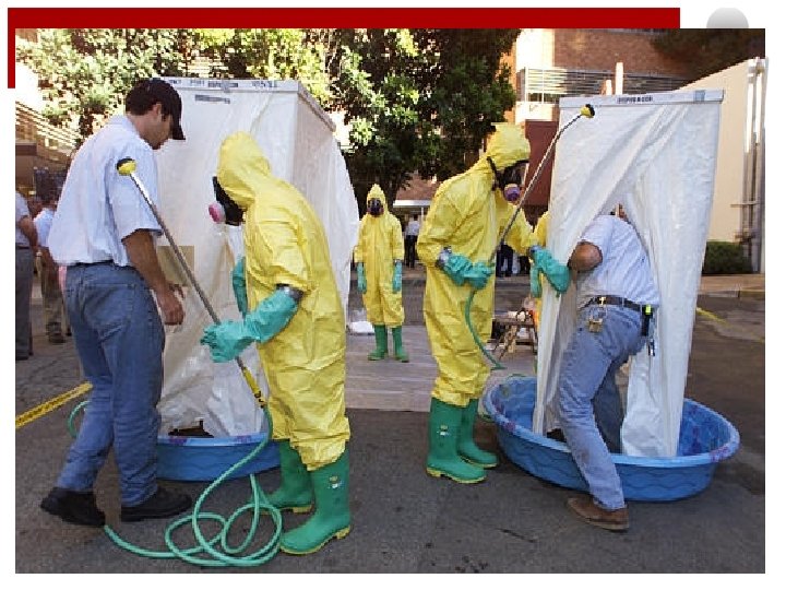
2 -Topical decontamination Ú Prior to the assessment of the patient, you MUST be Ú sure the toxin is not toxic to you. Ú If the patient needs to be decontaminated, do this PRIOR to your assessment. No need for 2 or more patients. Ú Generally; achieved by; undressing patients and washing them thoroughly with copious amounts of water Ú Should occur outside of ER (Pt should initially be in isolated area) Ú All towels and clothing should be put into hazardous waste bags 1 - Skin decontamination: -Rapid decontamination is needed specially if the poison is a corrosive or is easily absorbed from the skin. -Wash contaminated areas as well as exposed areas with warm water or saline , with careful washing of the skin, behind ears, under nails, and skin folds. 1 -Phenol: Can apply olive oil. 2 -Oxalic acid: Soak the affected area with solution of calcium gluconate. 3 -Organophosphorus: Soap water
• Cont. ; Topical decontamination • Gently rinse and wash the skin with copious amounts of water for at least 30 minutes, including the hair, fingernails and perineum. • Do not use forceful flushing in a shower, as it may cause deeper penetration of the toxic substance. Initially, the water should be slightly cool to avoid vasodilation and increased absorption. Use soap to remove oily substances. • Toxic substances such as organophosphorous compounds, organic metal compounds (e. g. lead), aniline, phenol, hydrocyanic acid may penetrate the intact skin and must be handled with proper protective equipment.
3 -Eye decontamination • • Irrigation of the eyes with large amounts of water. For at least 15 min. irrigation of eyes with normal saline or water. Alkaline or acid irrigating solutions should be avoided. Continue to irrigate the eye for as long as the p. H is abnormal. The eye is usually affected by a corrosive : apply anesthetic drops e. g. cocaine HCI (Xylocaine) After first aid treatment the patient should be referred to an ophthalmologist in order to asses and treat any inflammation. Alkaline corneal burns are requiring ophthalmic consultation. 4 -Demulcents • Many plants and chemicals cause oral and gastric mucosa irritation but no serious toxicity. Management for these acute ingestion may include ice cream or milk. Egg whites, which serve as a source of readily available protein, have been given for corrosive intoxications.
B-If the poison was ingested: Three general methods involve removing toxin from stomach via the mouth, binding it inside gut lumen, or mechanically flushing it through GIT. Each method has benefits and risks. However, we usually begin with dilution 1 - Dilution: - By using water & milk immediately after poisoning. - This reduce the gastric irritation induced by many ingested poisons. - Milk provides dilution and is also a demulcent.
Cont. ; Dilution • The initial procedure generally recommended when ever ingestion of a poison is suspected is dilution. In cases of ingestion of solid dosage forms such as tablets or capsules, dilution is not universally recommended. In this case dilution promotes dissolution of the medication and actually increases absorption. • A general rule is that nothing should be administered orally to an unconscious patient. Water is given to accomplish at least two functions 1 - It helps reduce the gastric irritation induced by many ingested poisons. 2 - It adds bulk to the stomach that may be needed later for emesis. Ipecacinduced emesis is more effective if there is fluid or bulk present in the stomach. Milk provides dilution and is also a demulcent.
2 - Delay gastric emptying: a)-Emesis Emetics are substances that cause regurgitation of gastric contents causing removal of stomach contents, thereby preventing the absorption. Emesis may occur by either locally irritating the stomach or centrally by stimulating the medullary chemoreceptor trigger zone. As Syrup of ipecac (Local irritation +stimulation of CTZ) and Apomorphine (Stimulation of CTZ). Advantages of Apomorphin over Syrup of Ipeca: 1 - Its action is reliable and more effective than syrup Ipecac. 2 - It can be used along with orally administered activated charcoal, whereas syrup of ipecac should be given at least 30 min before activated charcoal. Disadvantages of Apomorphin over Syrup of Ipeca: : 1 - It can cause CNS depression, respiratory depression, and hypotension. Apomorphine should be avoided with ingested poisons which produce CNS depression. To overcome the effect of protracted emetic action, respiratory depression, and sedative properties, some physicians administrate a narcotic antagonist, such as naloxone, shortly following onset of emesis. 2 -It must be freshly prepared as it is unstable in solution , and it must be given by injection. 3 - It is not readily available and it should only be administered by qualified medical personnel.
Cont. ; Delay gastric emptying: a)Emesis General conditions in which emesis should not be attempted. • • Do not induce vomiting if the poison is a: Convulsant. Hydrocarbon. Corrosive acid or alkali. Do not induce vomiting if the patient: Is Unconscious or comatose. Has severe cardiovascular disease or, emphysema, or extremely weakenedblood vessels. • Is under 6 months of age. -If the victim is unconscious danger exists that vomitus may be aspirated into the lungs. If the poison is a convalsant, forced emesis may precipitate seizures. -For petroleum, this substance can be readily aspirated into the lungs during emesis, and may cause chemical pneumonitis. -If the ingested poison is corrosive, emesis should be avoided because it may induce further damage to the esophagus as the substance is brought up. Children under 6 months of age should not receive syrup of ipecac unless supervised by a physician. Their gag reflex is poorly developed and emetic may cause choking with aspiration. • •
b)-Gastric lavage • • Gastric lavage is only effective when ingestion of the poison is discovered within 1 hours except salicytates may be within 4 -6 hours (it sticks to the mucous membrane). This procedure is often reserved for patients with impaired consciousness and uncooperative or when ipecac failed to produce emesis. Solutions used : sodium bicarbonate, saline, tannic acid or water. Indicated for – Heavy metals – Iron – Lithium – Sustained or delayed release formulations Risk The tube may be accidentally inserted into the trachea. Improperly used, lavage can incite a greater risk of aspiration of solution. Aspiration pneumonia secondary to emesis with unprotected airway. Laryngospasm with cyanosis. The tube may cause perforation of the stomach or hasten its emptying time into the intestine. If a cool lavaging solution is used too rapidly, the body temperature may be lowered.
3 -Adsorbents Activated charcoal is administered orally to adsorb or bind toxins and allows them to pass from the GIT without being absorbed into the systemic circulation. • Because of its almost universal binding of toxins, charcoal should be given to all overdoses in which there is not a specific contraindication. • The notable exceptions to charcoal administration are poisonings with heavy metals (do not bind to charcoal), caustic agents, hydrocarbons, and possibly cyanide Time Interval for MAXIMALeffect, is within the first 30 minutes of the poisoning EXCEPT in: a- Drugs that slow the gastric emptying, sedative, etc. Here charcoal can be given up to 6 -8 hours following poison ingestion. b- Following salicylate ingestion, it can be given 9 -10 hours later, as salicylate sticks to gastric mucous membrane c- Drugs that enterohepatic circulation. Adverse effects: 1. Constipation, can be prevented if given with cathartics. 2 - Distention of the stomach with potential risk of pulmonary aspiration. 3. Many new products add sorbitol to the mixture so repeated doses may lead to excessive diarrhea, dehydration in children and elderly. Contraindications: 1 - Absence of the bowel sounds. 2 - Intestinal obstruction. 3 - DO NOT give with syrup of ipecac because it will bind it. •
4 -Cathartic Agents Cathartics are substances that reduce the transit time of drugs in the gut. This reduces the contact time with the absorption sites and reduces toxicity. • Cathartics may be hyper-osmotic saline, bulk-forming stimulant, and lubricant laxatives. • Commonly used cathartics are classified saline (magnesium citrate) and saccharide (sorbitol). Saline cathartics are poorly absorbed salts and exert their effects by increasing of osmotic load and thereby stimulating persistalsis. Careful monitoring of the fluid and electrolyte status is necessary. Contraindications: • Catharsis should not be attempted when the patient has electrolyte disturbance or bowel sounds are absent. • Magnesium containing cathartics-should not be used in cases of renal failure…… because of the possibility of causing CNS depression due to accumulation of high concentration of magnesium in the serum. • Sodium containing cathartics are, likewise, best avoided by persons with congestive heart failure • Oil cathartics e. g. castor oil may increase the absorption of fat-soluble poisons such as pesticides. •
IV-Enhance Excretion
1 -Forced Diuresis: • • Forced diuresis is employed to remove chemicals from the blood after they have been absorbed. It is useful when the compound or its metabolite is eliminated in the urine and when diuresis enhances the excretion. The procedure is based on increasing the volume of flow of urine through renal tubules so the chemical may be more quickly eliminated. The urine output should be of 300 -500 m. L/h or 8 -14 L/day using either mannitol or furosemide. Increasing the urinary flow is not a mean to remove any chemicals from the blood. Chemicals which are not normally reabsorbed by the kidney will not be significantly lowered in concentration with forced diuresis. 2 -p. H alteration The principle of p. H alteration is to enhance renal excretion of weak acidic or basic drugs by increasing the degree of the ionization through p. H alteration. a-Acid Diuresis: with ammonium chloride can enhance the elimination of weak bases e. g. phencyclidine, amphetamine, strychnine and quinidine. b-Alkaline Diuresis: with Sodium bicarbonate can remove weak acids (e. g. salicylates and Phenobarbital).
3 -Extracorporeal techniques: Dialysis and Hemoperfusion • As adjuncts for management of severely intoxicated patients. • Dialysis and hemoperfusion should never replace more specific antidotes. • These procedures would be of little value in treating acute ingestions of cytotoxic poisons such as cyanide which produce toxic effects very rapidly. • Dialysis reserved for specific toxins: salicylates, methanol, ethylene glycol, lithium, theophylline, amanita (mushrooms) • Benefits: removal of toxins already absorbed by gut, ability to remove parent compound active metabolite
A. Peritoneal dialysis • It is the easiest method of dialysis and is associated with the lowest risk of complications. It involves the diffusion of toxins from mesenteric capillaries across the peritoneal membrane, which is utilized as the membrane of dialysis, into dialysate dwelling in the peritoneal cavity. • It involves the insertion of a tube through a small incision made in the midabdomen area into the peritoneum. • 1 - 2 L of warmed dialyzing solution is introduced into the peritoneal cavity over a period of 15 -20 minutes. The fluid is left in place for 45 -60 minutes till equilibrium occurs, and removed. • The dialyzable chemical will diffuse from the blood into the dialyzing fluid (from an area of higher to lower concentration).
Cont. ; Peritoneal dialysis The dialysis solution consists of balanced electrolyte solution which may contain 1. Dextrose to make the solution hypertonic and to ↑ recovery of watersoluble chemicals. 2. Albumin which increase the recovery of highly protein-bound drugs. 3. A substance to alter the p. H to the acidic or alkaline side to ↑ total drug recovery. e. g. In acute Phenobarbital ingestion, the use of alkaline solution increases the recovery. 4. Peanut oil to attract highly lipid soluble compounds. Indications: 1 - Acute ingestion in children because of their large peritoneal surface area in relation to body size and the ease of penetration of the abdominal wall. 2 - Treatment of patients with acute renal failure especially in patients with bleeding problems or venous access problems Adverse effects Abdominal pain, hemorrhage, peritonitis, volume overload or depletion, electrolyte imbalance, liability for infection.
B. Hemodialysis In this method a cellophane bag (artificial kidney) forms a semipermeable membrane similar to peritoneal membrane in the peritoneal dialysis. Two catheters are inserted into the patient’s femoral vein, about 2 inches apart. Blood is pumped from one catheter through the dialysis unit, across the semipermeable membrane and returned through the other catheter. The procedure continued for 6 -8 hours. Characteristics of drugs or substances for hemodialysis • Molecular weight below 500 daltons. • Low protein binding , High water solubility • Compounds with small volume of distribution (Vd) e. g salicylates are more easily removed by dialysis, because the plasma concentrations are greater in relation to the total amount of drug in the body. Conversely, if the Vd is greater than 250 -300 L, then less chemical per unit of blood is available for elimination by dialysis.
Cont. ; Hemodialysis Indications: • Clinical intoxication with vital sign abnormalities such as hypotension, apnea or hypothermia that don’t respond to supportive care. • Impairment of normal excretion routes or the presence of disease that ↓ drug clearance. • Aspiration pneumonia and Prolonged coma • Presence of diseases such as COPD. • Presence of a significant amount of a toxin that is metabolized to a toxic metabolite. Adverse reactions 1. Hypotension, electrolyte and osmolar imbalance (muscle cramps, confusion, convulsions or coma) hypoxemia, spontaneous bleeding. 2. Mechanical complications (air embolism, blood leak). 3. Sudden death during dialysis has resulted from machine malfunction or fistula rupture, complete heart block.
C. Hemoperfusion It is more effective than peritoneal dialysis and hemodialysis for removing intoxicating compounds, particularly the lipid soluble, protein bounded drugs or poorly dialyzable compounds. • The blood is withdrawn from the patient via an arteriovenous or venous shunt and passed directly over the adsorbing material contained in sterile columns to remove toxic materials. The adsorbing materials are: • Charcoal coated adsorbing material which removes both polar (salicylates, methotrexate) and non-polar drugs as well as metabolites. • Anion exchange resins as well as nonion resins such as Amberlite resin which clears nonpolar, lipid soluble drugs (e. g. ethchlorvynol) better than charcoal- coated cartridges. •
Cont. ; Hemoperfusion Indications • Severe intoxication with drugs which have small Vd and long half life, so the drug • • can be drawn from the tissues to the blood and consequently removed. e. g. Chloramphenicol toxicity associated with gray baby syndrome Phenytoin overdose. D. Hemofiltration • • • Hemofiltration is performed by a method similar to that in case of hemodialysis, except that the blood is pumped through a hemifilter. During continuous arteriovenous hemofiltration an arteriovenous pressure difference induces a convective transport of solutes through a hollow fiber or flat sheet membrane. This permits a substantial flow of the plasma water and a high permeability to compounds with a relative molecular weight less than or equal to 40, 000 daltons. Advantage of hemofiltration over hemoperfusion and hemodialysis: Is the ability to remove compounds with large (4, 500 to 40, 000) molecular weights as aminoglycoside antibiotics and metal chelat complexes such as aluminum or iron deferoxamine.
E. Plasmapheresis and Plasma exchange: Plasmapheresis separates cellular blood components from plasma. The cells are resuspended in either colloidal albumin or fresh frozen plasma and reinfused. The toxic agents soluble in plasma or bound to plasma proteins are removed by plasma exchange. The efficacy of the method depends on the number of plasma exchanges. A single plasmapheresis exchange sacrifices a part of a patient’s own plasma proteins. Indications: • Blood purification in autoimmune diseases. • Overdoses of toxic compounds of high protein binding that cannot be eliminated by hemoperfusion or hemodialysis. • Overdoses with vincristine, paraquate, inorganic mercury, thyroxin, amanita, theophylline, digoxin antibody complexes. The main complications are thrombocytopenia, nausea, hyopvolemia, hypertension, hypercoagulability, infection, anaphylaxis, seizures and arrhythmia. F. Exchange transfusion It involves the removal of the patient’s blood and replacement with fresh whole blood. In the double-exchange transfusion technique, an amount of blood equivalent to twice the patient’s total blood volume is exchanged. Indications: • Iron, quinine, meperidine, glutethimide, chloralhydrate, diphenhydramine and neonatal chloramphenicol overdoses. • Severe methemoglobinemia, in patients who do not respond to or cannot tolerate appropriate doses of the antidote, methylene blue. The main complications are Mismatches, chills, hypotension, infection, bleeding. G. Plasma perfusion It is a combination of plasmapheresis and hemoperfusion used in methylparathion poisoning.
V-Antidote
I. Chelator antidotes 1. Deferoxamine mesylate (desferal). It chelates iron to form ferrioxamine. The complex is water soluble excreted by the kidney. Used in Iron and Aluminum toxicity. 2. Dimercaprol (British antilewisite, BAL in Oil). BAL contains ligands that bind heavy metals in place of the body’s sulfhydryl groups. The resulting chelate mercaptide product is less toxic and more easily excreted from the body than the heavy metals. Used in arsenic toxicity but not arsine gas poisoning. Adjunctive therapy in acute lead encephalopathy. 3. Calcium disodium EDTA (Versenate). Divalent and trivalent heavy metals displace calcium in Ca. Na 2 EDTA, forming a stable complex that is excreted renally. Ca. Na 2 EDAT is preferred to Na. EDTA because the latter sequesters calcium leading to hypocalcemia. Used in acute lead, cadmium, zinc poisoning. Ocular exposure to calcium oxide, lime and cement. 4. Penicillamine (Cuprimine). It is non-bactericidal degradation product of all penicillins. The Disomer of penicillamine is an oral chelating agent. Used in copper, lead, mercury, arsenic bismuth poisoning. 5. Zinc trisodium Diethylene-triamine pentaacetic acid (DTPA). It increases the elimination of radionuclides & metals from the body by a process involving the exchange of organic compounds for inorganic ions to form a stable ring complex which is readily excreted by the kidney. The FDA approved its use for plutonium, It is used for chelation of rare earth metals as cerium. 6. Dimercaptosuccinic acid, Succimer, DMSA (Chemet). It provides two sulfhydryl groups that can chelate metals. It is an oral medication approved in 1991 by FDA. Used in severe lead poisoning in children.
II. Cyanide antidotes 1. Dicobalt Edetate. Cobalt salts form a stable nontoxic ion complex with cyanide. It is used in UK the treatment of Cyanide poisoning. 2. Cyanide antidote kit (Amylnitrite, sodium nitrite, and sodium thiosulphate). Cyanide binds to ferric ion, interferes with cytochrome oxidase and reduces cellular respiration, Nitrites exogenously given in cases of cyanide poisoning induce methemoglobin formation which in turn attracts cyanide of cytochrome oxidase. Thiosulfate enhances renal elimination of cyanide by forming thiocyanate when combined with cyanomethemoglobin. Amyl nitrite ampoules are first crushed in gauze and given to patients to inhale for 15 seconds. Use a fresh ampoule every 3 minutes. Sodium nitrite is given intravenously over 2 -5 minutes. Subsequently, sodium sulfate is administered intravenously over 10 minutes. 3. Hydroxocobolamin. It binds to cyanide to form cyanocobolamin (Vitamin B 12) which is excreted unchanged in urine. It does not interfere with tissue oxygenation. Used in treatment of cyanide poisoning developed from sodium nitroprusside infusions.
III. Calcium salts Calcium gluconate and calcium chloride. Calcium binds to the fluoride ion preventing further skin penetration in case of concentrated hydrofluoric acid skin burns. Calcium reverses the neuromuscular paralysis. Used in hydrofluoric acid burns of the skin and ingestion of fluoride salts. Calcium channel blocker overdose. In B-blocker overdose. IV calcium gluconate should be given slowly through. IV. Antivenins Different forms of antivenins are available against spiders and snake bites. These products are derived by immunizing healthy horses against the specific species for which the antivenin is produced and collecting venom neutralizing serum globulins. All patients should be skin tested for serum sensitivity before antivenin administration V. Antidotes to methyl alcohol and ethylene glycol • • Ethanol competitively inhibit alcohol dehydrogenase enzyme thus it reduces the formation of toxic metabolites produced after methanol and ethylene glycol ingestion. It delays the formation of formic acid in methanol poisoning and both glycolic and glyoxylic acid in ethylene glycol poisoning. Used in ingestion of methanol or ethylene glycol and in acidosis. Solutions of 10% ethanol in dextrose should be used for i. v. infusions because absolute alcohol is highly irritating to veins. 4 -methylpyrazole (4 -MP). It is an inhibitor of alcohol dehydrogenase enzyme. It is administered by the oral or intravenous route leading to the rapid excretion of free ethylene glycol in the urine. Patients must be treated soon after ingestion (ideally within 3 hours)
VI. Individual antidotes • • • 4 -aminopyridine (4 -AP). It is an antagonist of non-depolarizing neuromuscular blocking agents. 4 -AP enhances transmembrane calcium influx which results in facilitation of calcium influx. It also increases the release of acetylcholine at the neuromuscular junction. Pancuronium overdosage, Possible use in disorders of neuromuscular transmission as myasthenia gravis. Antihistamines (Benztropine and diphenhydramine). They reverse drug induced dystonias through competitive inhibition of muscarinic receptors and blockade of dopamine uptake. Used in the toxicity of neuroleptic drugs as haloperidol, phenothiazines and metoclopramide. Atropine antagonizes cholinergic stimuli at muscarinic receptors. Used in the toxicity of anticholinesterase pesticides (organophosphorous compounds). Physostigmine excess. Antimyasthenic agents as pyridostigmine. Nerve gas agents. Synthetic choline esters. Treatment of bradyarrhythmias induced by different medications. Dantrolene. It decreases the release of calcium from the sarcoplasmic reticulum of skeletal muscle cells resulting in rapid treatment of hyperthermia, dysrrhythmia, muscle rigidity, tachycardia, and hypercapnia. Used in the treatment of catabolic syndromes as malignant hyperthermia or neuroleptic malignant syndrome. Treatment of hypermetabolic state associated with rhabdomyolysis secondary to theophylline poisoning. Flumazenil. It is a competitive inhibitor to benzodiazepine at the GABA-benzodiazepine receptor complex. Used in the treatment of symptoms of benzodiazepines overdose.
Cont. ; Individual antidotes • • • Folinic acid (leucovorin) and folic acid. Folic acid is the precursor of the active form tetrahydrofolic acid which is produced by the action of the folic acid reductase enzyme. tetrahydrofolic acid is required for nucleoprotein synthesis and normal erythropoiesis. Therefore, folic acid is not the metabolically active form. Folic acid antagonists (e. g. methotrexate) inhibit folic acid reductase and prevent the formation of tetrahydrofolic acid. Therefore, overdoses of folic acid antagonists require leucovorin which is a derivative of tetrahydrofolic acid rather than folic acid. Used in folic acid antagonist overdose e. g. methotrexate, trimethoprime & pyrimethamine. Folic acid accelerates the detoxification of the toxic metabolite, formic acid, in methanol intoxication. Glucagon. Stimulation of the production of c. AMP by increasing cardiac c. AMP, glucagon increases the chronotropic and inotropic activity of the heart. Stimulation of the release of glucose from liver stores, and the release of catecholamines. Used in the treatment of hypoglycemia or hypoglycemic agent overdoses. In symptomatic B-adrenergic blocker poisoning. Correcting the hemodynamic instability associated with calcium channel blocker poisoning. Hyperbaric oxygen (HBO). Oxygen displaces carbon monoxide from hemoglobin, myoglobin and cytochrome oxidase enzyme. The half-life of carboxyhemoglobin may be reduced to 23 minutes at 3 atmospheres with 100% oxygen. Used in CO poisoning, Cyanide and hydrogen sulfide poisoning, CCl 4 poisoning.
Cont. ; Individual antidotes • Methylene blue. It acts as an electron carrier for the hexose monophospahte pathway which reduces methemoglobin to hemoglobin. In patients with methemoglobinemia, the erythrocytes methemoglobin reductase reduces methylene blue to leukomethylene blue (colorless form) which in turn reduces methemoglobin to hemoglobin. It is used to treat patients who are exposed to methemoglobin forming compounds as aniline dyes, nitrates, local anesthetics as benzocaine, dapson, naphthalene, nitrobenzene and nitrophenol. • Pyridoxine hydrochloride (Vitamin B 6). Pyridoxine is essential in the synthesis of GABA within the CNS. It controls isoniazide induced seizures which are due to decreased GABA levels because isoniazid inhibits brain pyridoxal-5 -phosphate activity. • Sodium bicarbonate. It dissociated to produce bicarbonate which neutralizes hydrogen ions and raises p. H. Sodium bicarbonate can reverse QRS prolongation in antidepressant overdose. It also reverses cardiac conduction defects caused by quinidine-like effects of caridotoxic drugs presumably by antagonizing sodium channel inhibition. and increases protein binding of tricyclic antidepressants
Thank You