Chapter 63 Management of Patients With Neurologic Trauma






- Slides: 37
Chapter 63 Management of Patients With Neurologic Trauma Spinal Cord Injury Dr. Maha Subih Copyright © 2010 Wolters Kluwer Health | Lippincott Williams & Wilkins
Spinal Cord Injury (SCI) Ø A major health problem Ø Persons live with disability from SCI Ø Risk factors: Young Caucasian adult, ages 16– 30 account for more than half of all new SCIs Ø male gender Ø alcohol and drug use. Ø Motor vehicle crashes Ø falls Ø violence (gun shot wounds) Ø recreational sporting activities Copyright © 2010 Wolters Kluwer Health | Lippincott Williams & Wilkins
ØA victim with a spinal injury has an increased risk of permanent neurologic damage, Paraplegia (paralysis of the lower body) and tetraplegia (formerly quadriplegia —paralysis of all four extremities). Copyright © 2010 Wolters Kluwer Health | Lippincott Williams & Wilkins
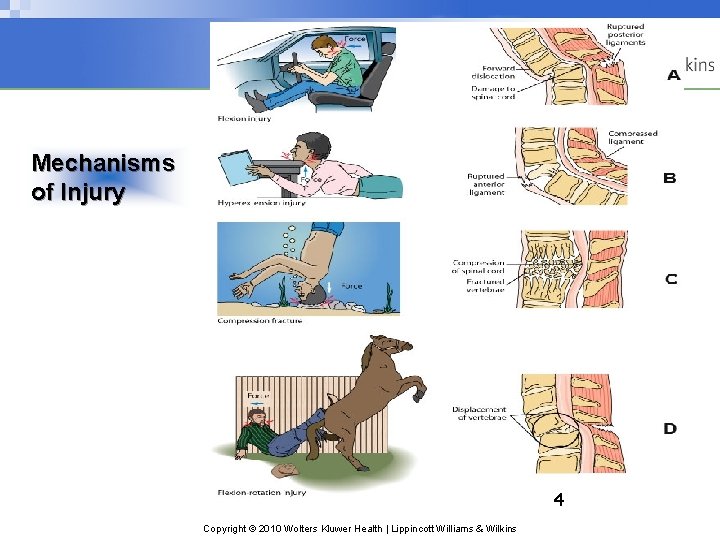
Mechanisms of Injury 4 Copyright © 2010 Wolters Kluwer Health | Lippincott Williams & Wilkins
Pathophysiology ØDamage in SCI ranges from transient concussion (from which the patient fully recovers), to contusion, laceration, and compression of the SC substance, to complete transection of the spinal cord. ØThe vertebrae most frequently involved are (C 5 to C 7), (T 12), and (L 1), because there is a greater range of mobility in the vertebral column in these areas. ØSCIs two categories: primary injuries and secondary injuries. Copyright © 2010 Wolters Kluwer Health | Lippincott Williams & Wilkins
Pathophysiology Ø Primary injuries are the result of the initial trauma and are permanent. Ø Secondary injuries are the result of a contusion or tear injury, in which the it damage nerve. produces ischemia, hypoxia, edema, and hemorrhagic lesions. it is reversible during the first 4 to 6 hours after injury. Ø Early treatment to prevent partial damage from becoming total and permanent Copyright © 2010 Wolters Kluwer Health | Lippincott Williams & Wilkins
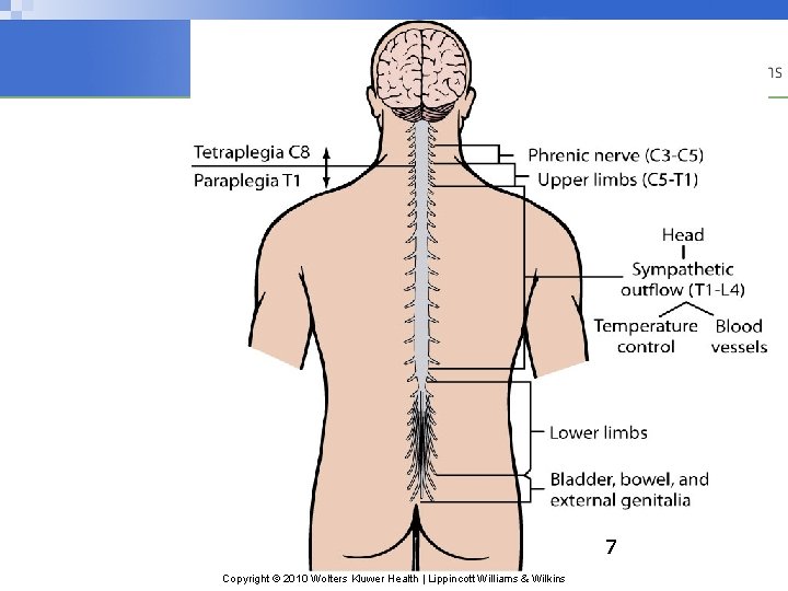
7 Copyright © 2010 Wolters Kluwer Health | Lippincott Williams & Wilkins
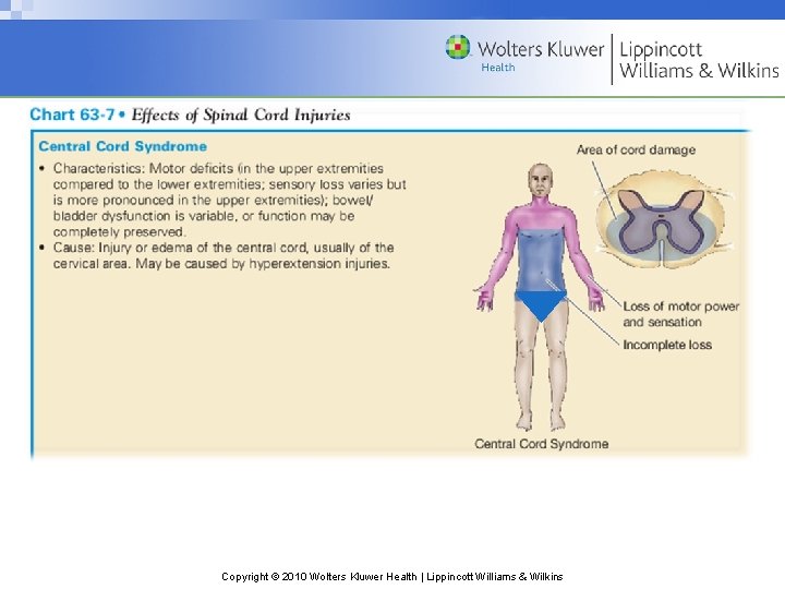
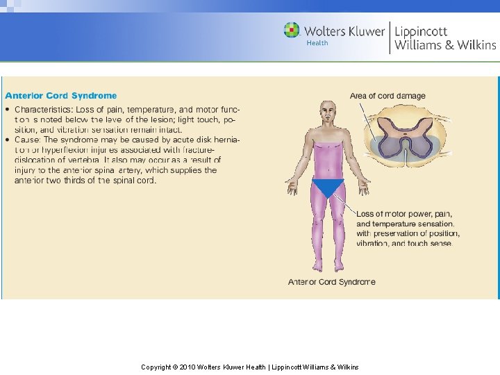
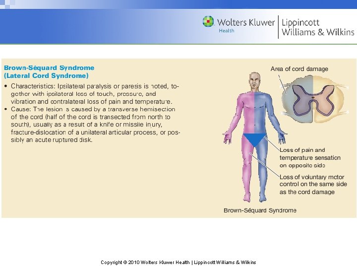
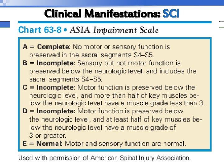
Clinical Manifestations: SCI Ødepend on the type and level of injury (Chart 63 -7) ØIncomplete spinal cord lesions (sensory or motor fibers, or both, are preserved below the lesion) are classified according to the area of SC damage: q Central, lateral, anterior, or peripheral. (Chart 63 -8). Ø“Neurologic level” : the lowest level at which sensory and motor functions are normal. Below it, total sensory and motor paralysis, loss of bladder and bowel control, loss of sweating and vasomotor tone, and reduction of BP Copyright © 2010 Wolters Kluwer Health | Lippincott Williams & Wilkins
Copyright © 2010 Wolters Kluwer Health | Lippincott Williams & Wilkins
Copyright © 2010 Wolters Kluwer Health | Lippincott Williams & Wilkins
Copyright © 2010 Wolters Kluwer Health | Lippincott Williams & Wilkins
Clinical Manifestations: SCI Copyright © 2010 Wolters Kluwer Health | Lippincott Williams & Wilkins
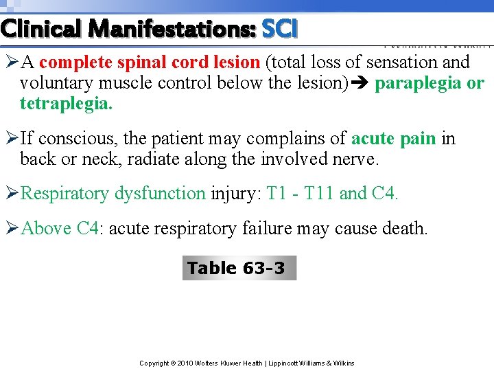
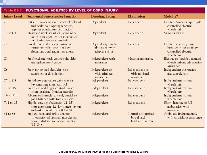
Clinical Manifestations: SCI ØA complete spinal cord lesion (total loss of sensation and voluntary muscle control below the lesion) paraplegia or tetraplegia. ØIf conscious, the patient may complains of acute pain in back or neck, radiate along the involved nerve. ØRespiratory dysfunction injury: T 1 - T 11 and C 4. ØAbove C 4: acute respiratory failure may cause death. Table 63 -3 Copyright © 2010 Wolters Kluwer Health | Lippincott Williams & Wilkins
Copyright © 2010 Wolters Kluwer Health | Lippincott Williams & Wilkins
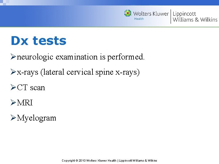
Dx tests Øneurologic examination is performed. Øx-rays (lateral cervical spine x-rays) ØCT scan ØMRI ØMyelogram Copyright © 2010 Wolters Kluwer Health | Lippincott Williams & Wilkins
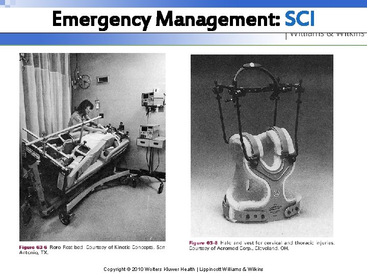
Emergency Management: SCI Ø Initial care must include a rapid assessment, immobilization, and stabilization or control of life-threatening injuries, and transportation to the most appropriate medical facility. Ø the patient immobilized on aback board, with the head and neck maintained in a neutral position, to prevent an incomplete injury from becoming complete. Ø One of the team must control patient’s head by placing hands on both sides of the patient’s head at about ear level while a spinal board or cervical immobilizing device is applied Copyright © 2010 Wolters Kluwer Health | Lippincott Williams & Wilkins
Emergency Management: SCI ØOnce the extent of the injury has been determined, the patient may be placed on a rotating specialty bed or in a cervical collar. ØIf a specialty bed is not available, the patient should be placed in a cervical collar and on a firm mattress. Øif SCI have been ruled out, the patient may be moved to a conventional bed or the collar may be removed Copyright © 2010 Wolters Kluwer Health | Lippincott Williams & Wilkins
Emergency Management: SCI Copyright © 2010 Wolters Kluwer Health | Lippincott Williams & Wilkins
Medical Management (Acute Phase): SCI ØThe goals: q to prevent secondary injury q to observe symptoms of progressive neurologic deficits q to prevent complications. Copyright © 2010 Wolters Kluwer Health | Lippincott Williams & Wilkins
Medical Management (Acute Phase): SCI ØResuscitation and oxygenation and maintain cardiovascular stability ØPharmacologic Therapy q high-dose IV corticosteroids in the first 24 or 48 hours ØRespiratory Therapy q Oxygen to maintain a high Pa. O 2, because hypoxemia worsen neurologic deficit q If ETT is necessary, extreme care to avoid flexing or extending the neck q In high cervical spine injuries, diaphragmatic pacing to stimulate the diaphragm to help the patient breathe Copyright © 2010 Wolters Kluwer Health | Lippincott Williams & Wilkins
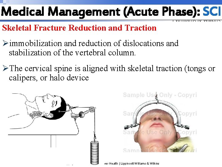
Medical Management (Acute Phase): SCI Skeletal Fracture Reduction and Traction Øimmobilization and reduction of dislocations and stabilization of the vertebral column. ØThe cervical spine is aligned with skeletal traction (tongs or calipers, or halo device Copyright © 2010 Wolters Kluwer Health | Lippincott Williams & Wilkins
Skeletal Fracture Reduction and Traction ØTraction is applied to the skeletal traction device by weights, the amount depending on the size of the patient and the degree of fracture displacement. ØThe traction is then gradually increased by adding more weights. As the amount of traction is increased, vertebrae are given a chance to slip back into position. ØThe weights should hang freely so as not to interfere with the traction. Copyright © 2010 Wolters Kluwer Health | Lippincott Williams & Wilkins
Surgical Management: SCI Ø Surgery is indicated in any of the following situations: Ø • Compression of the cord is evident. Ø • The injury results in a fragmented or unstable vertebral body. Ø • The injury involves a wound that penetrates the cord. Ø • Bony fragments are in the spinal canal. Ø • The patient’s neurologic status is deteriorating. Ø The goal of surgical treatment are to preserve neurologic function by removing pressure from the SC and to provide stability Copyright © 2010 Wolters Kluwer Health | Lippincott Williams & Wilkins
Complications/Spinal and ØSpinal shock. Neurogenic Shock q A sudden depression of reflex activity below the level of spinal injury(a reflexia) q Muscular flaccidity, paralyzed, lack of sensation ØNeurogenic shock q Due to loss of function of the ANS q BP, heart rate, and CO decrease q Venous pooling due to peripheral vasodilation Copyright © 2010 Wolters Kluwer Health | Lippincott Williams & Wilkins
Complications ØWith injuries to the cervical and upper thoracic spinal cord, innervation to the major accessory muscles of respiration is lost and respiratory problems develop. Ødecreased vital capacity, retention of secretions, increased (Pa. CO 2) levels and decreased oxygen levels, respiratory failure, and pulmonary edema. Copyright © 2010 Wolters Kluwer Health | Lippincott Williams & Wilkins
Complication of SCI ØDeep Vein Thrombosis related to immobility Ørisk for PE ØLow-dose anticoagulation therapy is initiated ØAntiembolism stockings ØPermanent indwelling filters Copyright © 2010 Wolters Kluwer Health | Lippincott Williams & Wilkins
Autonomic Dysreflexia ØAcute emergency! ØOccurs after spinal shock has resolved and may occur years after the injury. ØOccurs in persons with a SC lesion above T 6. ØANS responses are exaggerated. ØSymptoms: severe pounding headache, sudden increase in BP, diaphoresis, nausea, nasal congestion and bradycardia. Copyright © 2010 Wolters Kluwer Health | Lippincott Williams & Wilkins
Nursing Diagnosis ØIneffective breathing pattern ØIneffective airway clearance ØImpaired physical mobility ØDisturbed sensory perception ØRisk for impaired skin integrity ØImpaired urinary elimination ØConstipation ØAcute pain Copyright © 2010 Wolters Kluwer Health | Lippincott Williams & Wilkins
Nursing Interventions Promoting Adequate Breathing and Airway Clearance ØMonitor carefully to detect potential respiratory failure q Pulse oximetry, ABGs and Lung sounds ØEarly and strong pulmonary care to prevent and remove secretions ØSuctioning with caution? ? stimulating the vagus nerve and producing bradycardia and cardiac arrest ØBreathing and coughing exercises ØHumidification and hydration Copyright © 2010 Wolters Kluwer Health | Lippincott Williams & Wilkins
Nursing Interventions Improving Mobility Ø Maintain proper body alignment Ø Turn only if spine is stable and as indicated by physician Ø Monitor BP with position changes (Patients with lesions above the midthoracic level have loss of sympathetic control of peripheral vasoconstrictor activity, leading to hypotension). Ø Passive ROM at least four times a day to prevent contractures Ø Use neck collar, as prescribed, when patient is mobilized Ø Move gradually to erect position Copyright © 2010 Wolters Kluwer Health | Lippincott Williams & Wilkins
Nursing Interventions Promoting Adaptation to Sensory and perceptual alteration ØAssist to compensate for sensory and perceptual alterations ØStimulate intact senses above the level of the injury by touch, aromas, flavorful food and music. ØProvide prism glasses to see from supine position Øuse of hearing aids ØProvide emotional support and teaching Copyright © 2010 Wolters Kluwer Health | Lippincott Williams & Wilkins
Nursing Interventions Maintaining Skin Integrity ØPressure ulcers are a significant complication of SCI may begin within hours of an acute SCI Øpatients who wear cervical collars for prolonged periods may develop breakdown from the pressure of the collar under the chin, on the shoulders, and at the occiput. ØTurning and position is changed at least every 2 hours. Øskin should be kept clean and dry. Copyright © 2010 Wolters Kluwer Health | Lippincott Williams & Wilkins
Nursing Interventions Maintaining Urinary Elimination(neurogenic bladder) ØThe urinary bladder becomes a tonic and cannot contract by reflex activity. ØUrinary retention due to loss of sensation of bladder distention and overstretching of the bladder ØIntermittent catheterization q record fluid intake, voiding pattern, amounts of residual urine after catheterization, characteristics of urine, and any unusual sensations Copyright © 2010 Wolters Kluwer Health | Lippincott Williams & Wilkins
Nursing Interventions Improving Bowel Function Øa paralytic ileus usually develops ØNGT to relieve distention and to prevent vomiting and aspiration. ØBowel activity usually returns within the first week. ØAfter BS return give a high-calorie, high-protein, high-fiber diet, with the amount of food gradually increased. Østool softeners Copyright © 2010 Wolters Kluwer Health | Lippincott Williams & Wilkins
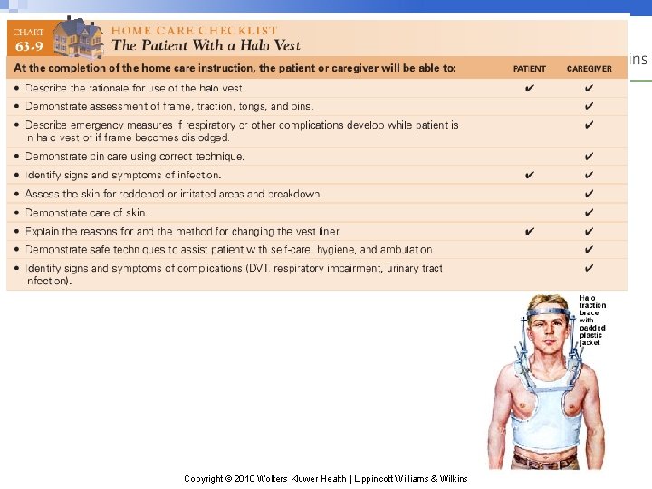
Nursing Interventions Providing Comfort Measures: ØHalo Traction + pins, tongs for cervical stabilization ØAppearance of these devices and being caged in Ø noise created by the steel frame of a halo device Øclean areas around the pin sites and observe for redness, drainage, pain and loosening ØThe skin under the halo vest is inspected Copyright © 2010 Wolters Kluwer Health | Lippincott Williams & Wilkins
Copyright © 2010 Wolters Kluwer Health | Lippincott Williams & Wilkins
Nursing Interventions Monitoring and Managing Potential Complications Ø Thrombophlebitis. ØOrthostatic hypotension q Close monitoring of vital signs before and during position changes q Vasopressor medication q Anti-embolism stockings q Abdominal binders ØAutonomic dysreflexia Copyright © 2010 Wolters Kluwer Health | Lippincott Williams & Wilkins