Gastrointestinal pathology esophagus and stomach lecture 1 Dr


- Slides: 47
Gastrointestinal pathology esophagus and stomach lecture 1 Dr Heyam Awad FRCPath
Pathology lectures in GI pathology subject Number of lectures lecturer Esophageal diseases 2 Dr H Awad Gastric disease 2 Dr H Awad Small intestine 2 Dr M Salihi Large intestine 2 Dr M Salihi liver 5 Dr M Shomaf pancreas 1 Dr M Shomaf Gall bladder 1 Dr M Shomaf
Types of diseases that can affect the esophagus • Obstruction inflammation
Esophageal diseases 1. 2. 3. 4. Obstruction Vascular diseases Inflammation Tumors
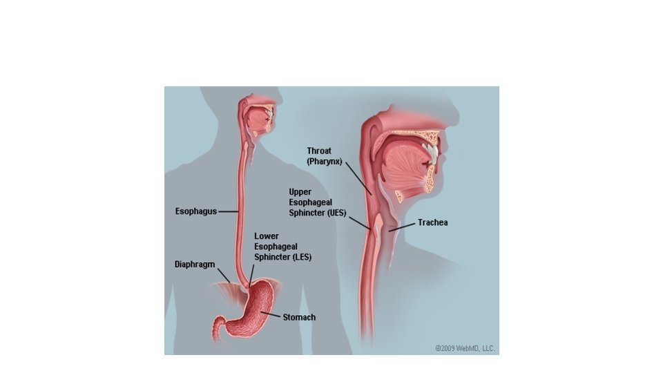
Esophageal obstruction • Can be mechanical or functional obstruction • Mechanical: the esophagus is obstructed due to developmental abnormality: atresia, fistula, or duplication. • Functional: caused by several conditions that affect normal motility.
Mechanical obstruction • Agenesis. • Atresia • Fistula • Duplication
ﺍﻟﻕ • Agenesis = no esophagus. extremely rare = AGENESIS
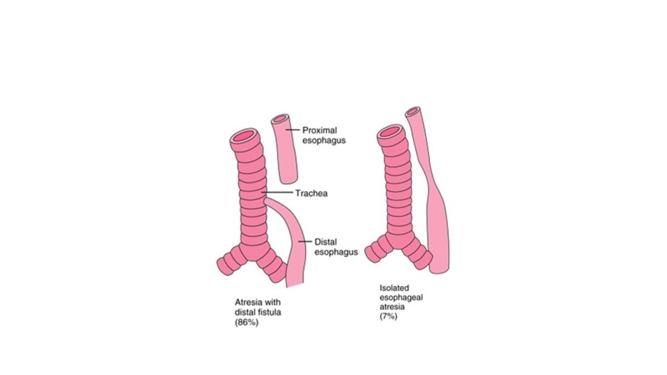
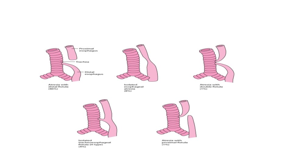
Atresia= ﺍﻧﺴﺪﺍﺩ= ﺍﻟﺮﺗﻖ • = Absence or closure of a natural passage or channel of the body. • Atresia of esophagus: thin non-canalized cord replaces a segment of esophagus • Atresia occurs mostly at or near tracheal bifurcation • Usually associated with fistula connecting upper or lower esophageal pouches to a bronchus or the trachea • This abnormal connection cause aspiration, suffocation, pneumonia or severe fluid and electrolyte imbalance
Atresia • Discovered shortly after birth because it causes regurgitation during feeding • Must be corrected surgically
Stenosis • Stenosis: thickening due to fibrous thickening of the submucosa, atrophy of muscularis propria, and secondary epithelial damage • Stenosis is usually due to inflammation and scarring which could be due to reflux, irradiation or caustic injury • These patients have dysphagia which is progressive. Difficulty in eating solids occurs long before problems with liquid
Caustic injury= corrosive injury • An injury of muco-cutaneous surfaces—e. g. , eyes, esophagus, skin— with tissue destruction due to direct contact with a strong acid or with a strong base.
Caustic injury
Functional obstruction • Efficient delivery of food to the stomach requires coordinated peristalsis of the muscles • Esophageal dysmotility interferes with these peristaltic contractions • Achalasia is the most important cause of functional obstruction.
achalasia
Achalasia= ﻻﺍﺭﺗﺨﺎﺋﻴﺔ • Caused by failure of the LES muscles to relax • = Incomplete LES relaxation, increases LES tone and aperestalsis • Can be Primary and secondary • Primary: failure of the distal esophageal inhibitory neurons. Idiopathic • Secondary= Chagas disease: Trypanosoma cruzi infection destroys myenteric plexus neurons
symptoms • Achalasia is characterized by difficulty in swallowing, regurgitation, and sometimes chest pain.
note • Achalasia like disease can occur in any disease that can affect the neuronal innervation of the esophageal muscles Examples: 1. diabetic autonomic neuropathy, 2. infiltrative disorders: malignancy, amyloidosis, sarcoidosis 3. lesions of dorsal motor nuclei like in polio or surgical ablation
Treatment
Diseases of blood vessels • The most important is : esophageal varices.
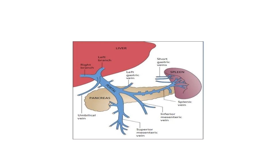
Normal blood flow to the GIT • Venous blood from GIT is delivered to the liver via the portal vein before reaching inferior vena cava. • So drugs and other materials absorbed in the intestines are processed by the liver before entering the systemic circulation • If this flow is impaired: portal hypertension develops. • Portal hypertension causes esophageal varices
Splanchnic circulation
Esophageal varices • One of the sites where the splanchnic and systemic circulation can communicate is the esophagus. • That’s why when portal hypertension increases, collateral vascular channels develop in the esophageal veins to allow blood to shunt from the portal to caval system (inferior vena cava) • These collateral veins (varices) enlarge and can rupture.
Causes of esophageal varices • Any disease that causes increased portal hypertension will result in esophageal varices • Liver cirrhosis is the most common cause worldwide, especially alcoholic liver disease • Hepatic schistosomiasis is the second most common cause.
morphology • Varices appear as tortuous dilated veins within the submucosa of distal esophagus and proximal stomach
Esophageal varices: note the dilated veseels
Clinical features • Varices are usually asymptomatic. • But, can rupture and cause hemorrhage • If hemorrhage is severe: can result in death • Half patients die from the first bleed • Those who survive: more than 50% will have another bleed that can be fatal • Variceal rupture is the most common cause of death associated with advanced cirrhosis.
Bleeding due to varices
Esophageal inflammations= esophagitis • Lacerations • Chemical esophagitis • Infectious esophagitis • Reflux esophagitis • Eosinophilic esophagitis • Barrett esophagus
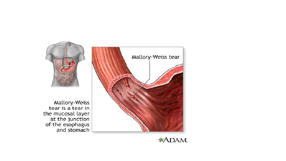
Esophageal lacerations vomiting • Most common esophageal laceration: Mallory Weiss tears • Associated with severe vomiting or with acute alcohol intoxication • Normally there is a reflex relaxation of the gastroesophageal muscles before antiperistaltic contractile wave associated with vomiting • This reflex fails during prolonged vomiting resulting in esophageal wall stretch and tear. • Patients present with hematemesis ( bloody vomit) • The tears are longitudinal, superficial , cross the gastroesophageal junction and healing is usually rapid and complete… no surgical intervention is needed.
Mallory Weiss lacerationseiss
lacerations
Chemical esophagitis • Inflammation of the esophagus can result from chemicals like: alcohol, acids, bases, hot fluid, heavy smoking. • Pill induced esophagitis: medicinal pills lodge into the esophagus and dissolve there • Chemotherapy and radiotherapy can also cause esophagitis
infections • Herpes simplex: causes punched out ulcers • CMV: causes shallow ulcers • Candida: pseudo membrane
Ulcer. . This can be infectious
Herpes inclusions
candida
Reflux esophagitis • Is inflammation of the lower esophagus due to reflux of gastric contents, which are acidic, from the stomach to the esophagus. • It is the mot common cause of esophagitis Also called: gastroesphageal reflux disease GERD
pathogenesis • Reflux of gastric juices into esophagus causes mucosal injury in the esophagus Causes of this reflux • 1. Decreased LES tone will cause reflux: alcohol, smoking • 2. increased abdominal pressure: obesity, pregnancy, • 3. delayed gastric emptying and increased gastric volume • 4. in many cases, no cause is known !!
morphology • Hyperemia: redness… seen macroscopically or during endoscopy • Microscopically: eosinophils, neutrophils, basal zone hyperplasia, elongation of lamina propria papillae
Inflammation in esophagus GERD is the most common cause
Clinical features • Occurs in adults older than 40 • Symptoms: heartburn, dysphagia, regurgitation • Rarely: severe chest pain that can mimic heart disease • Complications: ulceration, strictures, Barrett's mucosa