
DISORDERS OF ESOPHAGUS Dr S Jaya Sandeep Congenital



- Slides: 52
DISORDERS OF ESOPHAGUS Dr S Jaya Sandeep
Congenital Infectious Traumatic Inflammatory Perforation Diverticula Narrowing Motility disorders Neoplasms Miscellaneous
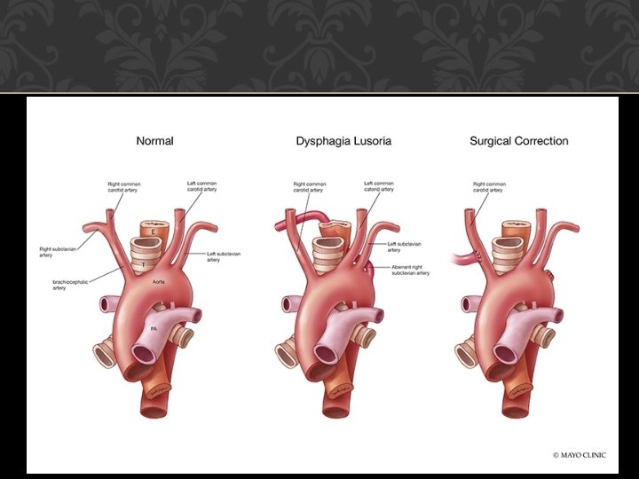
CONGENITAL Tracheoesophageal fistula Esophageal Atresia Stricture Dysphagia lusoria
ACUTE ESOPHAGITIS (i) ingestion of hot liquids, (ii) ingestion of caustic or corrosive agents, (iii) laceration due to swallowed foreign body, or trauma of esophagoscopy, (iv) monilial infection of esophagus from thrush in the oral cavity (v) systemic disorder like pemphigus
TRAUMATIC (INJURY) CORROSIVE BURNS Acids, alkalies or other chemicals may be swallowed accidentally in children or taken with the purpose of suicide in adults. Pathology Severity : nature and duration Extent of lesion : Alkalis are more destructive and penetrate deep into the esophagus
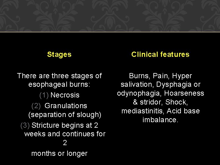
Stages Clinical features There are three stages of esophageal burns: (1) Necrosis (2) Granulations (separation of slough) (3) Stricture begins at 2 weeks and continues for 2 months or longer Burns, Pain, Hyper salivation, Dysphagia or odynophagia, Hoarseness & stridor, Shock, mediastinitis, Acid base imbalance.
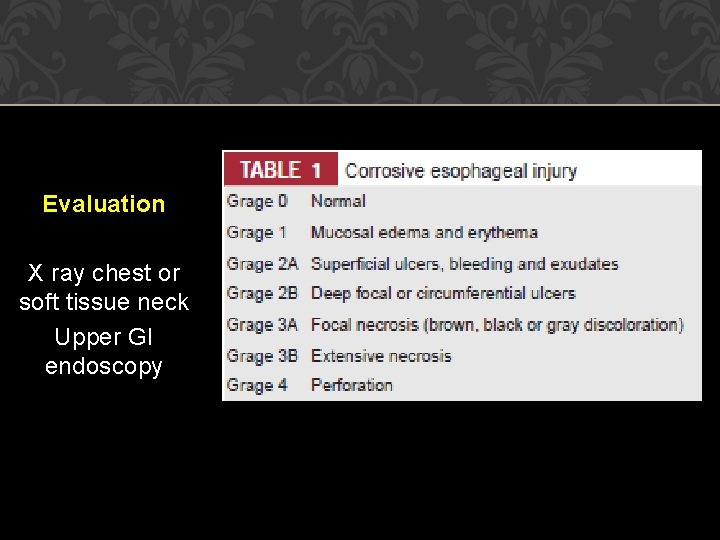
Evaluation X ray chest or soft tissue neck Upper GI endoscopy
Treatment Admit in ICU Wash & irrigation Feeding IV fluids & electrolytes Maintain i/o chart Tracheotomy or intubation Analgesics Antibiotics & steroids
MALLORY WEISS SYNDROME Mucosal tear is caused at squamocolumnar junction (SCJ) by repeated vomiting or retching. Clinical features: Hematemesis after an episode of emesis or retching. �Diagnosis: EGD will show the site and extent of tear. Barium swallow is not indicated. �Treatment : This self-limiting condition usually responds to conservative measures. Angiographic embolization or vasopressin may be used in refractory cases.
INFLAMMATORY Gastroesophageal reflux disease refers to damage of esophageal mucosa due to abnormal reflux of gastric content. Etiology Inappropriate function of LES permits reflux of gastric contents into esophagus. �Highlevels of progesterone in pregnancy Sliding hiatus hernia �Fatty foods, chocolate and peppermints Tobacco smoking and alcohol �Drugs , which relax the smooth muscle: Anticholinergic, betaadrenergic drugs and calcium channel blockers.
Clinical features Typical : heartburn, dysphagia, angina like chest pain Atypical : non-productive cough, hoarseness of voice and asthma-like symptoms and dental erosions. Diagnosis Upper GI endoscopy Ambulatory p. H monitoring
Treatment The aims of the treatment include decreasing reflux, improving esophageal clearance and protecting esophageal mucosa. Life style modification Medication : Antacids. , H 2 receptor antagonists, proton pump inhibitors, Prokinetic drugs. Antireflux surgery: In Nissen’s fundoplication, fundus of
PERFORATION Early diagnosis is essential, as mediastinitis can rapidly prove fatal. Perforation of thoracic esophagus is more serious than cervical esophagus. Etiology Instrumentation Spontaneous rupture: This occurs due to vomiting and usually involves lower third esophagus. In Boerhaave’s syndrome, there occurs postemetic rupture of all the layers of esophagus. �Pathological : Malignancy. �Penetrating injuries.
Features of cervical oesophageal rupture pain, fever, difficulty to swallow and local tenderness, along with signs of surgical emphysema in the neck. Features of thoracic oesophageal rupture pain, referred to the interscapular region, fever, signs of shock, surgical emphysema in the neck, crunching sound over the heart (Hamman’s sign, because of air in the mediastinum) and pneumothorax
Diagnosis �X-rays of the chest and neck findings Widening of the mediastinum and retrovisceral space Surgical emphysema, pneumothorax, or gas under the diaphragm Pleural effusion Treatment �Nil by mouth Intravenous fluids for nutrition Antibiotics: To combat infection Management of shock
Perforations of cervical esophagus Early cases may be managed conservatively. Drainage is required in cases of suppuration. Retrovisceral space and upper mediastinum are drained. � Rupture of thoracic esophagus Perforation is surgically repaired and pleural cavity drained within 6 hours. Repair is not possible after 6 hours but infected area needs drainage.
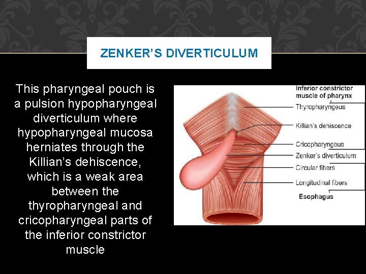
ZENKER’S DIVERTICULUM This pharyngeal pouch is a pulsion hypopharyngeal diverticulum where hypopharyngeal mucosa herniates through the Killian’s dehiscence, which is a weak area between the thyropharyngeal and cricopharyngeal parts of the inferior constrictor muscle
Etiology Spasm of cricopharyngeal sphincter or its incoordinated contractions during the act of deglutition is considered to be an important predisposing factor. Pathology Herniation of pharyngeal mucosa, which extends behind the esophagus, begins in the midline and later on pouch extends to lie on the left. Mouth of the sac becomes wider than the opening of esophagus. Food usually enters into the sac.
Clinical features Most common symptoms are halitosis, dysphagia (difficulty initiating swallowing or may increase after a few swallows, which fill the pouch with the food and then presses on the esophagus). Gurgling sound during swallowing. Regurgitation of undigested food at night (due to recumbent position) results in coughing and choking. �Lossof weight and malnourishment. �Aspiration pneumonia
Diagnosis �Bariumswallow will show the site and size of diverticulum. �EGD and nasogastric intubation are contraindicated because of the risk of perforation of the pouch. Treatment �Diverticulectomy (Excision of pouch) or cricopharyngeal myotomy or both. �Dohlman’s procedure: Endoscopic diathermy of the partition wall between esophagus and pouch is preferred in poor risk debilitated patients.
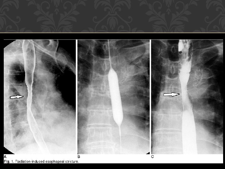
STRICTURES The esophageal strictures occur when its muscular layer is damaged. Causes �Intrinsic Congenital: Common in the lower third. Burns: Corrosives or hot fluids. Trauma: Impacted FBs or external injuries. Iatrogenic: Pill-induced, postnasogastric, sclerotherapy, radiation-induced, and sites of surgical anastomosis. Ulcers: Reflux esophagitis, diphtheria and typhoid. �Extrinsic
Clinical Features �Dysphagiafirst with solid and then with liquids �Regurgitation and coughing �Malnourishment. Diagnosis �Bariumswallow: Shows number, extent and severity of strictures. �Esophagoscopy: Diagnostic and therapeutic
Treatment Esophagoscopy and prograde dilatation with bougies under direct vision: Patients usually require repeated dilatation. Gastrostomy: It provides feeding to the patient and offers rest to the inflamed mucosa above the strictures. Prograde dilatation may be restored once inflammation subsides and lumen becomes visible. Excision and reconstruction: Strictured segment is
MOTILITY DISORDERS Hypermotility disorder Cricopharyngeal spasm, diffuse esophageal spasm, nutcracker esophagus, and hypertensive lower esophageal sphincter Hypomotility disorders Cardiac achalasia, connective tissue disease (scleroderma and amyotrophic lateral sclerosis) and hypoperistalsis—CREST syndrome
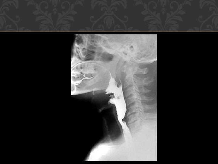
CRICOPHARYNGEAL SPASM There occurs incoordination between relaxation of the UES and simultaneous contraction of the pharynx. UES fails to relax properly. Causes �. Cerebrovascular strokes �. Parkinson’s disease �. Bulbar polio �. Multiple sclerosis �. Muscular dystrophies
DIFFUSE ESOPHAGEAL SPASM In this motility disorder of smooth muscle, there occur spontaneous strong nonperistaltic contractions of the body of esophagus. There occurs degeneration of nerve processes but sphincter relaxation is normal. Clinical Features �. Sudden dysphagia and substernal chest pain simulating angina pectoris in adults. �. Symptom improves with sublingual nitroglycerin
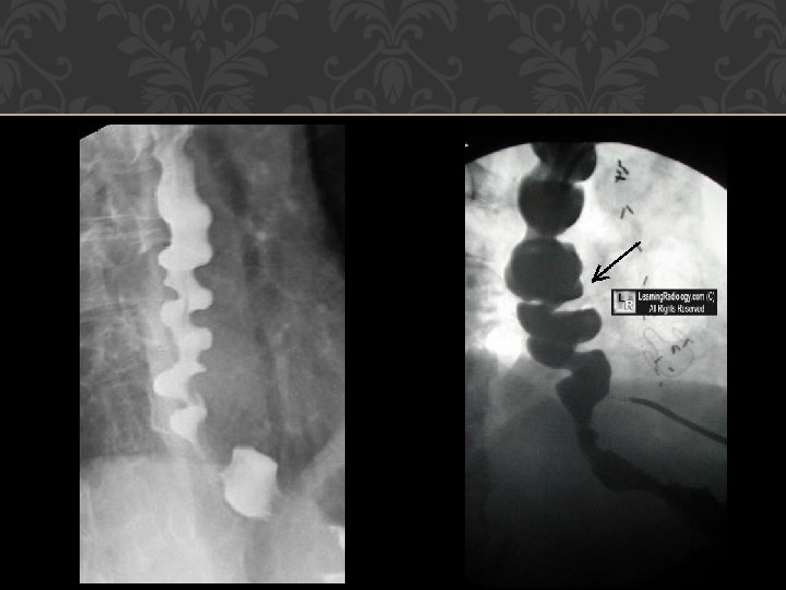
Diagnosis �Bariumswallow: Shows segmented esophageal spasms, which appear like a rosary bead or a corkscrew. �Manometry: Normal relaxation of the sphincter on swallowing and strong nonperistaltic uncoordinated esophageal contractions. Treatment �Medical : Calcium channel blockers and nitrates usually give good response. �Surgical : Dilatation of lower esophagus and myotomy of esophagus extending from the arch of aorta to lower sphincter is done in severe refractory cases.
NUTCRACKER ESOPHAGUS There occur strong and high amplitude peristaltic esophageal contractions, which can be seen in manometric studies. � Patients present with dysphagia and substernal pain like diffuse esophageal spasm (DES). � Treatment is also similar to DES.
CARDIAC ACHALASIA The characteristic features of this smooth muscle motility disorder are absence of peristalsis in the body of esophagus and high resting pressure in LES, which does not relax during swallowing. Pathology Loss of ganglion cells. �Myenteric neural fibrosis Selective loss of postganglionic inhibitory neurons in LES results in insufficient LES relaxation.
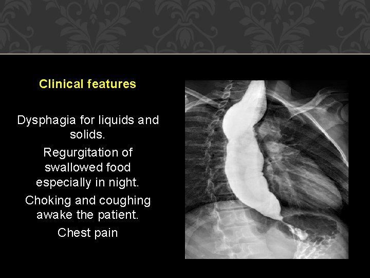
Clinical features Dysphagia for liquids and solids. Regurgitation of swallowed food especially in night. Choking and coughing awake the patient. Chest pain
Diagnosis (i) radiography (barium swallow shows dilated oesophagus with narrowed rat tail lower end), sometimes also called bird-beak appearance; (ii) manometric studies (low pressure in the body of oesophagus and high pressure at lower sphincter and failure of the sphincter to relax) (iii) endoscopy to exclude benign stricture or any development of carcinoma which is a common complication of this disorder.
Treatment Endoscopic pneumatic dilatation: It tears LES muscle fibers and thus reduces LES pressure. Perforation occurs in 5% of patients. �Modified Heller’s operation: Myotomy (incision of circular muscle fibers) of narrowed lower portion of esophagus is the definitive treatment. �Botulinumtoxin injection in LES: It block’s cholinergic nerves and needs to be repeated every 2 years. � Medical: Calcium channel blockers and nitrates
SCLERODERMA Scleroderma affects mid and distal esophagus. The atrophy and fibrosis of esophageal smooth muscle result in decrease or absence of peristalsis (similar to achalasia) and incompetent LES (opposite to achalasia). Clinical features Progressive dysphagia to solids and liquids like malignancy. �Featuresof GERD due to decreased LES tone. �Cutaneouslesions
Diagnosis �Bariumswallow: Dilation and absence of peristalsis in distal two-third of the esophagus. �Motility studies: Decreased smooth muscle contraction. Treatment No effective treatment except antireflux therapy.
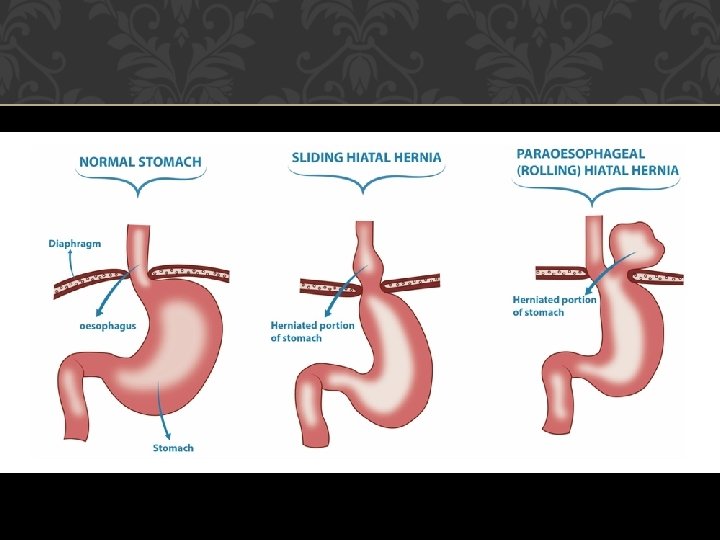
HIATUS HERNIA It refers to displacement of stomach into the chest through the diaphragm. It is common in elderly patients above 40 years of age. Sliding hiatus hernia: Raised intra-abdominal pressure can push the stomach into the thorax in the line of esophagus. Clinical features: Features of reflux esophagitis can cause ulceration, stenosis and hematemesis. Paraesophageal or rolling hiatus hernia: A part of the stomach and its peritoneal covering enters into the thorax by the side of esophagus.
BARRETT’S ESOPHAGUS Pathogenesis In Barrett’s esophagus, the normal stratified squamous epithelium of distal esophagus is replaced by intestinal columnar metaplasia. It predisposes to the development of esophageal adenocarcinoma which is more likely seen in �. Barrett is more than 8 cm long �. Presence of dysplasia �. Smoking. Diagnosis : Upper GI endoscopy & Ba swallow
Treatment �Antireflux therapy �EGD surveillance: These patients need regular endoscopic surveillance to detect dysplasia and adenocarcinoma at an earlier and curable stage. No dysplasia: Every 3 years. Low-grade dysplasia: Every 1 year until no dysplasia. High-grade dysplasia: Focal: Every 3 months. Multifocal: Esophagectomy, ablation therapy, or esophageal mucosal resection.
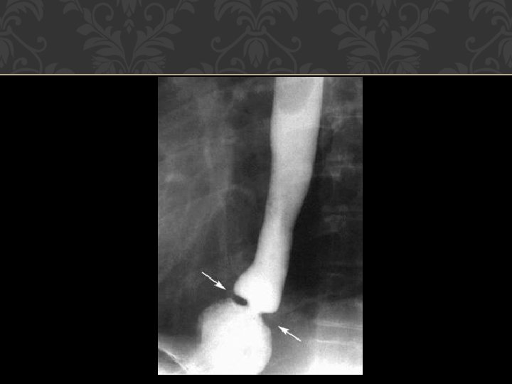
SCHATZKI'S RING It is a web-like mucosal ring, which is seen at the SCJ or proximal to LES. � Clinical features: It remains asymptomatic in about 10% people. Young patient presents with episodic dysphagia to solids and sometimes, liquids. � Diagnosis: Barium swallow will confirm the diagnosis. � Treatment: Pneumatic dilation is done in symptomatic
PLUMMER-VINSON (PATTERSON BROWN-KELLY) SYNDROME Plummer-Vinson (PV) syndrome predominantly affects females past 40 years. it consists of atrophy of the mucous membrane of the alimentary tract, subepithelial fibrosis in lower part of laryngopharynx and iron deficiency anemia Clinical Features �. Dysphagia immediately after trying to swallow foo �. Iron-deficiency anemia �. Glossitis �. Angular stomatitis �. Koilonychias (spooning of nails)
GLOBUS HYSTERICUS PHARYN GEUS Functional disorder with no true dysphagia. �Patient complains of “lump” in the throat. The feeling of lump is more marked between the meals when patient voluntarily and consciously swallows the saliva. �Thereis no difficulty in swallowing food. �Patient usually has fear of throat cancer. �Clinical examination of the pharynx and larynx is normal. �Management : Rule out any organic cause and reassure the patient.
BENIGN NEOPLASMS Neoplasms of esophagus are rare and those found are usually malignant. �Leiomyoma arises from the smooth muscle of esophagus wall. It accounts for two-third of all benign neoplasms. �Otherbenign tumors include mucosal polyps, lipomas, fibromas and hemangiomas. �Theyare usually pedunculated and seen in the esophageal lumen. � Treatment: Endoscopic removal is not done because of the fear of perforation. They need surgical excision with external approach
CARCINOMA ESOPHAGUS Incidence of esophageal malignancy is high in China, Japan, USSR and South Africa. It constitutes 3. 6% (in affluent class) and 9. 13% (in poor class) of all body cancers in India. Squamous cell carcinoma: Usually occurs in proximal two third of esophagus. Black males, Smoking and alcohol consumption, Chewing of Paan, Sopari and tobacco, Diet rich in nitrates and pickled vegetables. Adenocarcinoma: Usually occurs in distal one-third esophagus and GEJ. White males, GERD and Barrett’s esophagus, Hiatus hernia.
Spread of Carcinoma : Direct, Lymphatic, Blood borne. Clinical Features 1. Early symptoms 2. Progressive dysphagia and emaciation 3. Pain 4. Aspiration problem
Diagnosis 1. Barium swallow. It shows narrow and irregular oesophageal lumen, without proximal dilatation of the esophagus. 2. Esophagoscopy. Useful to see the site of involvement, extent of the lesion and to take biopsy. Flexible fiber-optic esophagoscopy obviates the need for general anesthesia and gives a magnified view. 3. Bronchoscopy. It helps to evaluate any extension of growth into the trachea and bronchi. 4. CT scan. It is useful to assess the extent of disease and nodal metastases
Treatment Surgery of upper two-thirds of esophagus is difficult. Radiotherapy is the treatment of choice Surgery is the preferred method of treatment for cancer of lower one-third. In advanced lesions, only palliation is possible. An alternative food channel can be provided by: 1. A by-pass operation. 2. Oesophageal intubation with Celestin or Mousseau- Barbin or a similar tube. 3. Permanent gastrostomy or a feeding jejunostomy. 4. Laser surgery: Oesophageal growth is burnt with Nd: YAG laser to provide a food channel. Chemotherapy is palliative.
THANK YOU