Essentials of Human Anatomy Physiology Unit 4 Nervous


")
 · Act to increase surface")

 Area a.")


")


- Slides: 35
Essentials of Human Anatomy & Physiology Unit 4: Nervous System CNS & the Brain
Protection of the Central Nervous System · Bones: Skull & Vertebral Column · Membranes: Dura Mater, Arachnoid Mater, Pia Mater · Fluid: Cerebrospinal Fluid
Meninges · membranes around the brain and spinal cord are called meninges · they offer protection to the soft tissue of the brain and spinal cord · Three distinct membranes · dura mater, arachnoid mater, pia mater · Subdural hematoma · Meningitis
https: //www. youtube. com/watch? v=Ji 7 foh. BEPn. M
Brain Meninges · Dura mater · Outermost membrane attached to periosteum of skull · Double-layered external covering · Tough, dense white fibrous CT · Contains many blood vessels and nerves · Splits into 2 layers where it encloses the dural sinuses (structure that collects venous blood from the brain) · Periosteal layer – attached to surface of the skull · Meningeal layer – outer covering of the brain · Folds inward in several areas · Separate cerebral hemispheres; cerebrum from cerebellum
Brain Meninges · Arachnoid layer · Middle layer · Between dura mater and pia mater · Thin, net-like membrane · Beneath the arachnoid mater lies a wide space called the sub-arachnoid space · Filled with Cerebrospinal Fluid (CSF) · Cushions the brain
Brain Meninges · Pia mater · Internal membrane that clings to the brain surface · Very thin, delicate CT · Contains many nerves and blood vessels · nourish cells of brain and spinal cord · Dips into grooves and contours of the brain surface
Spinal Cord Meninges · Spinal cord · The dura mater is NOT attached to the bone of the vertebra · The space between the dura mater and the bone is called the epidural (subdural) space · Filled with Areolar CT and Adipose CT · CSF fills the subarachnoid space and the central canal
Ventricles and Cerebrospinal Fluid · In addition to filling the arachnoid space, CSF fills the ventricles (interconnected chambers) within the cerebral hemispheres and the brain stem · Continuous with central canal of spinal cord · Contain cerebrospinal fluid · Lined by ependymal cells
Secretion and Circulation of CSF · CSF is secreted by specialized capillaries in choroid plexuses into the lateral ventricles · Lateral ventricles (ventricles 1 & 2) extend into cerebral hemispheres · CSF circulates down interventricular foramina into the 3 rd ventricle, through the cerebral aqueduct & then 4 th ventricle and then into either: · The central canal of spinal cord · The subarachnoid space · CSF is reabsorbed back into the bloodstream through arachnoid villi (arachnoid granulations) that project into dural sinuses
Cerebrospinal Fluid · Total Volume · 150 m. L · 1 L secreted daily to replenish the circulating 150 m. L every 3 -4 hours · Functions · Mechanical protection: watery cushion to protect the brain (absorbs forces, maintain stable ion concentration, constant pressure) · Chemical protection: regulates ions and hormones · Characteristics · CSF secreted by the choroid plexus · Choroid plexus - specialized capillaries from the pia mater · Most CSF made in lateral ventricles · Circulation interference increases pressure and chance of brain injury · hydrocephalus · Lumbar puncture (spinal tap) measures CSF pressure
• What are three layers of the meninges from superior to deep? – Dura mater, arachnoid mater, pia mater • Which layer(s) contain nerves and blood vessels? – Dura and Pia • What structure makes CSF? – Choroid plexus • What are the two layers of the dura mater? – Periosteal and meningeal • What is the cavity deep to the arachnoid mater? – Subarachnoid space • How does the dura mater differ in the spinal cord region? – Not attached to bone (subdural/epidural space • How much CSF is secreted daily? How much circulates at one time? – 1 L; 150 ml • Trace the path of the CSF. – 1 st and 2 nd ventricles 3 rd ventricle cerebral aquaduct 4 th ventricle subarachnoid space central canal and subdural space • What is used to measure CSF pressure – Spinal tap (lumbar puncture)
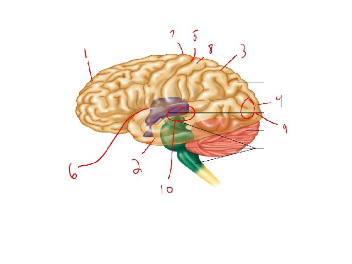
Regions of the Brain · Four major portions · Cerebrum – largest portion; higher mental functions · Diencephalon – process sensory information · Brain stem – connects different parts of nervous system; regulation · Cerebellum – muscular coordination
Cerebrum · Largest portion of the brain; · 2 cerebral hemispheres (right and left) · Connected by a deep bridge of nerve fibers corpus callosum · Each hemisphere is divided into lobes · Frontal · Parietal · Temporal · Occipital
Cerebrum · Surface ridges are called convolutions gyrus (gyri) · Act to increase surface area · Convolutions are separated by two types of grooves · Shallow grooves sulcus (sulci) · Central sulcus · separate frontal & parietal lobes · Lateral sulcus · separate temporal & other lobes · Deep grooves fissure (fissures) · Longitudinal fissure · separates R&L hemispheres · Transverse fissure · separates cerebrum & cerebellum
Cerebrum · Bulk of cerebrum is white matter · Contains bundles of myelinated nerve fibers (oligiodendrocytes) · Cerebral cortex · · Outermost portion of cerebrum Thin layer of gray matter Contains bundles of unmyelinated cell bodies Responsible for all conscious behavior by containing · motor (cortex) areas · sensory (cortex) areas · association (cortex) areas
Cerebral Cortex a. Motor Areas – frontal cortex 1. Primary motor (cortex) Area a. Initiates all voluntary muscles movements b. Located in gyrus just anterior to the central sulcus (pre-central gyrus) 2. Broca’s Area a. Motor speech area b. Located in left frontal lobe, above temporal lobe
Cerebral Cortex b. Sensory Areas – concerned with conscious awareness of sensations; parietal, occipital, and temporal cortex 1. Primary somatosensory (cortex) Area a. Receives information from general receptors (touch, temp, pressure, pain) a. Located in post-central gyrus of parietal cortex 2. Visual (cortex) Area a. Receives incoming information from vision receptors a. Located in occipital cortex 3. Auditory (cortex) Area a. Receives incoming information from hearing receptors a. Located in temporal cortex 4. Gustatory (cortex) Area a. Receives incoming information from taste receptors in taste buds b. Located in parietal cortex just above temporal lobe
Cerebral Cortex c. Association areas 1. General: a. Include areas that are not involved in motor or sensory function b. Are involved in many traits c. Are usually interconnected d. Involve all four lobes 2. Association traits include: a. Analyzing and interpreting sensory experiences b. Providing memory, reasoning, verbalizing, judgment & emotions
Hemisphere Dominance · a. k. a. Brain Lateralization · Basic functions (sensory & motor) are equally controlled by L&R hemispheres corpus callosum · Association functions have a: · dominant hemisphere (typically left) controls language-related activities: · speech, writing, reading, mathematics, logic · non-dominant hemisphere (typically right) controls nonverbal functions: · Music, art, emotions, intuition
Memory · Memory is the consequence of learning · Learning is the acquisition of new knowledge · Memory is the persistence of that learning, with the ability to access it at a later time · Two types of memory: · Short-term · Long-term
Memorize this list: • read, pages, letters, school, study, reading, stories, sheets, cover, pencil, magazine, paper, words
Which words do you remember? • house, pencil, apple, shoe, book, flag, rock, train, ocean, hill, music, water, glass, school
• Many tend to say that “book” was on list 1, even though only pencil and school were on list 1
Basal Nuclei · Masses of gray matter located deep within the white matter of the cerebral hemispheres (islands in an ocean) · Cell bodies act as relay stations for outgoing motor impulses from the brain · Modify pattern of instructions from primary motor cortex in frontal cortex to basal nuclei and then through brain stem, down spinal cord to skeletal muscles · Respond to inhibitory neurotransmitter dopamine · Parkinson’s – less dopamine; overactive basal nuclei; inhibit movement · Huntington's – basal nuclei deteriorate; unrestrained movement Examples: caudate nucleus, putamen, globus pallidus, caudate lentiform
Diencephalon the “interbrain” · Enclosed by the cerebral hemispheres · Above brain stem · Comprised of two important areas of gray matter · Thalamus · Central relay station for incoming sensory impulses (except for smell) that directs the impulses to the appropriate area of the cerebral cortex for interpretation · Hypothalamus · Main visceral control center of the body (regulates homeostasis) by linking nervous system and endocrine system · Structures to know: · Optic tracts and optic chiasma · Formed by optic nerves · Infundibulum · behind optic chiasma · Pituitary gland · Hangs from floor of hypothalamus · Mamillary bodies · Two rounded structures behind infundibulum · Pineal gland - cone-shaped endocrine gland (part of epithalamus)
Diencephalon · The Hypothalamus regulates the following: 1. 2. 3. 4. 5. 6. 7. Heart rate and blood pressure Body temperature Water and electrolyte balance Hunger and body weight Digestive movements and secretions Sleep-awake cycles Controls endocrine functioning
Diencephalon · Involved in Emotional Response / Limbic System: 1. Includes structures in the frontal and temporal cortex, basal nuclei and deep nuclei 2. Controls emotional experience and expression 3. Can modify the way a person acts 4. Produces feelings of fear, anger, pleasure, sorrow 5. Recognizes life-threatening upsets in a person’s physical or psychological condition and counters them 6. Involved in sense of smell
Brain Stem · The brain stem is composed of three major parts including: · Midbrain · Pons · Medulla oblongata · The brain stem serves as a pathway for fiber tracts running to (sensory impulses) and from (motor impulses) the cerebrum and houses many cranial nerves of the PNS
Midbrain 1. Between diencephalon and pons 2. Corpora quadrigemina = • 4 dome-like protusions (dorsal midbrain) 3. Gray matter within white matter • • Mostly composed of tracts of nerve fibers Joins lower parts of brainstem with higher parts of brain 4. Cerebral peduncles = • convey ascending(sensory) and descending(motor) impulses 5. Acts in reflex actions that are visual and auditory • Pupil dilation/ startle reflex 6. Contains areas associated with reticular formation • Sleep/awake cycles
Pons · Rounded bulging center of the brain stem · Separates midbrain from medulla oblongata · Bridge of conduction fiber tracts · Location of pneumotaxic area (regulation of breathing rate) of respiratory center · Contain areas associated with reticular formation · Sleep/awake cycles
Medulla Oblongata · Inferior portion of the brain stem which blends into the spinal cord at its base · Contains autonomic reflex centers involved in maintaining homeostasis of important visceral organs 1. Cardiac center – adjusts force & rate of heart contraction 2. Vasomotor center – regulates blood pressure by acting on smooth muscle in walls of peripheral arterioles a. Vasoconstriction = BP increases b. Vasodilation = BP decreases 3. Respiratory center – controls rate and depth of breathing 4. Other: a. Vomiting b. Hiccuping c. Swallowing d. Coughing e. Sneezing
Reticular Formation · Diffuse network of nerve fibers along the brain stem · Join centers of the hypothalamus, basal nuclei, cerebellum & cerebrum · Controls brain alertness; inhibited sleep (insomnia); CNS depressants (alcohol & tranquilizers); CNS stimulants (caffeine) · When sensory impulses reach reticular formation, it activates the cerebral cortex · Decreased reticular activity results in sleep
Cerebellum · Large, cauliflower-like structure located dorsally to the pons and medulla and inferiorly to the occipital lobe of the cerebrum · Separated from cerebrum by transverse fissure · White matter (within gray matter) creating a pattern called “arbor vitae” · Coordinates all voluntary muscle movements (subconsciously), skilled movements, posture, and equilibrium (balance)