Digestion part II Stomach Anatomy The stomach is


 Acts as a storage tank for food 2) Site of food")


 Attached to the stomach 2) Curves")


 simple columnar epithelium – make up the")


 Bile")


 Absorption of water to “dry out” indigestible food")
- Slides: 31
Digestion part II
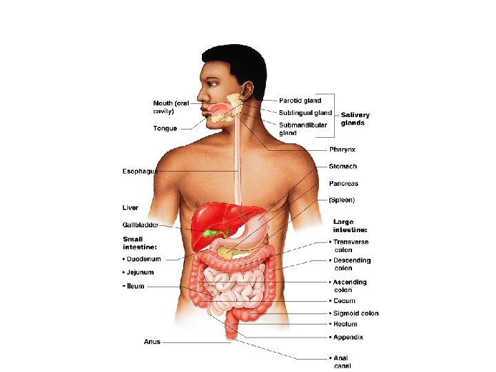
Stomach Anatomy § The stomach is located on the left side of the abdominal cavity and is about 10 inches long § Cardioesophageal sphincter – smooth muscle that opens to allow food into the stomach from the esophagus § Regions of the stomach § Cardiac region – superior; near the heart; entrance from esophagus § Fundus – superior region of stomach § Body – middle region of stomach § Pylorus – funnel-shaped terminal end; inferior region of stomach § Pyloric sphincter – Ring of smooth muscle that opens to allow small amounts of food into the small intestine
Stomach Anatomy § Rugae – internal folds of the mucosa; can expand to hold about a gallon of food § External regions 1) Lesser curvature – concave medial surface 2) Greater curvature – convex lateral surface § Oblique Muscle Layer - an additional third layer of muscle under the longitudinal and circular muscle layers of the muscularis externa § Allows the stomach to churn, mix, and pummel the food (besides moving it along)
Stomach Anatomy Figure 14. 4 a
Stomach Anatomy § Layers of peritoneum attached to the stomach § Lesser omentum – attaches the liver to the lesser curvature § Greater omentum – attaches the greater curvature to the posterior body wall § Contains fat to insulate, cushion, and protect abdominal organs
Stomach Anatomy
Stomach Functions 1) Acts as a storage tank for food 2) Site of food breakdown 3) Chemical breakdown of protein begins 4) Chyme (processed food) is delivered to the small intestine
Structure of the Stomach Mucosa Figure 14. 4 b–c
Structure of the Stomach Mucosa § Gastric pits formed by folded mucosa § Glands and specialized cells are in the gastric gland region
Specialized Mucosa of the Stomach § Simple columnar epithelium § Mucous neck cells – produce a sticky alkaline mucus (what is up with that spelling Mucous or mucus? ? ? ) § Gastric glands – secrete gastric juice with 3 types of cells 1) Chief cells – produce protein-digesting enzymes (pepsinogens) 2) Parietal cells – produce hydrochloric acid 3) Enteroendocrine cells – produce gastrin http: //www. youtube. com/watch? v=6 hquz. CXYl. Ng&NR=1 http: //www. youtube. com/watch? v=Kn 1 dt 8 TG 1 v. M&feature=relate
Small Intestine § The body’s major digestive organ; approximately 20 ft in length. § Site of nutrient absorption into the blood § Muscular tube extending form the pyloric sphincter to the ileocecal valve § Mesentery – shiny connective tissue that suspends the small intestine from the posterior abdominal wall
Subdivisions of the Small Intestine § Duodenum 1) Attached to the stomach 2) Curves around the head of the pancreas 3) First 10 – 12 inches § Jejunum 1) Attaches anteriorly to the duodenum 2) About 8 ft. § Ileum 1) Extends from jejunum to large intestine and attaches at the ileocecal valve 2) Last 12 ft (approx)
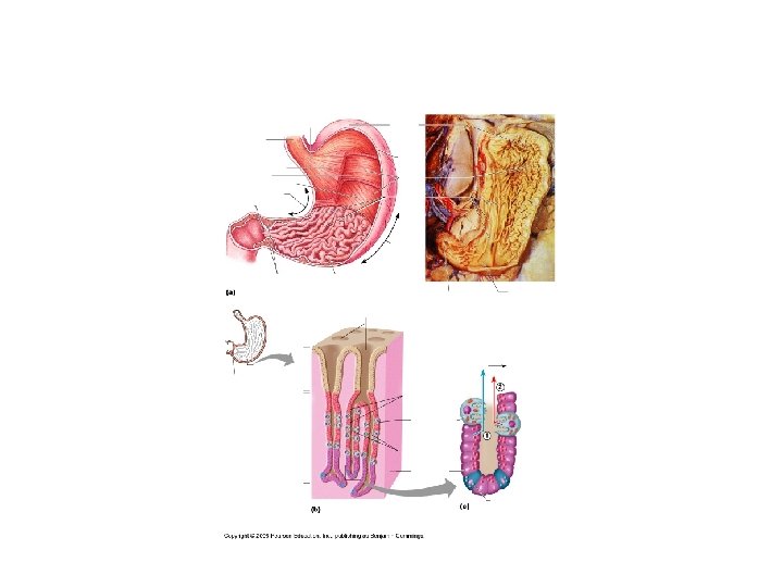
Villi of the Small Intestine § Villi are fingerlike structures formed by the mucosa § Villi give the small intestine more surface area Figure 14. 7 a
Microvilli of the Small Intestine § Microvilli - Small projections of the plasma membrane § Further increases the surface layer for absorption of nutrients Figure 14. 7 c
Structures Involved in Absorption of Nutrients 1) simple columnar epithelium – make up the Absorptive cells of the small intestine 2) Blood capillaries 3) Lacteals (specialized lymphatic capillaries) http: //www. youtube. com/watch? v=P 1 s. DOJM 65 Bc& feature=related Figure 14. 7 b
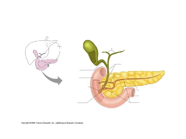
Chemical Digestion in the Small Intestine Figure 14. 6
Chemical Digestion in the Small Intestine § Source of enzymes that are mixed with chyme § Pancreas - Produces a wide spectrum of digestive enzymes that break down all categories of food § Enzymes are secreted into the duodenum § Alkaline fluid introduced with enzymes neutralizes acidic chyme § Endocrine products of pancreas – Homeostatic Mechanism § Insulin – carries blood sugar (glucose) into cells. Also stimulates the storage of glucose as glycogen in the liver. § Produced by Beta Cells in Pancreatic Islets § If a person lacks Beta Cells or “Burns them out” by eating too much sugar Diabetes results. § http: //www. youtube. com/watch? v=qzjj. W--I-2 Q § https: //www. youtube. com/watch? v=NZ 4 zcr. Tz. Uj. A
Chemical Digestion in the Small Intestine § Glucagons – stimulate the breakdown of glycogen into glucose when sugar levels are low – releases glucose § Produced by Alpha Cells in Pancreatic Islets § Mainly happens in the Liver § One molecule of glucagon can release 100 million molecules glucose!
Liver § Largest gland in the body – about 3 lbs § Located on the right side of the body under the diaphragm § Consists of four lobes suspended from the diaphragm and abdominal wall by the falciform ligament § Connected to the gall bladder via the common hepatic duct § Diseases § Cirrhosis – chronic inflammation of the liver usually due to alcohol abuse – causes “beer gut” § Hepatitis – inflammation of the liver due to viral infection. § Hepatitis B – transmitted by needles/sexual contact much like AIDS. Can easily lead to Liver Cancer
Bile § Produced by cells in the liver § Made up of: 1) Bile salts 2) Bile pigment (mostly bilirubin from the breakdown of hemoglobin) 3) Cholesterol, Phospholipids, Electrolytes
Chemical Digestion in the Small Intestine Figure 14. 6
Gall Bladder § Sac found in hollow fossa of liver § Stores bile from the liver by way of the cystic duct – much more concentrated in gall bladder § Bile is introduced into the duodenum in the presence of fatty food § Bile emulsifies fats – makes them watery § Gallstones can cause blockages which are caused by the crystallization of cholesterol § http: //www. youtube. com/watch? v=Uzl 6 M 1 Yl. U 3 w&feature=related
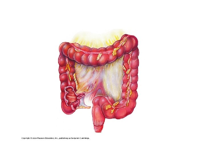
Large Intestine § Larger in diameter, but shorter than the small intestine § Frames the internal abdomen
Structures of the Large Intestine § Cecum – saclike first part of the large intestine § Ileocecal Valve – structure that allows undigested food into the large intestine (works like the sphincters) § Appendix § Accumulation of lymphatic tissue that sometimes becomes inflamed (appendicitis) pain in the right side of your body § Hangs from the cecum § Colon § Ascending (up) § Transverse (across) § Descending (down) § S-shaped sigmoid (between descending colon and rectum) § Rectum § Anus – external body opening § Internal anal sphincter – smooth muscle and involuntary
Large Intestine § Larger in diameter, but shorter than the small intestine § Frames the internal abdomen
Modifications to the Muscularis Externa in the Large Intestine § Smooth muscle is reduced to three bands (teniae coli) § Muscle bands have some degree of tone § Haustra - pocketlike sacs that form the walls of the large intestine.
Functions of the Large Intestine 1) Absorption of water to “dry out” indigestible food 2) Eliminates indigestible food from the body as feces § Fiber is extremely important in this phase of the digestion process § Fiber helps increase the movement of feces through the L. Intestine so that carcinogens don’t have a chance to “sit around” § Should be eliminating feces at least once a day. § Foods high in fiber: whole grains (like oatmeal etc. ), fruit, vegetables) § http: //www. gutsense. org/constipation/frequency. html § https: //www. youtube. com/watch? v=mh 90 RPA-C 10 § http: //www. youtube. com/watch? v=YMB 44 VKKVLQ&feature=related 3) Does not participate in digestion of food 4) Goblet cells produce mucus to act as a lubricant