Pharynx and Esophagus Deglutition Moves a bolus from

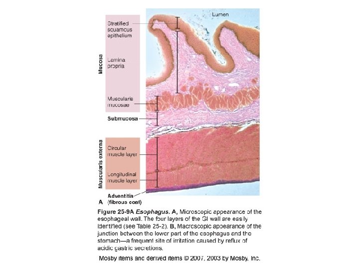
Pharynx and Esophagus • Deglutition – Moves a bolus from the mouth to the stomach • Mouth • Fauces • Oropharynx – Second division of the pharynx • Esophagus – Pierces diaphragm – First segment of digestive tube proper – Normally flattened – Stratified squamous epithelium – Striated, mixed, and smooth muscle • Stomach
 • Size and position of the stomach – Digestive tube")
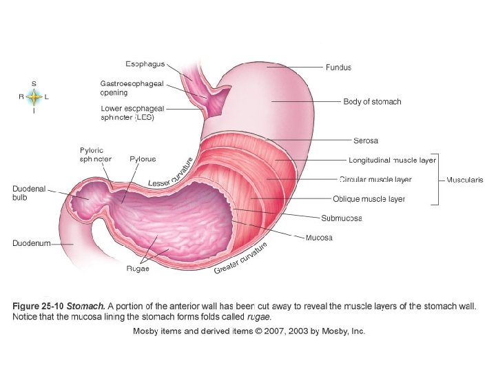
Stomach (Figure 25 -10) • Size and position of the stomach – Digestive tube dilates into an elongated pouchlike structure – Various factors affect size • Gender • Amount of distension • When empty, it is about the size of a large sausage • In adults, capacity ranges from 1. 0 to 1. 5 liters – Located in the upper part of abdominal cavity • Under the liver and diaphragm • Primarily left of the median line • Epigastrium and left hypochondrium (Figure 1 -14) • Can also crowd the heart and diaphragm after large meals
 • Divisions of the stomach – Fundus • Enlarged portion")
Stomach (Figure 25 -10) • Divisions of the stomach – Fundus • Enlarged portion • To the left and above the opening of esophagus into stomach – Body • central portion of stomach – Pylorus • Lower part of stomach • Curves of the stomach – Lesser curvature • Upper right curve of stomach – Greater curvature • Lower left curve of stomach
 • Sphincter muscles – Guard both stomach openings – Circular")
Stomach (Figure 25 -10) • Sphincter muscles – Guard both stomach openings – Circular fibers • Open when relaxed • Closed when contracted – Lower esophageal sphincter (LES) or cardiac sphincter • Controls opening of esophagus into stomach – Pyloric sphincter • Controls outlet of pyloric portion of stomach into duodenum

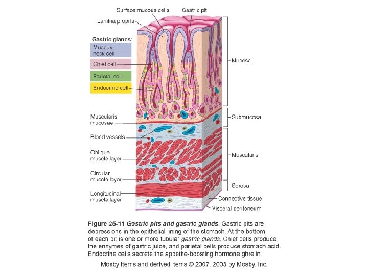
Stomach • Stomach wall – Gastric mucosa • Epithelial lining has rugae marked by gastric pits (Figures 25 -11 and 25 -12) • Gastric glands – Found below level of the pits – Secrete most of the gastric juice • Chief cells – Secretory cells found in gastric glands – Secrete the enzymes of gastric juice • Parietal cells – Secretory cells found in gastric glands – Secrete hydrochloric acid – Thought to produce intrinsic factor needed for vitamin B 12 absorption • Endocrine cells – Secrete gastrin and ghrelin – Gastric muscularis • Thick layer of muscle with three distinct sublayers

Stomach • Functions of the stomach – Reservoir food • Once partially digested, food moves further along GI tract – Secretes gastric juice • Acids and enzymes aid in digestion of food – Breaks food into small particles and mixes them with gastric juice • Contractions of the muscular coat – Secretes intrinsic factor – Limited absorption • Certain drugs, water, alcohol, short-chain fatty acids – Produces gastrin • Regulates digestive functions – Helps protect body from pathogenic bacteria

Small Intestine • Size and position of the small intestine – 2. 5 cm in diameter – 6 m in length – Coiled loops fill most of abdominal cavity • Divisions of the small intestine – Duodenum • Uppermost division • Approximately 25 cm long • Shaped roughly like the letter C – Jejunum • Approximately 2. 5 m long – Ileum • Approximately 3. 5 m long
 – Intestinal lining")
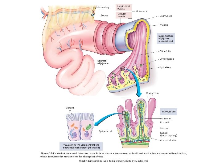
Small Intestine • Wall of the small intestine (Figure 25 -14) – Intestinal lining has plicae (folds) with villi (projections) – Villi—important modifications of mucosal layer • Each villus contains an arteriole, venule, and lacteal • Covered by a brush border made up of 1, 700 ultrafine microvilli per cell • Villi and microvilli increase surface area of small intestine hundreds of times – Main site of digestion and absorption – Mucus-secreting goblet cells – Intestinal crypts areas of rapid mitotic divisions – Enzymes inhibit bacterial growth

Large Intestine • Size of the large intestine – Average diameter, 6 cm – Approximately 1. 5 to 1. 8 m long
 – Cecum •")
Large Intestine • Divisions of the large intestine (Figure 25 -16) – Cecum • First 5 to 8 cm of large intestine • Blind pouch located in lower right quadrant of abdomen – Colon • Ascending colon – Vertical position on right side of abdomen – Ileocecal valve prevents material passing from large intestine into ileum • Transverse colon – Passes horizontally across abdomen – Above small intestine – Extends from hepatic flexure to splenic flexure • Descending colon – Vertical position on left side of abdomen • Sigmoid colon joins descending colon to rectum • Rectum – Last 7 or 8 inches of intestinal tube – Terminal inch is anal canal with opening called the anus (Fig 25 -17)
 – Intestinal mucous")
Large Intestine • Wall of the large intestine (Figure 25 -19) – Intestinal mucous glands • Produce lubricating mucus • Coats feces as they are formed – Uneven distribution of fibers in the muscle coat
- Slides: 16