Congenital Hearing Loss Ashley Starkweather MD UCLA Head
- Slides: 63
Congenital Hearing Loss Ashley Starkweather, MD UCLA Head and Neck Surgery February 25, 2009
Etiology § Congenital HL § 50% Genetic § 50% Acquired § Childhood Onset HL § 50% Genetic § 25% Acquired § 25% Unknown
Genetic HL § 75% non-syndromal § 25% syndromal § § 75% autosomal recessive (AR) 25% autosomal dominant (AD) 1 -2% X-linked Rare mitochondrial
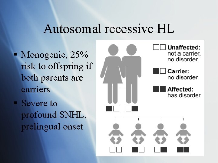
Autosomal recessive HL § Monogenic, 25% risk to offspring if both parents are carriers § Severe to profound SNHL, prelingual onset
Autosomal recessive syndromal HL § § Usher syndrome Pendred Jervel and Lange Nielsen Goldenhar (Oculoauriculoverterbral spectrum)
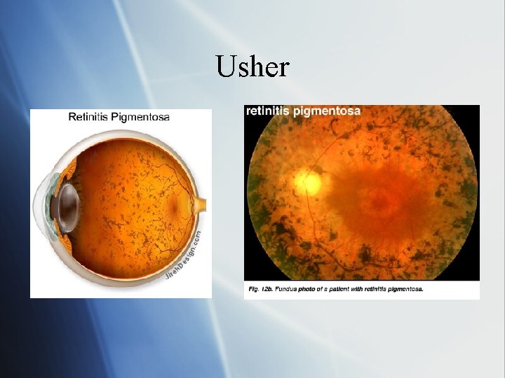
Usher Syndrome § Retinitis pimentosa and SNHL § Night blindness > field cut > central blindness § Most common cause of congenital deafness § Dx: electroretinography
Usher Types § Type I (most common): § Profound SNHL, no vestibular fxn § RP onset in early childhood § Atypical myosin (myosin 7 A): interferes with mechanoelectrical transduction in labyrinthine hair cells § Type II: § Congenital sloping SNHL § Normal vestibular fxn § RP onset in teens
Usher Types § Type III: § Progressive SNHL and vestibular dysfunction § Vestibulocerebellar ataxia § Type IV: § Mental retardation and hypotonia
Usher
Pendred Syndrome § Defect in tyrosine iodination § Gene mutation: affects pendrin, molecule involved in chloride-iodine transport § Sx: severe to profound SNHL, multinodular goiter in childhood § Assoc with Mondini malformation and enlarged vestibular aqueduct § Dx: (+) perchlorate test § Tx: thyroid hormone to suppress goiter
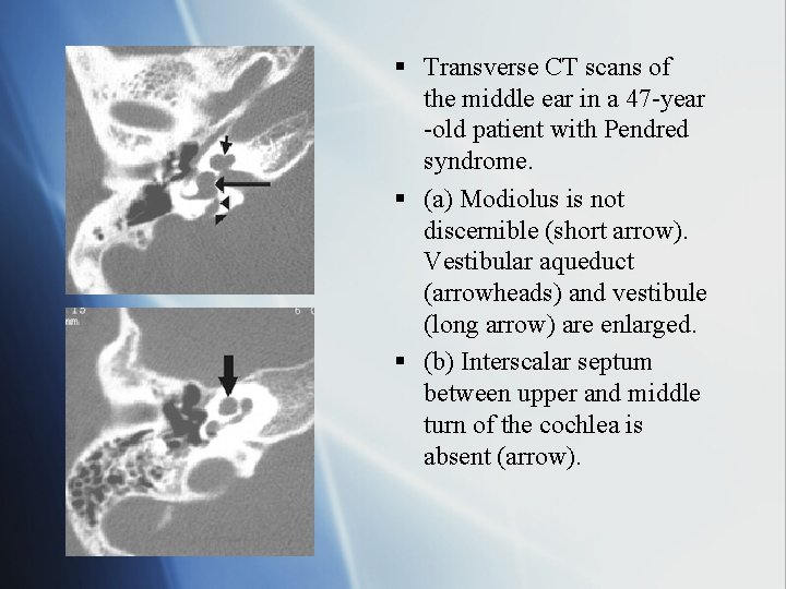
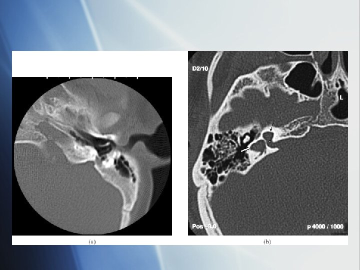
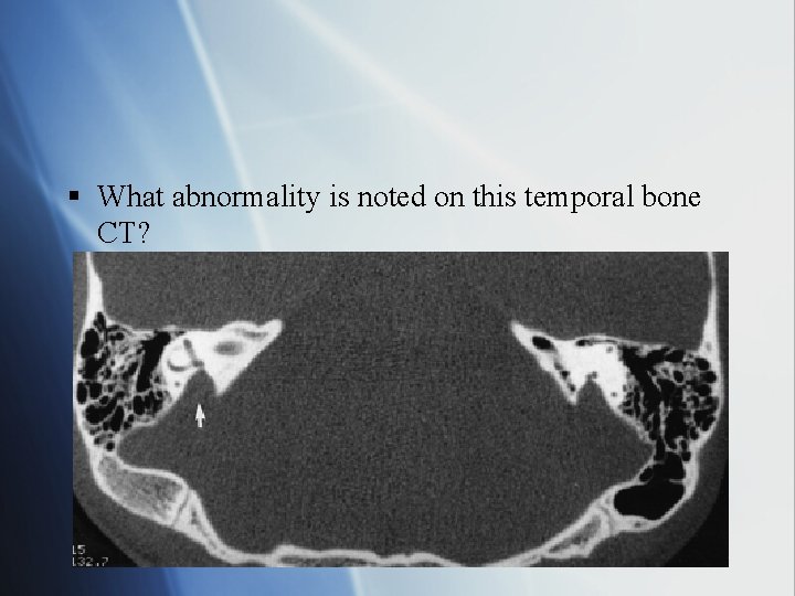
§ Transverse CT scans of the middle ear in a 47 -year -old patient with Pendred syndrome. § (a) Modiolus is not discernible (short arrow). Vestibular aqueduct (arrowheads) and vestibule (long arrow) are enlarged. § (b) Interscalar septum between upper and middle turn of the cochlea is absent (arrow).
Jervell and Lange Nielsen § Congenital profound SNHL § Prolonged QT interval with syncope, sudden death § Gene mutation: KVKQT 1 = abnormal K+ channel § Dx: EKG § Tx: Beta blockers, hearing aids
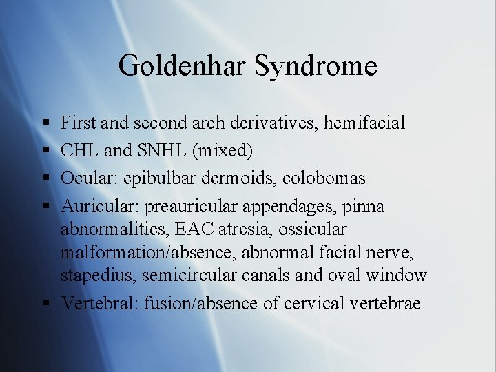
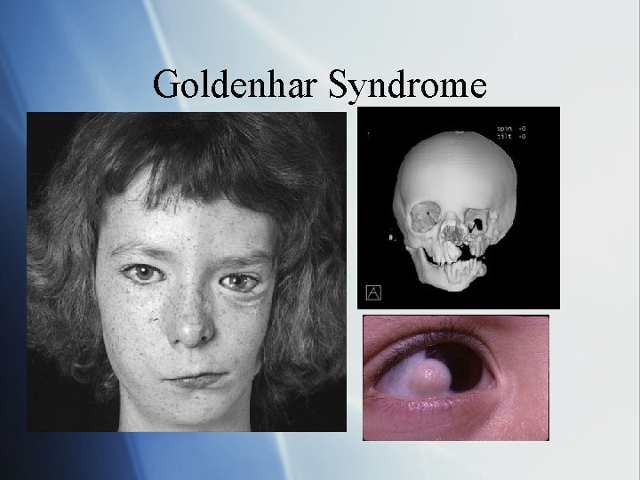
Goldenhar Syndrome § § First and second arch derivatives, hemifacial CHL and SNHL (mixed) Ocular: epibulbar dermoids, colobomas Auricular: preauricular appendages, pinna abnormalities, EAC atresia, ossicular malformation/absence, abnormal facial nerve, stapedius, semicircular canals and oval window § Vertebral: fusion/absence of cervical vertebrae
Goldenhar Syndrome
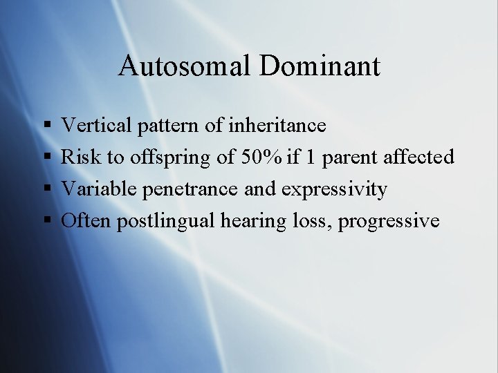
Autosomal Dominant § § Vertical pattern of inheritance Risk to offspring of 50% if 1 parent affected Variable penetrance and expressivity Often postlingual hearing loss, progressive
AD Syndromes § § § § Waardenburg Treacher Collins Apert Crouzon Stickler Neurofibromatosis Brancio-oto-renal
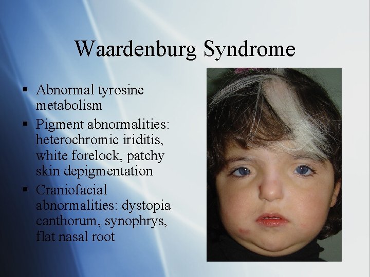
Waardenburg Syndrome § Abnormal tyrosine metabolism § Pigment abnormalities: heterochromic iriditis, white forelock, patchy skin depigmentation § Craniofacial abnormalities: dystopia canthorum, synophrys, flat nasal root
Waardenburg Types § Type I: § Dystopia canthorum, pigment and craniofacial abnormalities, 20% with SNHL § Mutation in PAX 3 gene § Type II: § No dystopia canthorum, 50% with SNHL but not as severe § MITF mutation
Waardenburg Types § Type III (most severe): § Unilateral ptosis and skeletal abnormalities § PAX 3 mutation § Type IV: § Type II plus Hirschsprung’s disease (aganglionic megacolon)
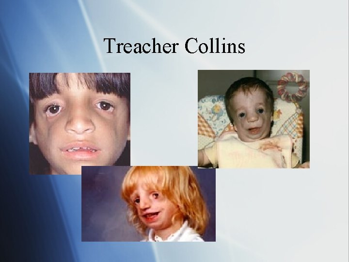
Treacher Collins (Mandibulofacial dysostosis) § § § Hypoplasia of mandible and facial bones Downsloping palpebral fissures, colobomas Atretic external and middle ear Mixed HL Cleft palate (35%) Gene mutation on chr 5 q: TCOF 1 codes for a cell transport protein (treacle) § Tx: BAHA, bone conduction HA, surgical correction of aural atresia
Treacher Collins
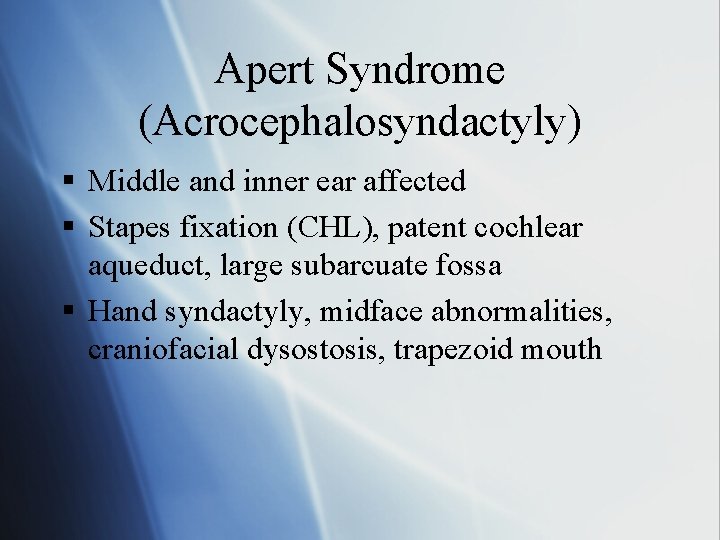
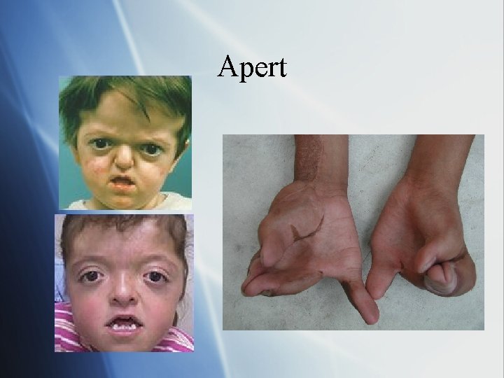
Apert Syndrome (Acrocephalosyndactyly) § Middle and inner ear affected § Stapes fixation (CHL), patent cochlear aqueduct, large subarcuate fossa § Hand syndactyly, midface abnormalities, craniofacial dysostosis, trapezoid mouth
Apert
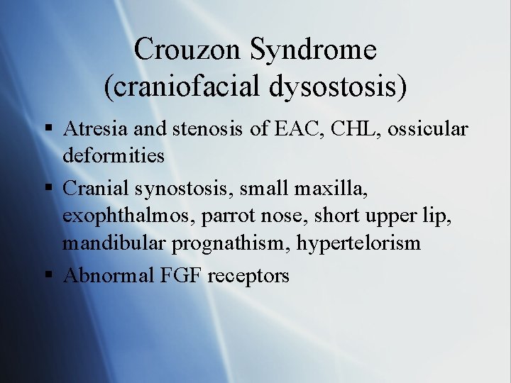
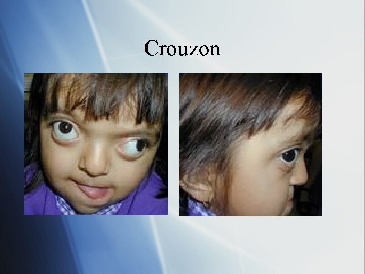
Crouzon Syndrome (craniofacial dysostosis) § Atresia and stenosis of EAC, CHL, ossicular deformities § Cranial synostosis, small maxilla, exophthalmos, parrot nose, short upper lip, mandibular prognathism, hypertelorism § Abnormal FGF receptors
Crouzon
Stickler Syndrome § § § § Progressive Arthro-Ophthalmopathy Progressive SNHL (80%) Marfanoid body habitus Severe myopia, retinal detachment Flat midface Hypermobile joints Pierre Robin sequence: micrognathia, glossoptosis, cleft palate
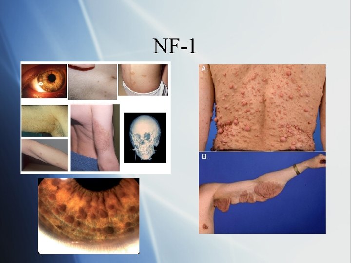
Neurofibromatosis § NF-1 (Von Recklinghausen Disease) § Café au lait spots, neurofibromas, Lisch nodules, 5% risk of unilateral acoustic neuroma § NF-1 gene on Chr 17 § NF-2 (central neurofibromatosis) § Bilateral acoustic neuromas or unilateral with 1 st degree relative with NF-2 or multiple central schwannomas § NF-2 gene Chr 22 q 12 (tumor suppressor gene mutation)
NF-1
Branchio-oto-renal (Melnick Fraser Syndrome) § Renal abnormalities: mild hypoplasia to bilateral aplasia § Branchial cleft cyts § Preauricular pits § EYA 1 on Chr 8 q 13 § Hearing loss: § § Penetrance: 80% Mixed: 50% Conductive: 30% SNHL: 20%
X-linked Disorders § Alport’s syndrome § Otopalatal-digital § Norrie syndrome
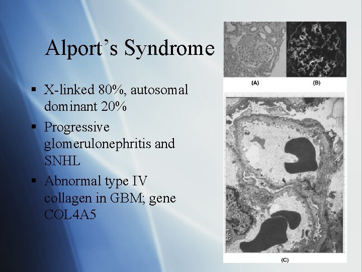
Alport’s Syndrome § X-linked 80%, autosomal dominant 20% § Progressive glomerulonephritis and SNHL § Abnormal type IV collagen in GBM; gene COL 4 A 5
Alport’s Syndrome § Bilateral degeneration of organ of Corti and stria vascularis § Ocular disorders (myopia, cataracts) § Dx: UA, BUN, Cr § Tx: dialysis, renal transplant
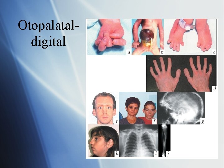
Otopalatal-digital § § Ossicular malformation (CHL) Palate defects Digital abnormalities: broad fingers and toes Hypertelorism, short stature, mental retardation
Otopalataldigital
Norrie Syndrome § Blindness § Progressive mental retardation § Hearing loss
Mitochondrial Disorders § § § Follows maternal line Postlingual HL Associated with systemic metabolic disorders Increased sensitivity to aminoglycoside ototoxicity Ex: § MELAS: mitochondrial encephalopath, lactic acidosis, and strokelike syndrome § MIDD: maternally inherited diabetes and deafness
Acquired Congenital HL § Prenatal: infections, teratogens § Perinatal: NICU admission § Postnatal: infections, neoplasms
Prenatal Infections § TORCHS: § § § Toxoplasmosis Rubella CMV HSV encephalitis Syphilis
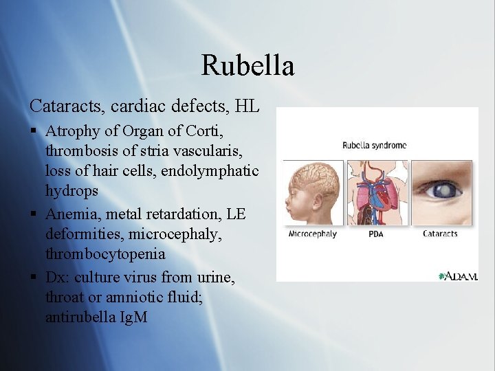
Rubella Cataracts, cardiac defects, HL § Atrophy of Organ of Corti, thrombosis of stria vascularis, loss of hair cells, endolymphatic hydrops § Anemia, metal retardation, LE deformities, microcephaly, thrombocytopenia § Dx: culture virus from urine, throat or amniotic fluid; antirubella Ig. M
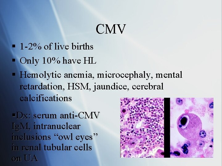
CMV § 1 -2% of live births § Only 10% have HL § Hemolytic anemia, microcephaly, mental retardation, HSM, jaundice, cerebral calcifications §Dx: serum anti-CMV Ig. M, intranuclear inclusions “owl eyes” in renal tubular cells on UA
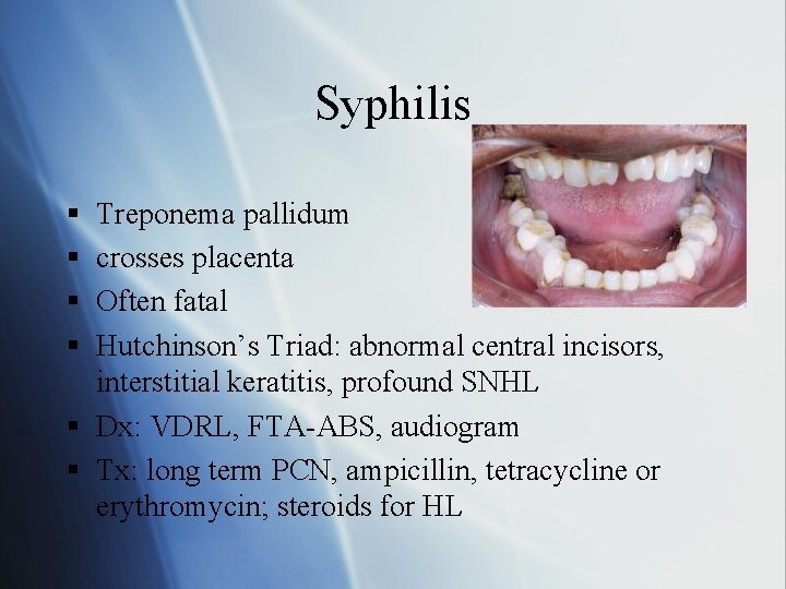
Syphilis § § Treponema pallidum crosses placenta Often fatal Hutchinson’s Triad: abnormal central incisors, interstitial keratitis, profound SNHL § Dx: VDRL, FTA-ABS, audiogram § Tx: long term PCN, ampicillin, tetracycline or erythromycin; steroids for HL
Prenatal Teratogens § § Et. OH Thalidomide Radiation Aminoglycosides
Perinatal Causes of HL § Hypoxia § Kernicterus § Persistent fetal circulation
Postnatal Causes of HL § Meningitis (suppurative labryrinthitis) § Ossification of labryinth § Steroids help prevent HL § Most common postnatal cause of HL § § § Viral infection: mumps Ototoxins/Chemotherapy Trauma (acoustic, blunt, penetrating) Perilymph fistula Neoplasm: medulloblastoma, AN, fibrous dysplasia, histiocytosis) § Autoimmune (rare in children)
Inner Ear Dysmorphologies § § § § Michel’s aplasia Mondini aplasia Scheibe aplasia Alexander aplasia Bing Siebenmann Enlarged vestibular aqueduct Absence of CN VIII
Michel’s aplasia § § AD or thalidomide exposure Complete aplasia of inner ear Anacusis, normal middle and outer ear Dx: CT shows hypoplastic petrous pyramid, absent cochlea and labyrinth
Mondini Aplasia § § AD Most common cochlear abnormality Progressive or fluctuating HL risk of perilymphatic gusher and meningitis from dilated cochlear aqueduct § Dx: CT reveals single turned cochlea, no interscalar septum § Tx: HA, cochlear implant
Schiebe Aplasia § AR § Partial or complete aplasia of pars inferior (cochlea and saccule), normal pars superior (SCC and utricle) § Defect of membranous labyrinth only, therefore can not diagnose on CT
Alexander Aplasia § § AR Abnormal cochlear duct/ basal turn High frequency SNHL Cannot diagnose on CT
Enlarged vestibular aqueduct § Defined by diameter of duct >2 mm at midpoint § Progressive cochleovestibular loss § No treatment
Thanks for listening!
Questions § What % of patients with NF-1 have acoustic neuromas? a) b) c) d) 5% 20% 50% 95%
§ What % of patients with NF-2 have acoustic neuromas? a) 5% b) 20% c) 50% d) 95%
§ What is the basic defect that causes Alport syndrome? a) abnormal renal tubules b) abnormal collagen IV in glomerulus c) abnormal collagen I in glomerulus d) abnormal renal arteries
§ What is the primary inheritance pattern for Alport’s syndrome?
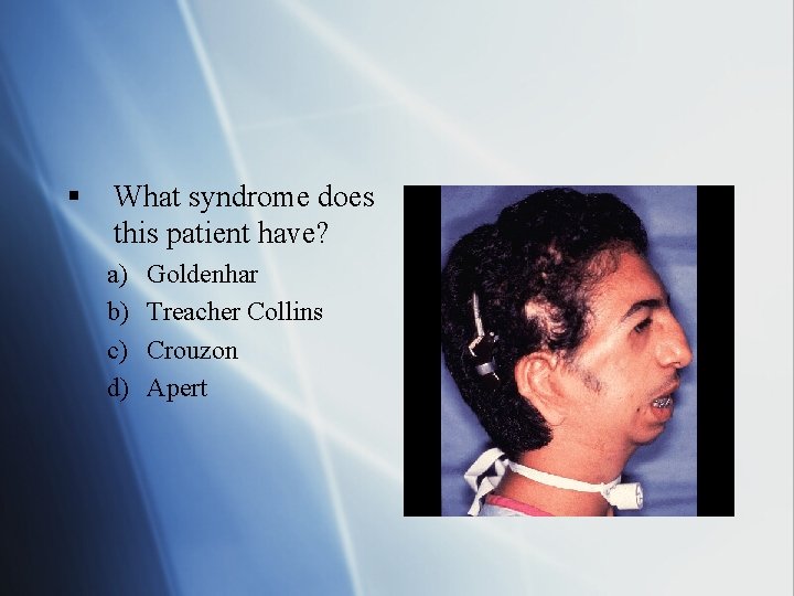
§ What syndrome does this patient have? a) b) c) d) Goldenhar Treacher Collins Crouzon Apert
§ What inner ear aplasia will not allow for cochlear implants or amplification aids? a) b) c) d) Mondini aplasia Michel’s aplasia Enlarged vestibular aqueduct Alexander aplasia
§ Which inner ear dyplasia is characterized by a lack of septae in the cochlea and only a basal turn? a) b) c) d) Mondini aplasia Michel’s aplasia Enlarged vestibular aqueduct Alexander aplasia
§ What is the inheritance pattern of MIDD and MELAS?
§ What abnormality is noted on this temporal bone CT?
§ What genetic mutation is responsible for type I and III Waardenburg syndrome? a) b) c) d) COL 2 A 1 Pendrin PAX 3 Chr 22 q 12
Thanks for listening!