Approach to Hematuria and Proteinuria in Children Adi


")


 • Pathology: Ig. A nephropathy • Clinical: - purpuric rash")
: E. coli O 157: H 7")
 • Homozygous mutation in genes encoding type IV collagen in")
 • • • Autosomal dominant Hetrozygous mutation")
 1. Gross hematuria: onset, duration, progression, aggravating,")

 • • • Negative Trace 1+ 2+ 3+ 4+ < 10")
 Non- nephrotic Nephrotic • Urine prot/cr: > 20 mg/mmol • Urine prot/cr")


: 585 -9.")
 •")


- Slides: 52
Approach to Hematuria and Proteinuria in Children Adi Alherbish
Objectives • To be able to define and recognize hematuria and proteinuria • To be able to generate a differential diagnosis of the commonest and most serious causes of hematuria and proteinuria • To have a clinical approach to both conditions.
Case 1 • 14 year old boy presenting with red urine since last night. Otherwise healthy. Normal BP, no flank pain, no ankle edema. • What’s the next step?
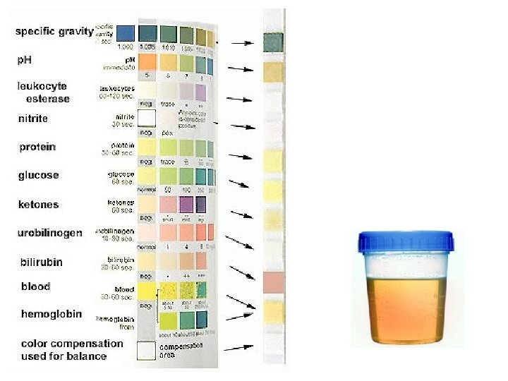
Case 1 • Urine dipstick: negative
Case 2 • 5 year old boy presenting with pallor, and shortness of breath. Urine dip: SG 1. 015, Hg 2+, Prot neg, Urinalysis: RBC 0, WBC 0
Case 2 • CBC: Hg 80, WBC 5, Plt 180 • Retics: 3% Hemolytic Anemia • Send blood for: Hg electrophoresis, peripheral smear, Coombs test, G 6 PD
Case 3 • 14 year old girl, healthy • Regular check up: Urine dip: SG 1. 035, Hg 2+, Prot trace Urinalysis: RBC 5 - 10 /HPF WBC 0 - 5 / HPF
Case 3 • Repeat urinalysis after drinking a bottle of water: Urine SG: 1. 015 RBC: 1 - 5 /HPF WBC: 0 - 5 / HPF
HPF= x 400
Case 5 • 9 year old girl, presenting with fever, rash, coryza, conjuctivitis, and dark urine. • Urine dip: SG 1. 015, Hg +3, Prot trace • Urinalysis: RBC > 100/ HPF WBC 10 - 25/ HPF
Case 5 • Urine positive for adenovirus
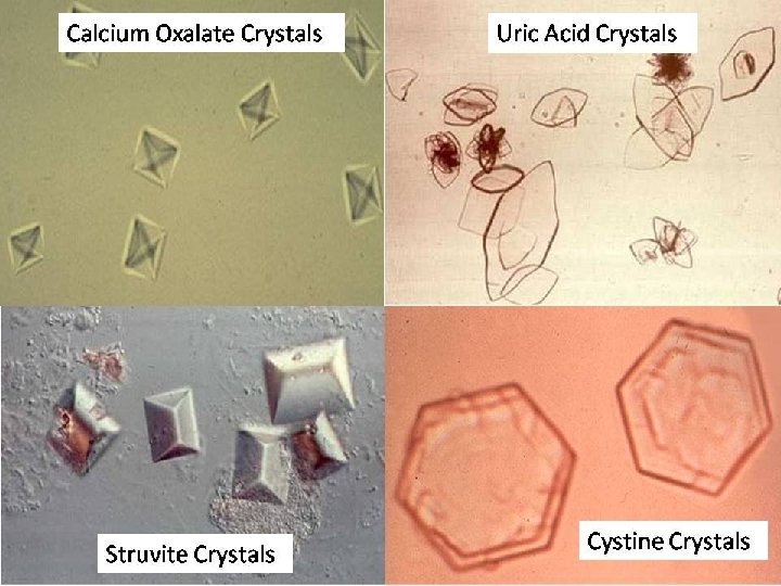
Case 6 • 14 year old girl, presenting with intermittent, sudden onset left flank pain and dark urine. • Urine SG: 1. 015, Hg 3+, Prot neg • Urinalysis: RBC 100/ HPF, WBC 0 Crystals present
Case 6 • In clinic: send urine for Ca/ Cr ratio, citrate, oxalate, uric acid, cystine
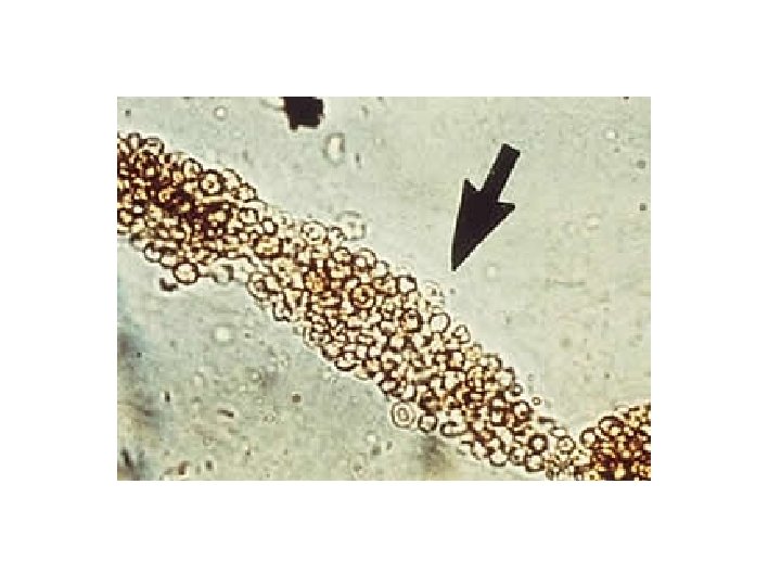
Case 7 • 14 year old girl, with hypertension, left knee arthritis, dark urine, malar rash • Urine dip: SG 1. 010, Hg 2+, Prot 2+ • Urinalysis: RBC 10 - 25/ HPF WBC 0 RBC casts
Case 7 • Send blood for: C 3, C 4, ANA, anti-ds DNA
Hematuria • Presence of > 5 RBC/ HPF, on more than two occasions, in the context of a normal urine specific gravity
The 3 Vital Questions 1 2 3 Is it true hematuria? Is it serious (urgent)? What is the cause?
Is it serious? Nephritis • • Hematuria Hypertension Oliguria Increased Cr Nephrosis • Edema • Nephrotic range proteinuria • Low albumin • Hypercholestrolemia
Rapidly Progressive Glomerulonephritis (RPGN)
RPGN Immune Complex • • Post- strep GN Ig. A nephropathy Lupus HSP Pauci- immune Anti- GBM • Wegner’s granulomatosis • Microscopic polyangiitis • Polyartritis nodosa • Goodpasture’s disease
RPGN Immune Complex • • Post- strep GN Ig. A nephropathy HSP Lupus • ASO, anti-DNase • Immunoglobulins • ANA, anti-ds DNA, C 3, C 4 Pauci- immune Anti- GBM • Wegner’s granulomatosis • Microscopic polyangiitis • Polyartritis nodosa • Goodpasture’s disease • ANCA • Anti- GBM
Post strep Glomerulonephritis • Strep pharyngitis, or strep skin infection, followed 10 to 14 days by microscopic hematuria, nephritis, or nephrosis • Diagnosis: positive ASO low C 3 which normalize in 8 weeks • Management: supportive • Prognosis: Excellent (Vog et. Al: 137 cohort. ESRD: none, high Cr 10%)
Ig. A nephropathy • Typical presentation: intermittent gross hematuria that happen during colds • Other: gross hematuria microscopic hematuria nephritis nephrotic syndrome ESRD
Ig. A nephropathy • Diagnosis: clinical suspicion Ig. A level 20% sensitivity! Kidney biopsy- Ig. A in Immunoflorecence • Treatment: supportive in mild cases ACEI in proteinuria Steroids
Henoch Schonlein Purpura (HSP) • Pathology: Ig. A nephropathy • Clinical: - purpuric rash - arthritis - intestinal edema (intussusception) - hematuria/ nephritis/ nephrosis
Hemolytic Uremic Syndrome • Pathogenesis: - typical (d+): E. coli O 157: H 7 shiga toxin 1 induced vascular injury - atypical (d-): alternative complement pathway defect • Clinical: triad of microangiopathic hemolytic anemia, thrombocytopenia, ARF
Alport Syndrome (Hereditary Nephritis) • Homozygous mutation in genes encoding type IV collagen in basement membrane • Genetics: 80% X-linked AR, AD • Clinical: persistent microscopic hematuria, hearing loss, lenticonus
Benign familial hematuria (thin basment membrane nephropathy) • • • Autosomal dominant Hetrozygous mutation in type IV collagen Microscopic hematuria Screen the parents’ urine Benign course
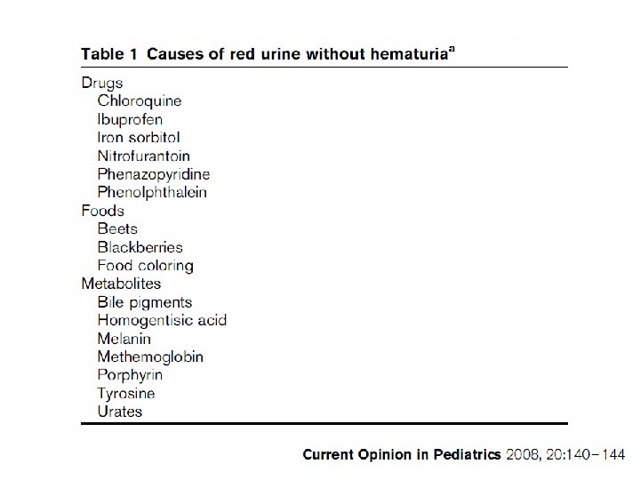
Work up for hematuria (History is important!) 1. Gross hematuria: onset, duration, progression, aggravating, relieving factors, associated symptoms 2. UTI symptoms: dysuria, frequency, urge incontinence 3. Food intake: beet 4. Drugs: rifampin, nitrofurantoin, ibuprofen 5. Ig. A: gross hematuria onset while having colds 6. post strep: history of sore throat, tonsillitis, skin infection 7. HUS: diarrhea, pallor, fatigue, SOB 8. HSP: pupuric skin rash over legs and buttocks (palpable), join swelling/pain, abdominal pain/bloody stools 9. Goodpasture/Wegners: hemoptysis, cough, SOB 10. SLE/ vasculitis: butterfly rash, discoid rash, mouth ulcers, photosensitivity, CNS seizures/psychosis, join swelling 11. Kidney stones: renal colic, radiation to groins, past history or family history of stones 12. Anatomic: antenatal U/S, single umbilical artery, abdominal swelling 13 Hereditary: family history of deafness, family member with hematuria 14. Bleeding diathesis: hemarthrosis, epistaxis, petechaie, heavy periods in older girls 15. Problems with high blood pressure 16. Family history: nephritis, kidney failure, transplant, deafness, stones, hematuria, consanguinity
Work up for hematuria • Nephritis: ASO, C 3, C 4, anti-ds DNA, ANCA, anti- GBM • Kidney and bladder U/S • Stone work up: urine Ca, Cr, oxalate, citrate, cystine, uric acid • Urinalysis in both parents • Bleeding tendency: PT, PTT, INR
Proteinuria (Urine dip) • • • Negative Trace 1+ 2+ 3+ 4+ < 10 mg/dl 10 - 20 mg/dl 30 mg/dl 100 mg/dl 300 mg/dl 1000 mg/dl
Proteinuria (Quantitative) Non- nephrotic Nephrotic • Urine prot/cr: > 20 mg/mmol • Urine prot/cr > 200 mg/mmol • 24 h urine collection: > 100 mg/m 2/day > 4 mg/m 2/hr • 24 h urine collection: > 1 g/m 2/ day > 40 mg/m 2/hr
The 3 Vital Questions 1 2 3 Is it persistent? Is it nephrotic? What is the cause?
Case 1 • 15 year old, athletic boy • Regular check up: Urine dip: Prot 2+ Urine prot/Cr ratio: 50 mg/mmol • What next?
Case 1 • 8 am: urine prot/Cr ratio- 10 mg/mmol • 4 pm: urine prot/Cr ratio- 50 mg/mmol Orthostatic proteinuria
Non Persistant Proteinuria • • • Fever Strenuous exercise Cold exposure Epinephrine infusion Orthostatic
Case 2 • 1 year old infant with failure to thrive. Both height and weight are below the 3 rd percentile. He has sings of rickets in exam. • Urine dip: Prot 3+ , Glu 2+
Derakhshan Ali et al. Saudi J Kidney Dis Transpl. 2007 Oct-Dec; 18(4): 585 -9.
Fanconi Synrome • PCT defect • Proximal renal tubular acidosis (type II RTA) • Glucosuria • Aminoaciduria • Phosphaturia • hypokalemia
Proteinuria Glomerular • Congenital: -Finish- type - TORCH infection • Nephritis: - postinfectious GN - lupus - Wegner - HUS - Goodpasture • Nephrotic: - Minimal change - FSGS - MPGN • Drugs: captopril • Neoplasia • Renal vein throbosis • • • Tubualr absorption Protein overload ATN Fanconi Syndrome Cystic/dysplastic Interstial nephritis Pyelonephritis • Hemolysis • Rhabdomyolysis • Light chain
Proteinuria Glomerular • Congenital: -Finish- type - TORCH infection • Nephritis: - postinfectious GN - lupus - Wegner - HUS - Goodpasture • Nephrotic: - Minimal change - FSGS - MPGN • Drugs: captopril • Neoplasia • Renal vein throbosis • • • Tubualr absorption Protein overload ATN Fanconi Syndrome Cystic/dysplastic Interstial nephritis Pyelonephritis • Hemolysis • Rhabdomyolysis • Light chain Urine electrophoresis: • Glomerular: albumin • Tubular: other proteins. .
Case 3 • 5 year old boy, presenting with puffy eyes, enlarged tummy, and feet swelling. • Exam: normal BP, ascites, pitting edema • Urine dip: Prot 4+ • What’s the next step?
Case 3 • Urine prot/cr 1500 mg/mmol • Serum albumin 15 g/l • High cholesterol
Nephrotic Syndrome • • Urine Prot/Cr > 200 mg/mmol Serum albumin < 25 g/l Edema Hyperlipedemia
Nephrotic Syndrome • • • Minimal change disease Focal segmental glomerulosclerosis Membranoproliferative Membranous GN Infection: HIV, hepatits, syphilis Lupus, Ig A, HSP, post strep
Initial therapy • • Supportive: albumin 25% and lasix prn Salt restriction Fluid restriction while nephrotic Prednisone 60 mg/m 2/day for 6 weeks followed by 40 mg/m 2/day for 6 weeks then wean. .
Indications for biopsy • Steroid resistant: fail to enter remission after 8 weeks of therapy • Steroid dependent: intially enter remission, but develping relapse while on therapy, or within 2 weeks of steroid discontinuration • • Hematuria Increased Cr (when intravasculary repleted) Low complement Positive lupus serology