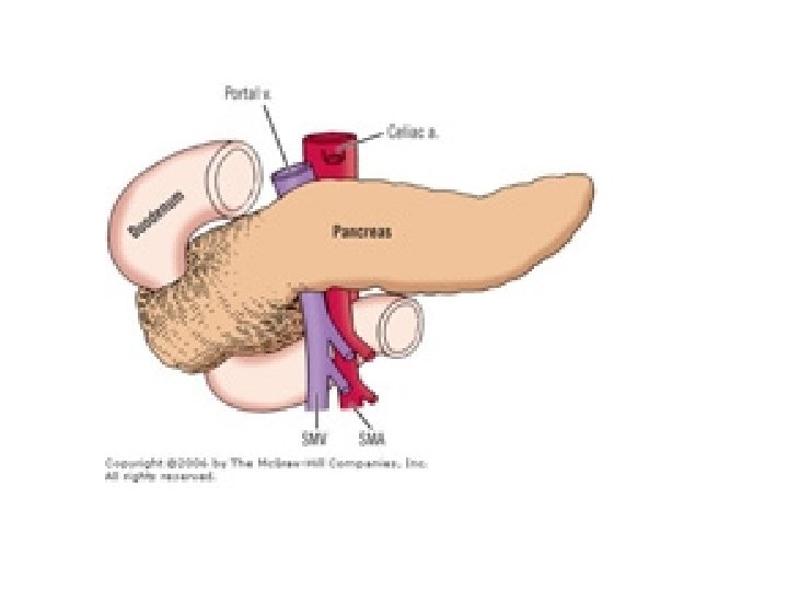
Anatomy Retroperitoneal Organ Weighs 75 To 100 G


 G: Gallstone E: Ethanol T: Trauma S: Steroid M: Mump A:")
 •")

 • CBC • Nonspecific ( elevated")


 14")


, 20% • Other, 10% • •")


. CT scan showing multiple, calcified, intraductal stones in")


 • Head-of-Pancreas")


 Ductal adeno carcinoma (arising in the exocrine part of")


 Ultrasound Scan C. T. Scan MR Imaging Scan")


 For tail of the pancreas (Distal pancreatectomy) Body")

 Percutaneous coeliac")


: 1 2")


 The tumour arising from the islets cell of langhans in the")


. Peptic Marked peptic ulcer disease following in acid reducing operation ulcer")


- Slides: 70
Anatomy Retroperitoneal Organ Weighs 75 To 100 G 15 To 20 Cm Long Head Neck Body Tail 2
What is Pancreatitis? Inflammation or infection of the pancreas Normally digestive enzymes secreted by the pancreas are not active until they reach the SI. When the pancreas is inflamed, the enzymes attack and. damages the tissue that produce them 2 types: 1. Acute Pancreatitis 2. Chronic Pancreatitis 3
Acute Pancreatitis 4
• Pancreatitis>> activation of pancreatic enzymes by the effect of the alkaline media ( bile ) • The normal pressure in the pancreatic duct 62 mm. Hg , while normal pressure in the biliary tree 17 mm. Hg , so the presence of stones increase the pressure in the biliary tree , so reflux of bile to the pancreas 5
Definition and Incidence Inflammatory disease with little or no fibrosis. Initiated by several factors: 90% of acute pancreatitis is secondary to acute cholelithiasis or ETOH abuse Develop additional complications 300, 000 cases occur in the united states each year leading to over 3000 deaths. 6
Etiology: (GET SMASHED) G: Gallstone E: Ethanol T: Trauma S: Steroid M: Mump A: Autoimmune S: Scorpion bits H: Hyperlipidemia ( triglyceride more than 1000 ) E: ERCP ( iatrogenic ) D: Drugs 7
Clinical Presentation • Abdominal pain • Epigastric ( relieved by bending forward ) • Radiates to the back • Worse in supine position • Nausea and vomiting • Garding • Tachycardia ( due to third space loss) , Tachypnea ( ARDS) , Hypotension ( third space loss ) Hyperthermia • Elevated Hematocrit & Pre renal azotemia • Cullen's sign • Grey Turner's sign 8
Hemorrhagic pancreatitis Grey Turner sign Cullen’s sign 9
Diagnosis: Biochemical • serum amylase ( sensitive ) • CBC • Nonspecific ( elevated with • Increased Hb ( with dehydration ) mumps , bowel ischemia … ) Low Hb >> with bleeding • Returns to normal in 3 -5 days • Thrombocytosis • Normal amylase does not • Leukocytosis exclude pancreatitis • Level of elevation does not predict disease severity • Liver Function Test • Urinary amylase • Serum Bilirubin elevated ( • P-amylase obstructive jaundice ) • Serum Lipase (more specific ) • Alkaline Phosphatase elevated No relation with severity of the • Aspartate Aminotransferase disease elevated 10
Assessment of Severity • Criteria • 1. ranson • 2. APACHE-2 • Biochemical Markers • Computed Tomography Scan 11
Ranson Criteria for acute gallstone pancreatitis • Admission • • • Age > 55 WBC > 16 Glucose > 200 LDH > 350 AST > 250 • During first 48 hours Hematocrit drop > 10 Pao 2 less than 60 mm Hg Serum calcium < 8 Base deficit > 4 Increase in BUN (blood urea nitrogen ) >5 • Fluid sequestration > 6 l • • • 12
CT scans of normal kidneys and pancreas Pa nc r Stomach ea s Liver V A R Kidney L Kidney Spleen 13
Pancreatic Necrosis dirty fatty planes ( inflammation) 14
Treatmaent of Mild Pancreatitis • Pancreatic rest • Supportive care • fluid resuscitation – watch BP and urine output • Pain Control • NG tubes and H 2 blockers or PPIs are usually not helpful • Refeeding (usually 3 to 7 days) If: • Bowel Sounds Present • Patient Is Hungry • Nearly Pain-free (Off IV Narcotics) • Amylase & Lipase Not Very Useful 15
Treatment of Severe Pancreatitis • Pancreatic Rest & Supportive Care • Fluid Resuscitation – may require 5 -10 liters/day • Careful Pulmonary & Renal Monitoring – ICU • Maintain Hematocrit Of 26 -30% • Pain Control – PCA pump • Correct Electrolyte Derangements (K+, Ca++, Mg++) • Contrasted CT scan at 48 -72 hours • Prophylactic antibiotics if present • Nutritional support • May be NPO for weeks • TPN • NG tube >> with pancreatitis part of the small bowel ( ileum , duodenum) mask the site of inflammation >> called sentinel lobe >> lead to paralytic ileus 16
Complications • Local • Phlegmon, Abscess, Pseudocyst, Ascites • Involvement of adjacent organs, with hemorrhage, thrombosis, bowel infarction, obstructive jaundice, fistula formation, or mechanical obstruction • Systemic • A. Pulmonary: pleural effusions, atelectasis, hypoxemia, ARDS. • B. Cardiovascular: myocardial depression, hemorrhage, hypovolemia. • C. Metabolic: Hypocalcemia, hyperglycemia, Hyperlipidemia, coagulopa thy • D. GI Hemorrhage • E. Renal • F. Hematologic • G. CNS • H. Fat necrosis • DIC , pleural effusion … 17
Psedocyst • Fluid collection within cyst • Types of cysts : 1 - true : the covering is endothelium 2 - pseudo : fibrous If its 5 cm in size , persist more than 6 weeks >> indication for intervention ( by endoscopy , we make opening between the sac and stomach “ cysto gastrostomy “ or between sac and jejunum “ cysto jejunostomy “ 18
Management 19
Chronic Pancreatitis Recurrent inflammation and fibrosis , decreasing the percentage of viable pancreatic tissue available. 20
Definition and Prevalence • Defined as chronic inflammatory condition that causes irreversible damage to pancreatic structure and function. • Incurable • 5 To 27 Persons Per 100, 000 21
Etiology • Alcohol, 70% • Idiopathic (including tropical), 20% • Other, 10% • • Hereditary Hyperparathyroidism ( men 1 syndrome or by hypercalcemia ) Hypertriglyceridemia ( familial >> mainly type 5 ) Autoimmune pancreatitis Obstruction Trauma Pancreas divisum 22
Classification: 1. calcific pancreatitis 2. obstruction pancreatitis 3. inflammatory pancreatitis 4. auto immune pancreatitis 5. asymptomic fibrosis 6. tropical pancreatitis 7. hereditary pancreatitis 8. idiopathic pancreatitis 23
Signs and Symptoms • Steady And Boring Pain • Not Colicky • Nausea Or Vomiting • Anorexia Is The Most Common • Malabsorption And Weight Loss ( 90% of the pancreas inflamed ) • Diabetes ( 80% of the pancreas inflamed ) 24
Laboratory Studies Tests for Chronic Pancreatitis I. Measurement of pancreatic products in blood A. Enzymes B. Pancreatic polypeptide II. Measurement of pancreatic exocrine secretion A. Direct measurements 1. Enzymes 2. Bicarbonate B. Indirect measurement 1. Bentiromide test 2. Schilling test 3. Fecal fat, chymotrypsin, or elastase concentration 4. [14 C]-olein absorption III. Imaging techniques A. Plain film radiography of abdomen B. Ultrasonography C. Computed tomography D. Endoscopic retrograde cholangiopancreatography E. Magnetic resonance cholangiopancreatography 25
Pancreatic calcifications ( indicate chronicity ). CT scan showing multiple, calcified, intraductal stones in a patient with hereditary chronic pancreatitis Endoscopic retrograde cholangiopancreatography in chronic pancreatitis. The pancreatic duct and its side branches are irregularly dilated 26
CT features • The cardinal CT features of CP are pancreatic atrophy, calcifications, and main pancreatic duct dilation. 27
Treatment • Analgesia • Enzyme Therapy • Antisecretory Therapy • Neurolytic Therapy • Endoscopic Management • Surgical Therapy 28
Complications • Pseudocyst • Pancreatic Ascites • Pancreatic-Enteric Fistula ( good ) • Head-of-Pancreas Mass • Splenic and Portal Vein Thrombosis 29
Management 30
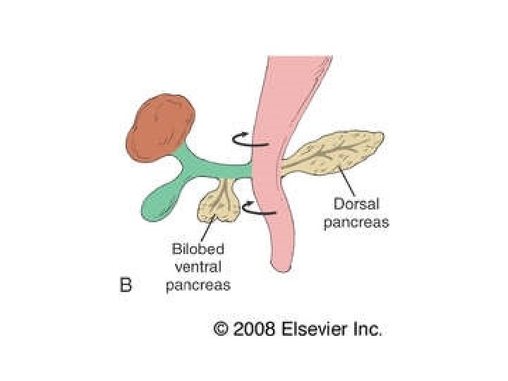
Annular Pancreas • When the ventral pancreatic anlage fails to migrate correctly to make contact with the dorsal anlage, the result may be a ring of pancreatic tissue encircling the duodenum. • Such an annular pancreas may cause intestinal obstruction in the neonate or the adult. • Symptoms of postprandial fullness, epigastric pain, nausea, and vomiting may be present for years before the diagnosis is entertained. • Treated by duodenostomy
• The radiographic findings are symmetric dilation of the proximal duodenum with bulging of the recesses on either side of the annular band, effacement but not destruction of the duodenal mucosa, accentuation of the findings in the right anterior oblique position, and lack of change on repeated examinations. • The differential diagnosis should include duodenal webs, tumors of the pancreas or duodenum, postbulbar peptic ulcer, regional enteritis, and adhesions. • Patients with annular pancreas have an increased incidence of pancreatitis and peptic ulcer.
Pancreas divisum • Occurs when the embryologic ventral and dorsal pancreatic anlagen fail to fuse, so that pancreatic drainage is accomplished mainly through the accessory papilla. • Is the most common congenital anatomic variant of the human pancreas. • Does not predispose to the development of pancreatitis in the great majority of patients. • However, the combination of pancreas divisum and a small accessory orifice could result in dorsal duct obstruction.
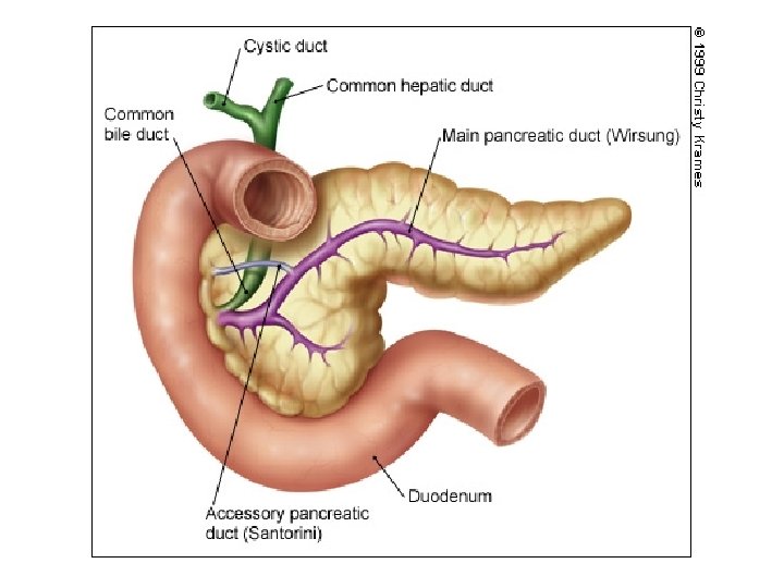
• Main pancreatic duct open proximal to CBD
MRCP of pancreas divisum
TUMOURS OF THE PANCREAS The tumours of the pancreas can be A. Non-Endocrine neoplasms B. Endocrine neoplasms 5 years survival only 5 %
Malignant non-endocrine neoplasms. exocrine The most common are: Ductal adenocarcinoma Cystadenocarcinoma NOTE: Periampullary carcinoma is term used for juxta-pancreatic carcinomas. ( good prognosis ) >> growth then sluffing cause blood per stool with fluctuant jaundice They are three forms: Carcinoma of the ampulla Carcinoma of the lower CBD Duodenal carcinoma 40
• Tumor in the tail of pancreas presented with symptoms like that of renal colic or early onset diabetes 41
ENDOCRINE NEOPLASMS: These are less common than non-endocrine tumours and generally benign and sometimes multiple. They includes: Insulinoma non men syndrome common Glucogonomas Others: - Gastrinomas - Somatostatatinomas - Vipomas (Vasoactive Intestinal Polypeptide) usually associated with men 1
EVALUATION OF PANCREATIC NEOPLASMS: History Clinical Examination Investigations The specific investigations: Ultrasound Scan CT Scan MR Imaging ERCP Histology & cytology Angiography Laparoscopy
NON-ENDOCRINE NEOPLASMS: (ADENOCARCINOMA OF PANCREAS) Ductal adeno carcinoma (arising in the exocrine part of pancreas) account for 90% of pancreatic tumour 2/3 rd located in the head of pancreas. (2/3 th treated by wipple procedure ) Most of pancreatic tumors are exocrine Cystadenocarcinoma and endocrine tumour account for most of the remains of malignancy.
The exact causative factors responsible are unknown. The peak incidence in the 6 th and 7 th decade and more in men than women The predisposing factors are: Diet (high protein & high fat) Smoking Exposure to industrial carcinogens
Spread of pancreatic tumours: A. Local Invasion B. Lymphatic C. Blood D. Via peritoneal & omental causing ascites
CLINICAL FEATURES: The diagnosis of pancreatic cancer varies from the simple and clinically obvious to the most difficult and almost impossible the initial symptoms and signs depend on the site and extent of the pancreatic cancer.
Modes of presentation: Weight loss Pain Jaundice Steatorrhoea Diabetes Mellitus Acute Pancreatitis Malignant Ascites Gastric Outlet Obstruction
Approach to Investigations: (Selective Investigations) Ultrasound Scan C. T. Scan MR Imaging Scan ERCP Histology & Cytology Angiography (Coeliac, Superior - Mesenteric) Laparoscopy
DELAY IN DIAGNOSIS: Over 90% of patient with pancreatic cancer present in the late stage of their disease. At time no chance of cure. The factors responsible for late diagnosis A. Tumour is asymptomatic in the early stage. B. Patient delay. C. Physician delay. D. The patient may not have ready and easy access to competent diagnostic center.
MANAGEMENT OF PANCREATIC CANCER: A. Surgical Treatment B. Non Surgical Treatment
SURGICAL TREATMENT: Pancreatic Cancer is essentially incurable since metastasis occurs at such early stage. Any treatment must be regarded as palliative.
Surgical Options: For curative surgical treatment of cancer in the head of pancreas the optims are available: A. Whipple operation (Pancreatico- B. Pylorus Preserving C. Total Pancreatectomy duodenectomy) Pancreaticoduodenectomy
Palliative Surgical Treatment (Surgical Bypass) For tail of the pancreas (Distal pancreatectomy) Body of the pancreas (Distal + removal of the body of the pancreas)
Pre-operative preparation of the patient for major surgery: 1. All jaundiced patients must be kept in hydration. 2. Blood clotting deficiencies must be good state of nutrition and corrected. 3. Cardio pulmonary functioning carefully assessed. 4. Drainage procedure consider in certain cases.
NON-SURGICAL TREATMENT: The following options available: (Pallative procedure for non operable cases) Percutaneous coeliac ganglion blockade. (For pain) Stent to compress bile duct. . Combination of chemotherapy andradiotherapy may become alterative in the future. h
FUNCTIONING ENDOCRINE TUMOURS OF THE PANCREAS: These are much less common than adeno carcinoma. The beta cell tumours secrete (Insulin) and called INSULINOMAS. Another functioning tumour secrete(Gastrin) called GASTRINOMA which come from the islets which cannot be classified into either alpha or be (non-beta). 57
Other tumours are: a. Vipoma (Werner-Morrison syndrome, b. Somastatinoma c. d. HP (Human Pancreatic cholera) Glucagonoma Polypeptide tumours) Slow growing and therefore carry much better prognosis.
INSULINOMA: The commonest islet cell tumour and arise from the beta cell and situated anywhere on the surface or within the substance of the pancreas. Most tumours are benign adenomas but 15% are low grade carcinomas and secrete (insulin). (C peptide >> to distinguish endogenous insulin from exogenous one )
CLINICAL FEATURES: Whipple described a triad of features which typify the (insulinomas): 1 2 . . 3 Fasting produces fainting. During these “attacks” there is . The attacks may be relieved by hypoglycaemia. ingestion of glucose.
INVESTIGATIONS: 1. Measurement of blood sugar in an attack. Overnight fasting serum glucose and 2. insulin level (before & after overnight). Insulin level are estimated by radio-immunoassay. 3. Pre-operative localization of the tumour very important identification at operatio can be difficult. [Combination CT Scan and selective angiography]
TREATMENT: 1. If the tumour localized surgical resection is the TR of choice also this apply to metastases. 2. If the tumours not localized during surgery (Intra operative USS can be done to localize the tumour) than resected. 3. Sub total distal resection for multiple tumours is appropriate.
4. With negative exploration it is appropriate to perform pancreatectomy distal to the superior mesenteric vessels. 5. The Hypoglycemic attacks may be relieved by diazoxide or streptazotocin.
GASTRINOMA: (Zollinger-Ellison Syndrome) The tumour arising from the islets cell of langhans in the pancreas and in the duodenal wall. The majority (60%) of these tumours are malignant. They may be associated with (MEN 1)which are Parathyroid Hyperplasia, and Pituitary Adenoma. Gastrinoma give rise to ZE Syndrome which consist of triad (hypersecretion of gastric acid, severe peptic ulceration and the presence of non-beta cell tumour of the pancreas or duodenum).
CLINICAL FEATURES: The disease present as peptic ulcer disease in over 90%. They have typical pain more severe and less response to medical treatment. Co-existing All diarrhoea. complications of peptic ulcer disease are present in (ZE-Syndrome) as acute haemorrhage, perforation and recurrent ulceration.
THE DIAGNOSIS OF ZE-SYNDROME: Severe peptic ulcer disease doesn’t respond to treatment. Multiple peptic ulcers or ulcers in unusual locations such as the Peptic ulcer disease associated with diarrhoea. distal duodenum or jejunum.
Recurrent (surgery). Peptic Marked peptic ulcer disease following in acid reducing operation ulcer is associated with MEN- 1 Syndrome. elevation of serum gastrin.
TREATMENT: Medical therapy for control of the acid hypersecretion in patient with ZE-Syndrome Omprazole considered the antisecretory drug of choice for all gastrinoma patients.
Surgical Treatment: Tumour excision. Total gastrectomy. Patient with metastases should have medical therapy if fail total gastrectomy. Gastrinoma patient with MEN 1 Syndrome and documented hyperparathyroidism should have parathyroid surgery performed prior to removal of gastrinoma.