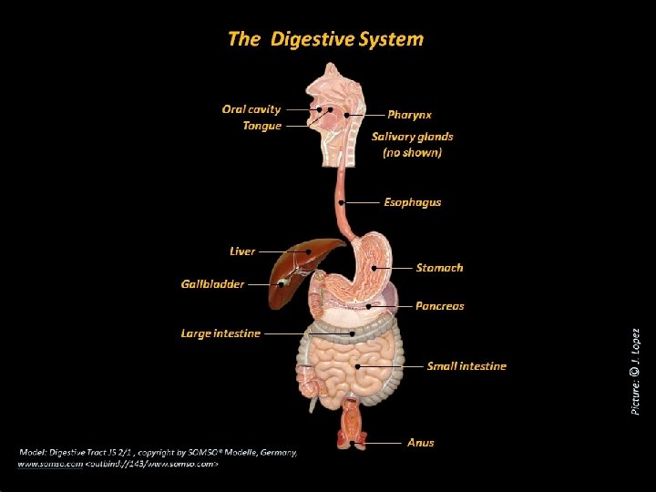
Lab 7 The Digestive System 1 The Teeth

Lab # 7 The Digestive System 1
 (Conical with a sharp")
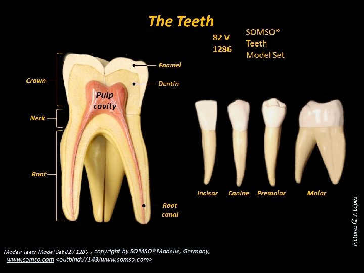
The Teeth Cuspid or canine (Blade-shape teeth: Clipping and cutting) (Conical with a sharp ridgeline and a pointed tip: Tearing or slashing) Molars Central incisor Lateral incisor Upper dental arch Bicuspids or premolars (Flattened crown with prominent ridges: Crushing, smashing and grinding) (Very large flattened crowns with prominent ridges: Crushing and grinding) Total: 32 permanent or secondary teeth Lower dental arch Total: 20 primary, temporary or deciduous teeth

Most of the digestive tract follows the basis structural plan with digestive tract wall consisting of the following tissue layers, in order from the inner to the outer surface: Cross section of the esophagus 1 - Mucosa: Stratified squamous epithelium Lamina propria Muscularis mucosae Stratified squamous epithelium 2 - Submucosa 3 - Muscularis externa: Inner circular layer Outer longitudinal layer Simple columnar epithelium 4 - Serosa Stratified squamous epithelium
 Stratified")
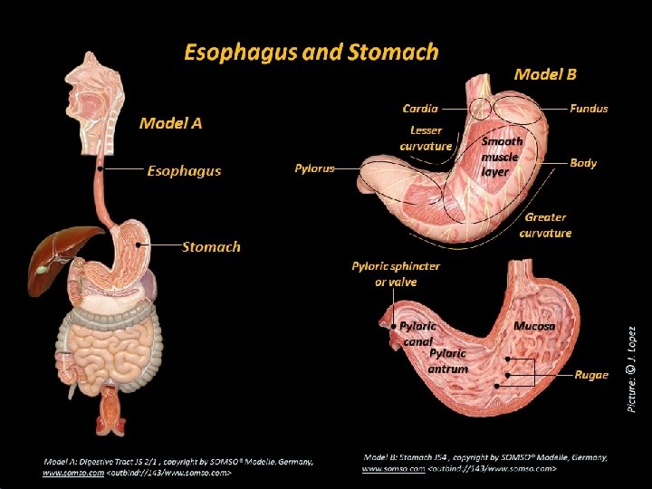
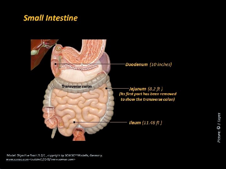
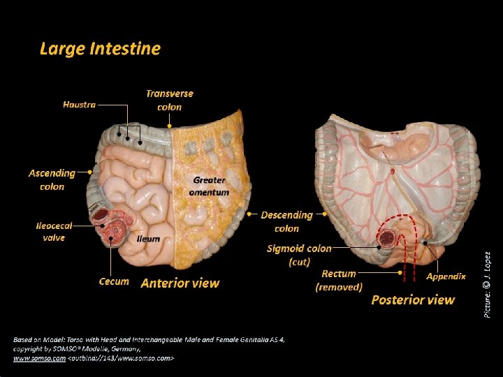
Diaphragm Stomach Esophagus Simple columnar epithelium (it contains gastric pits and gastric glands) Stratified squamous epithelium(also in oral cavity and pharynx. Three layers of smooth muscle in the muscularis externa: outer longitudinal, middle circular, and inner oblique. Two layers of smooth muscle in the muscularis externa: outer longitudinal and inner circular. Longitudinal folds of the mucosa that allow for expansion Small Intestine Simple columnar epithelium with microvilli (it contains crypts of Lieberkuhn and intestinal glands) Two layers of smooth muscle in the muscularis externa: outer longitudinal and inner circular. Transverse folds of the mucosa called plicae circulares, and fingerlike projections called villi. Folds of the mucosa called rugae. Taeniae coli Transverse folds of the wall called haustra. Large Intestine Simple columnar epithelium without villi (it is dominated by mucous cells) Two layers of smooth muscle in the muscularis externa: outer longitudinal reduced to the taeniae coli, and inner circular.

Histological Section of the Esophagus
 Epithelium Gastric pit Lamina propria Mucosa")
Microscopic Anatomy of Stomach Epithelium (Simple columnar glandular) Epithelium Gastric pit Lamina propria Mucosa Muscularis mucosae Gastric gland Submucosa Lamina propria Lymphatic nodule Muscularis externa Serosa Muscularis mucosae Obl ique Circ ular Lon gitu Gastric pit din laye r al la yer
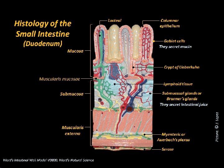

Copyright © The Mc. Graw-Hill Companies, Inc. Permission required for reproduction or display. Histological Section of the Duodenum Villi Intestinal crypts Muscularis mucosae Duodenal glands Muscularis externa Serosa 0. 5 mm © The Mc. Graw-Hill Companies, Inc. /Dennis Strete, photographer

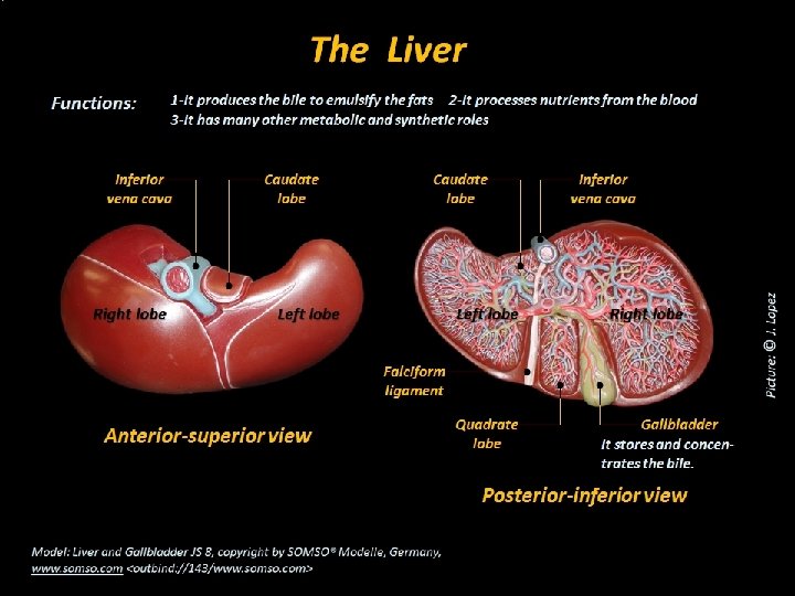
Gross Anatomy of Liver The liver is a reddish brown gland located immediately inferior to the diaphragm It is the body’s largest gland weighing about 1. 4 kg (3 pounds) It has a variety of functions, but only the secretion of bile contributes to digestion Caudate lobe Right lobe Inferior vena cava Left lobe Falciform ligament Round ligament (a) Anterior view Quadrate lobe Gallbladder (b) Posterior view

Macroscopic Anatomy of Liver Hepatic Lobule To the hepatic vein To the inferior vena cava Central vein Hepatic Triad: Branch of hepatic portal vein Branch of proper hepatic artery Bile ductule Blood from the intestine and stomach Hepatocytes Hepatic sinusoid Bile canaliculum To the right and left hepatic ducts

Histological Section of the Liver Copyright © The Mc. Graw-Hill Companies, Inc. Permission required for reproduction or display. Stroma Central vein Hepatic lobule Branch of hepatic portal vein Bile ductule Lymphatic vessel Branch of proper hepatic artery 0. 5 mm © The Mc. Graw-Hill Companies, Inc. /Dennis Strete, photographer

Bile canaliculum Bile ductule Right hepatic ducts Left hepatic ducts Common hepatic duct Cystic duct Bile duct Gallbladder It stores and concentrates bile Pancreatic duct Accessory pancreatic duct Pancreas Duodenum Minor duodenal papilla Hepatopancreatic sphincter Major duodenal papilla Jejunum Hepatopancreatic ampulla

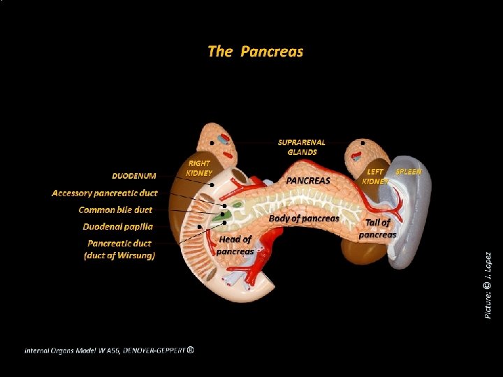
Pancreas It is a spongy retroperitoneal gland posterior to the greater curvature of the stomach The head of the pancreas is encircled by the duodenum It is both an endocrine and exocrine gland The endocrine portion consists of the pancreatic islets that secrete insulin and glucagon The exocrine portion is about 99% of pancreas and secretes 1200 to 1500 m. L of pancreatic juice per day Accessory pancreatic duct Body Head Pancreatic duct Tail

Histological Section of the Pancreas
- Slides: 21