An Intro To POCUS Point of Care Ultrasound
")


: • US Probes – What are they for? • Settings")


 2. Subcostal")
 • Longitudinal plane • Axillary Line • 7 -9 th intercostal")


: • Identify the 4 windows of the fast exam •")

- Slides: 36
An Intro To POCUS (Point of Care Ultrasound for Family Medicine)
Road Map • Lectures with breakout components • • POCUS and US Basics (15 min) Hands-On Ultrasound Time (15 min) FAST (15 min) Hands-On Ultrasound Time (40 min) • Evaluation (5 min)
Objectives • Gain confidence using US as a point-of-care tool • Demonstrate the basics of using an US machine to one of your colleagues • Preform an e. FAST exam on one of your friends
Shout to Contra Costa Thanks Guys!
Why POCUS? • Utilization by US is becoming more ubiquitous • Family Doctors are already using ultrasound • Underserved component to POCUS has been overlooked
The POCUS Philosophy • Quick bedside answers to focused questions • No diagnosis or procedure solely dependent on US • aid to diagnosis like an EKG or CXR or as an adjunct to doing a procedure • Bedside US is an extension of the PE STFM POCUS 2017
My Vision • Yearly POCUS training during resident school • Twice Monthly Trainings on the IPS • Fall 2017 Faculty Training • Expanded Yearly Resident Training? • More robust US applications in clinic?
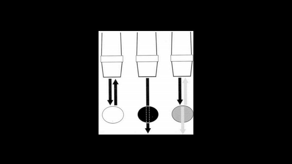
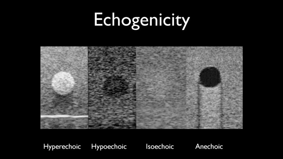
The Basics • Transmission • Probes • Orientation • Knobs and Buttons
Orientation • US Machine should be on the patient’s right • Indicator should point to • Head (longitudinal) • Patient’s right side (transverse) • Indicator on the screen should match the probe
• Modes • 2 D • Color • M Mode • Settings • Software changes the image based on the setting • Depth • Indicators on the side of the image • Gain • Brightness of the image • Near and far gain
Your Mission (15 min): • US Probes – What are they for? • Settings • Orientation of Machine and Indicators • Gain • Depth • If there’s time… try M Mode • Normal Lung VS Pneumothorax
The e. FAST Exam Focused Assessment with Sonography in Trauma
Santa Rosa, NM • 35 yo male headed to the Pecos River in VW Van • Head on collision with an elk • Exam – • HR 120, BP 120/70, RR 25, O 2 90% • Contusions to the left flank • Decreased left sided BS
FAST Overview • Clinical Questions • Relevant windows of the fast exam • Normal and Abnormal
2 questions • Is there fluid in the abdomen? • Is there fluid in the pericardium?
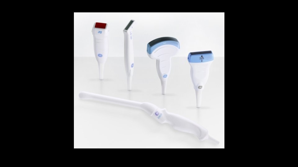
Windows… Use a Phase Array or Curvelinear Probe 1. Hepatorenal (Morison’s Pouch) 2. Subcostal 3. Perisplenic 4. Suprapubic
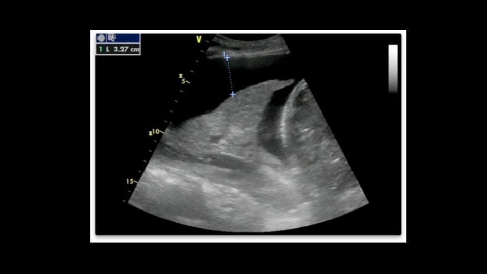
Hepatorenal (Morison’s Pouch) • Longitudinal plane • Axillary Line • 7 -9 th intercostal space
Morison’s Pouch
Perisplenic Recess • Posterior axillary line • 5 -7 th intercostal space • “knuckles to the bed”
Perisplenic
Suprapubic • Transverse View • Low in the pelvis • Look for fluid behind the bladder.
Longitudinal View • More sensitive for free fluid
Subxiphoid • Probe marker to right • Under the xiphiod process • Look to the left. • Real time subxiphoid view • Pericardial Effusion
e. FAST
e. FAST • Is there free fluid in the thorax? • Is there a pneumothorax?
Pleural Line • Look for sliding sign • Still above the pleura and movement below • Comet tails • M mode – Sands on the beach
A video… • Normal Lung VS Pneumothorax
Santa Rosa, NM • 35 yo male headed to the Pecos River in VW Van • Head on collision with an elk • Exam – • HR 120, BP 120/70, RR 25, O 2 90% • Contusions to the left flank • Decreased left sided BS
Your Mission (40 min): • Identify the 4 windows of the fast exam • Add the pulmonary exam to look for fluid in the thorax and pneumothorax
Evaluations please!