The Physics of Sound n n Sound travels
")


 created by throwing a rock into the")


 Air 330 Water 1480 Blood 1570 Fat 1460 Muscle 1580 Bone 3500")


 n n n Horizontal Axis: time in seconds Vertical")


 The")
 tissue depth at which 1/2 of the")


: area of the sound head that produces ultrasonic waves;")
; amount of energy being")

 : Sound Intensity")


 Why logarithms? n n To compress the very large range of")
 0 d. B is typically the softest volume that can be")


 n n Frequency Range: 20 - 20, 000")


 as it travel")


 I(transmit) I(reflect) Z 1 Z 2")


- Slides: 128
The Physics of Sound n n Sound travels through the air (and different media) in waves, called Sound Waves These waves cause the air to oscillate (vibrate) back and forth
Light 1. Does NOT require a medium 2. Transverse wave 3. Electromagnetic field Sound 1. REQUIRES a medium 2. Longitudinal wave 3. Mechanical vibration
Longitudinal vs. Transverse Waves Longitudinal waves – molecular displacement is along direction in which waves travel n Compression – regions of high molecular density (molecules in high pressure areas compress) n n n Rarefraction – regions of low molecular density (molecules in low pressure areas expand) Transverse waves – molecular displacement in direction perpendicular to wave (guitar string)
n Longitudinal waves – travel in solids & liquids Soft tissue – more like liquids n US primarily travels as longitudinal wave n n Transverse waves – cannot pass through fluids; found in the body only when ultrasound strikes bone
Not Just Any Wave The waves (ripples) created by throwing a rock into the pond are Transverse Waves n Sound waves are NOT transverse waves n Sound waves ARE Longitudinal Waves n
Transverse Waves Pretend like you are at a Canucks game. Everyone do the wave starting from the left to the right! n What direction is the wave traveling? n n n The wave travels to the right What direction is the displacement caused by the wave? n Displacement is vertical; Perpendicular to the travel direction.
Transverse Waves n NOT SOUND WAVES !!!
Longitudinal Waves Do the wave again, but this time, instead of moving your arms up and down, move them side to side. n What direction is the wave traveling? n n n The wave travels to the right What direction is the displacement caused by the wave? n Displacement is horizontal; Parallel to the travel direction
Sound Production n How does one make sound? n n Vocal cords, speakers, headphones etc. What do these all have in common? n They all vibrate the air!
Sound Waves compression rarefaction amplitude sin wave time
Sound Reproduction n Speakers take an electronic signal, and reproduce sound. By far most common type of speaker is the Dynamic Speaker. Other types of speakers include piezoelectric speakers, plasma arc speakers and electrostatic speakers.
The “Sonic” Spectrum Infrasound: < 20 Hz Sound: 20 Hz – 20, 000 Hz (20 k. Hz) Ultrasound: > 20 k. Hz (~1013 Hz maximum)
Human Hearing n n n Human range: 20 -20, 000 Hz Human ear is most sensitive in the 1, 000 4, 000 Hz range. Less sensitive in lower frequencies.
Hearing Range Hearing range in human is 20 Hz to 20 k. Hz n Infrasound is the sound the frequency of which is lower than 20 Hz n Ultrasound is the sound the frequency of which is higher than 20 k. Hz n
Nature Of Ultrasound The sound is no longer audible ︰� 20 k. Hz. n The medical ultrasound is in the 1~20 MHz region. n
Ultrasound n Ultrasound in Nature n Medical applications n. Diagnostic n. Treatment n Industrial Applications
Why Use Ultrasound? n Ultrasound is very safe. There is no firm evidence that it does any harm to the body (or the baby in the case of pregnancy scans). n X-rays are potentially dangerous, particularly to young children and pregnant women (they damage the unborn baby).
What is Ultrasound? Located in the Acoustical Spectrum n May be used for diagnostic imaging, therapeutic tissue healing, or tissue destruction n Thermal & Non-thermal effects n We use it for therapeutic effects n Can deliver medicine to subcutaneous tissues (phonophoresis) n
Ultrasound n Sinusoidal waveform n n Therapeutic ultrasound waves range from 750, 000 to 3, 000 Hz (0. 75 to 3 MHz) Displays properties of wavelength, n frequency, n Amplitude n
What is Ultrasound? n Ultrasound is simply sound that has a very high frequency. n Humans are not able to hear ultrasound, though some animals can hear them. n Sounds with frequencies above 20 000 hertz are called ultrasounds.
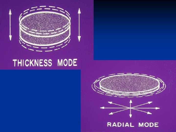
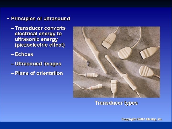
EQUIPMENT TRANSDUCERS ARE USED TO GENERATE THE ULTRASONIC ENERGY n THE MAJOR COMPONENT OF AN ULTRASOUND TRANSUCER IS THE PEIZOELECTRIC ELEMENT n
PIEZOELECTRIC MATERIAL n ARE CAPABLE OF CONVERTING ONE FORM OF ENERGY INTO ANOTHER n ABLE TO GIVE OFF AND RECIEVER SOUND ENERGY
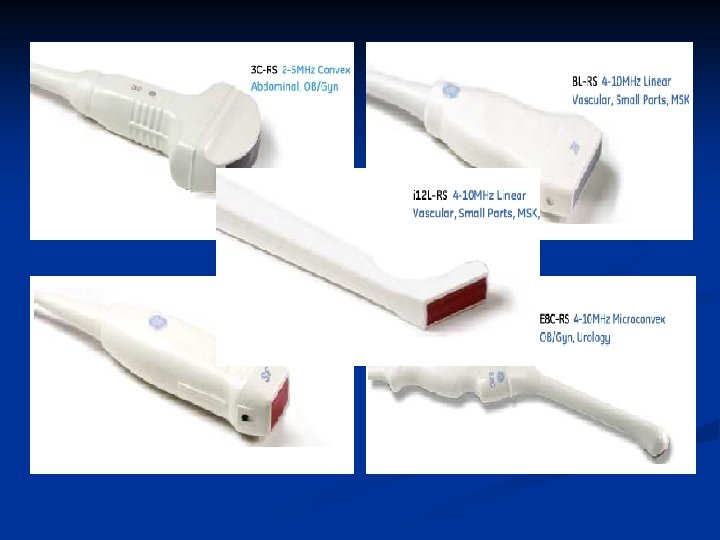
TRANSDUCERS n DIFFER IN SIZE, SHAPE & FREQUENCIES n SIZE AND SHAPE TO IMPROVE CONTACT WITH DIFFERENT BODY STRUCTURES
Transducer n n A device that converts one form of energy to another Piezoelectric crystal: a crystal that produces (+) and (-) electrical charges when it contracts or expands n n Reverse (indirect) piezoelectric effect: occurs when an alternating current is passed through a crystal resulting in contraction & expansion of the crystal n n n Crystal of quartz, barium titanate, lead zirconate, or titanate housed within transducer US is produced through the reverse piezoelectric effect Vibration of crystal results in high-frequency sound waves Fresnal zone (near field) – area of the ultrasound beam on the transducer used for therapeutic purposes
FREQUENCIES LOW FREQUENCY OF 1 MHz TO HIGHER FREQUENCIES OF 12 TO 20 MHz. n THE SMALLER THE OBJECT TO BE IMAGED, THE HIGHER THE TRANDUCER FREQUENCY n
Transducers It is not only used as a transmitter, but also a receiver. n For different applications, we need to choose the transducer resonant frequency, diameter and focal length. n
n Piezoelectric Effect— Certain substances change their dimensions when an electrical charges on their surfaces when deformed. Such substances are called piezoelectric. Ex. Quartz.
Metal case Transducer Matching layer Coaxial connector Backing Probe Acoustic insulator
n Backing— For imaging purpose, we are interested in generating short pulse of US and this is achieved by the transducer. Make sure that its acoustic impedance is the same as that of the transducer.
n Matching Layer— In order to improve the transmission from the transducer into the tissue efficiently, matching layer may be used. Ideal thickness is a quarter of a wavelength thick. d=λ/4
Metal case — The backing is enclosed within a metal case in order to provide a means of handling the transducer and to provide electrical shielding to prevent electrical interference. n Acoustic Insulator — EX. rubber or cork. To avoid transmission of US into the metal case. n
Generation of Ultrasound Pizoelectric effect - generated by pizoelectric crystals occurs when an electric current is passed through the crystal expands & contracts at frequencies that produce ultrasound pizoelectric crystal in transducer head ultrasound transducer Wavelength (l)
Generation of Ultrasound Properties of ultrasound higher the sound frequency, less the propagation wave diverges ultrasound beams are well collimated (straight line) like electromagnetic energy, ultrasound energy is… transmitted through a medium………or totally reflected back toward the point of generation……. . or refracted (bent)………or absorbed or attenuated (loose energy)
Generation of Ultrasound in tissues, ultrasound is transmitted, absorbed, reflected, or refracted absorption of ultrasound energy generates heat at higher F’s, more tissue friction must be overcome the more friction that must be overcome, the more heat is generated the more friction that must be overcome, less energy left for propagation higher frequencies of ultrasound penetrate less deep before being absorbed 3 MHz frequency used to treat tissues at depths of 1 cm to 2 cm 1 MHz frequency used to treat tissues > 2 cm from the surface
Transverse wave propagation
Longitudinal wave representation
Longitudinal wave propagation
Wave Properties l Wavelength: The distance between identical points on the wave. l Amplitude: The maximum displacement A of a point on the wave. l A wave varies in time and space. Wavelength y x A
Sound Wave Properties l Displacement: The maximum relative displacement s of a point on the wave. Displacement is longitudinal. l Maximum displacement has minimum velocity Molecules “pile up” where the relative velocity is maximum (i. e. , ds/dt = smax) Wavelength s DPmax=rvwsmax x smax
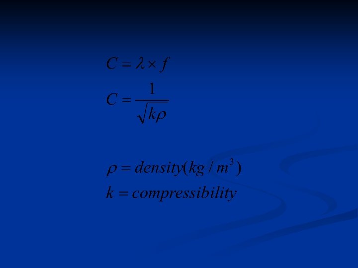
Speed of Sound V=λf =λ/T one wavelength period
Frequency n Frequency: number of times an event occurs in 1 second; expressed in Hertz or pulses per second Hertz: cycles per second n Megahertz: 1, 000 cycles per second n n In the U. S. , we mainly use ultrasound frequencies of 1, 2 and 3 MHz n 1 = low frequency; 3 = high frequency = depth of penetration n frequency = sound waves are absorbed in more superficial tissues (3 MHz) n
Frequency Pitch/Hz n n n Perceived as “Pitch”. Equal to the number of complete cycles that occur in one second. n one cycle = one compression and one rarefaction. Measured in Hertz (Hz).
Frequency A cycles is one complete variation in the acoustic variables n Frequency is the number of cycles that occurs in one second n The frequency is measured in unit of Herz (Hz, k. Hz and MHz) n Half of the cycles is rarefaction and the other half is compression. n
Period is the time that it takes for one cycle to occur n In ultrasound the unit for period is microsecond ( s ) n Period is the reciprocal of the frequency n
Velocity n The speed of sound wave is directly related to the density ( velocity = density) n Denser & more rigid materials have a higher velocity of transmission n At 1 MHz, sound travels through soft tissue is 1540 m/sec and 4000 m/sec through compact bone
Medium Velocity(m/s) Air 330 Water 1480 Blood 1570 Fat 1460 Muscle 1580 Bone 3500 Soft tissue(mean) 1540
Measurement of Dd & Dt
Amplitude n The Amplitude measures the displacement of the air molecules.
The Sine Wave (Pure Tone) n n n Horizontal Axis: time in seconds Vertical Axis: molecular movement Compression: upward movement Rarefaction: downward movement Amplitude: height of wave; intensity
Sound Wave Basics Two main components of a sound wave that affects what we hear are amplitude and frequency. n Amplitude determines how loud it is. n Frequency determines the “pitch”. n
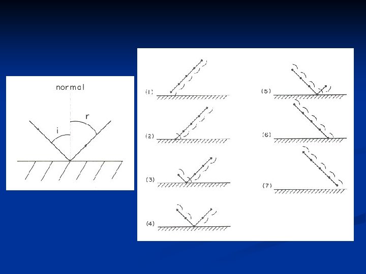
Wave Properties II 1. Reflection 2. Refraction 3. Interference 4. Diffraction
Influences on the Transmission of Energy n Reflection – occurs when the wave can’t pass through the next density n Refraction – is the bending of waves as a result of a change in the speed of a wave as it enters a medium with a different density n Absorption – occurs by the tissue collecting the wave’s energy
Reflection Flat surfaces Parabolic surfaces Ellipsoidal surfaces Ends of musical instruments
Reflection from flat surfaces Optical board Echo / Echolocation Acoustical Radar
Direction of Wave is Perpendicular to Wave Front
Source moving
The Doppler Effect Demonstration Graphs with description
Doppler Effect observer stationary
Doppler Effect source stationary
n If the source of sound is moving Toward the observer seems smaller n Away from observer seems larger n n If the observer is moving n n n Toward the source seems smaller Away from source seems larger If both are moving Doppler Example Audio n Doppler Example Visual Examples: police car, train, etc. (Recall: v
Frequency and Amplitude
Reflection of Ultrasound & Sonography Ultrasound is reflected at the interface of different tissues reflection amount & time until reflection returns to transducer can be charted w/ computer image construction: sonogram (depth, density, & position of tissue structures) Amount of Ultrasonic Reflection (Acoustic Impedance) Interface water-soft tissue - fat soft tissue - bone soft tissue - air Energy Reflected. 2% 1% 15 -40% 99. 9% highly reflective surfaces include: 1) muscle tendon junctions 2) intermuscular interfaces 3) soft tissue-bone
Attenuation n Decrease in a wave’s intensity resulting from absorption, reflection, & refraction n as the frequency of US is because of molecular friction the waves must overcome in order to pass through tissues n US penetrates through tissue high in water content & is absorbed in dense tissues high in protein n Absorption = Frequency (3 MHz) , and Penetration = Absorption (1 MHz) , so Penetration = Frequency + Absorption (1 MHz) n n Tissues water content = low absorption rate (fat) Tissues protein content = high absorption rate (peripheral nerve, bone) n Muscle is in between both
Attenuation: Acoustic Impedance n Determines amount of US energy reflected at tissue interfaces n n n n If acoustic impedance of the 2 materials forming the interface is the same, all sound will be transmitted The larger the difference, the more energy is reflected & the less energy that can enter the 2 nd medium US passing through air = almost all reflected (99%) US through fat = 1% reflected Both reflected/refracted @ m. interface Soft-tissue: bone interfaced = much reflected As US energy is reflected @ tissue interfaces with different impedances, intensity is increased creating a Standing Wave (hot spot)
Attenuation of Ultrasound The higher the tissue H 2 O content, the less the attenuation The higher the tissue protein content, the more the attenuation of 1 MHz beam Blood Fat Muscle Skin Tendon Cartilage Bone 3% / cm 13% / cm 24% / cm 39% / cm 59% / cm 68% / cm 96% / cm
Exponential Attenuation 1. 0 Quantity of Ultrasound (fraction of beam being further propagated) The quantity of the ultrasound beam decreases as the depth of the medium (tissue) increases. . 5 . 25. 125 1 st Half Value 2 nd Half Value Tissue depth 3 rd Half Value 4 th Half Value
Attenuation of Ultrasound Half value thickness (centimeters) tissue depth at which 1/2 of the sound beam of a given frequency is attenuated 1 MHz 2 MHz 3 MHz Fat 15. 28 5. 14 2. 64 Muscle 2. 78 1. 25. 76 Bone. 04. 01. 004
Physiological Effects of Ultrasound Non-thermal effects cavitations alternating expansion & compression of small gas bubbles may cause u cell membrane & vascular wall permeability (u nutrient and oxygen delivery) unstable cavitation may cause tissue damage unstable cavitation – large, violent changes in bubble volume microstreaming bubble rotation r fluid movement along cell membrane boundaries (u nutrient and oxygen delivery) changes in cell permeability & ion flux r d healing time Possible therapeutic benefits of non-thermal effects difficult to make distinction from thermal benefits u capillary density & u cell permeability u fibroblastic activity and associated collagen production u cortisol production around nerve bundles r d inflammation
Ultrasound Adverse Effects & Contraindications Adverse effects associated with ultrasound potassium leakage from red blood cells u platelet aggregation r d microscopic blood flow damage to tissue endothelium Contraindications to ultrasound thrombophlebitis or other blood clot conditions fractures ? (studies exist suggesting ultrasound may help) epiphyseal injuries in children vascular diseases (embolus formation - plaque rupture) spinal column injuries (treat low back pain with caution) cancer (danger of metastases) do not apply directly over heart (pacemaker concerns) do not apply to reproductive organs (pregnancy)
n Effective Radiating Area (ERA): area of the sound head that produces ultrasonic waves; expressed in square centimeters (cm 2) n n n n Represents the portion of the head’s surface area that produces US waves Measured 5 mm from face of sound head; represents all areas producing more than 5% of max. power output Always lesser area than actual size of sound head Large diameter heads – column beam Small diameter heads – more divergent beam Low frequency (1 MHz) – diverge more than 3 MHz Treatment Duration: time for total treatment
Intensity Output & Power n Power: measured in watts (W); amount of energy being produced by the transducer n Intensity: strength of sound waves @ a given location within the tissues being treated n Spatial Average Intensity (SAI): amount of US energy passing through the US head’s ERA; n n expressed in watts per square centimeter (W/cm 2) (power/ERA) Changing head size affects power density (larger head results in lower density) Limited to 3. 0 W/cm 2 of maximum output
Sound Level n I 0 is called the reference intensity It is taken to be threshold of hearing n I 0 = 1. 00 x 10 -12 W/ m 2 n I is the intensity of the sound whose level is to be determined n n b is in decibels (d. B) n Threshold of pain: I = 1. 00 W/m 2; b = 120 d. B n Threshold of hearing: I 0 = 1. 00 x 10 -12 W/ m 2 ; b = 0 d. B
Intensity of sounds n Some examples (1 pascal 10 -5 atm) : Sound Intensity Pressure Intensity amplitude (Pa) (W/m 2) level (d. B) Hearing threshold 3 10 -5 10 -12 0 Classroom 0. 01 10 -7 50 City street 0. 3 10 -4 80 Car without muffler 3 10 -2 100 Indoor concert 30 1 120 Jet engine at 30 m. 100 10 130
Lecture 22, Exercise 4 Plane Waves A: You are driving along the highway at 65 mph, and behind you a police car, also traveling at 65 mph, has its siren turned on. B: You and the police car have both pulled over to the side of the road, but the siren is f f’ still turned on. v In which case does the frequency of the siren seem higher to you? vo vs (A) Case A
Audio Fundamentals n Acoustics is the study of sound n. Generation, transmission, and reception of sound waves n. Sound wave - energy causes disturbance in a medium n Example is striking a drum n. Head of drum vibrates => disturbs air molecules close to head n. Regions of molecules with pressure above and below equilibrium
The Physics Of Sound Why do we hear what we hear? (Turn on your speakers)
Sound is made when something vibrates. The vibration disturbs the air around it. n This makes changes in air pressure. n These changes in air pressure move through the air as sound waves. n
The sound waves cause pressure changes against our ear drum sending nerve impulses to our brain.
Intensity Loudness/d. B n n Perceived as “Loudness”. Intensity is expressed as the sound pressure level (SPL), which is a function of distance the vibrating object is displaced (amplitude), which depends on energy applied. Measured in decibels (d. B). One d. B is 1/10 th of a bel. Decibels are logarithmic units. The reference used is. 0002 dynes/cm 2, roughly the smallest pressure that will move the TM.
Intensity (cont. ) Why logarithms? n n To compress the very large range of pressure our ears can hear in to a small range of numbers for convenience. 0 -140 d. B represents a sound pressure range of 1: 1, 000, 000 units (a ratio of 10 million to 1!)
Intensity (cont. ) 0 d. B is typically the softest volume that can be heard, but sound energy is also present below 0 d. B. n Human intensity range is 0 -140. n 140 d. B is the threshold of pain. n 170 -180 d. B causes tissue damage. n 180 d. B+ can cause death! n
INVERSE SQUARE LAW n Doubling the distance from a sound source decreases intensity by 6 d. B.
Doubling the Noise Source… n A combination of two different noise sources of equal loudness will increase the intensity by 3 d. B n For example, if noise source “A” is 93 d. BA and noise source “B” is 93 d. BA, the combined result of “A” and “B” is 96 d. BA.
Duration Time n n Perceived as “Time”. Can last from thousandths of a second, to several hours or all day! Occupational noise exposure varies over time. Can be constant or intermittent with continuous (steady-state) or impulse noise.
Sending/Receiving n Receiver n. A microphone placed in sound field moves according to pressures exerted on it n. Transducer transforms energy to a different form (e. g. , electrical energy) n Sending n. A speaker transforms electrical energy to sound waves
Signal Fundamentals Pressure changes can be periodic or aperiodic n Periodic vibrations n ncycle - time for compression/rarefaction ncycles/second - frequency measured in hertz (Hz) nperiod - time for cycle to occur (1/frequency) n Frequency ranges nbarametric pression is 10 -6 ncosmic rays are 1022 Hz Hz nhuman perception [0, 20 k. Hz]
Wave Lengths n Wave length is distance sound travels in one cycle n 20 Hz is 56 feet n 20 k. Hz is 0. 7 inch Bandwidth is frequency range n Transducers cannot linearly produce human perceived bandwidth n n. Frequency range is limited to [20 Hz, 20 k. Hz] n. Frequency response is not flat
Hazardous Noise Levels Defined As n Continuous or steady state noise > 84 d. BA n n Impulse/Impact noise > 140 d. B peak SPL n n Generator, Aircraft Noise, etc. Explosions or weapons fire Two or more objects hitting together Intensity and duration are the two main factors that determine if a particular sound is hazardous If it is loud enough for long enough, most people will suffer hearing loss. Often takes many years!
Sensitivity of the Human Ear (Review) n n Frequency Range: 20 - 20, 000 Hz Intensity Range: 0 - 140 d. B SPL n n n Primary speech frequencies: 500 - 4000 Hz n n Referred to as the dynamic range Sounds >140 d. B lose tonal quality Frequencies above and below add quality to speech, but little intelligibility Consonant Sounds – Primarily high freq’s, convey 80% of meaning of speech Vowel Sounds – Primarily low freq’s, convey 80% of energy of sounds Threshold = The lowest intensity that the human ear can hear
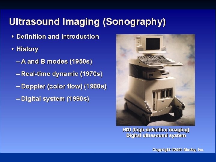
Introduction n Sonography comes from the LATIN sonus ( Sound( n Graphein come from the GREEK (to write( n Ultrasoundsonography, Ultrasonography , means imaging with ultrasound. n Diagnostic sonography is medical
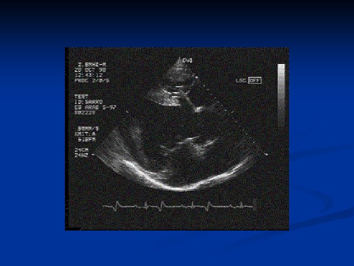
Ultrasound provides a windows into the body
One pulse of ultrasound generates a single scan line(series of echoes) as it travel through tissue.
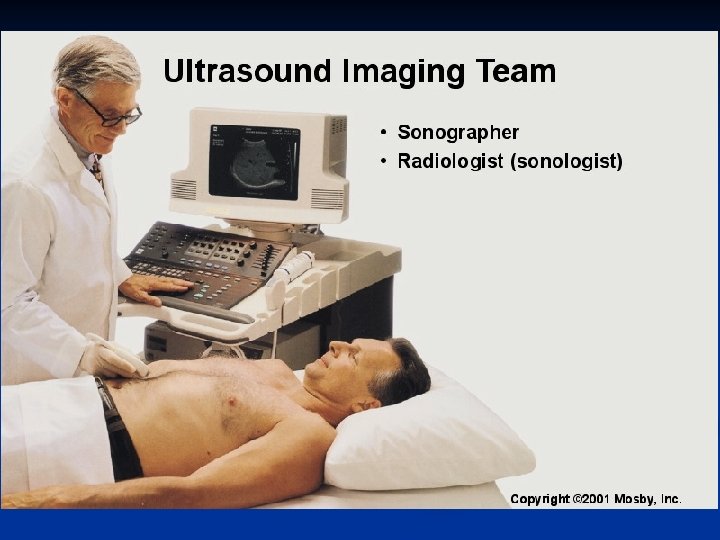
Sonography Ultrasonography means imaging with ultrasound n Diagnostic sonography is medical two dimensional cross-sectional and three dimensional anatomic and flow imaging using ultrasound n An interactive process nivolving the sonologist, patient, instrument, transducer, sonographer. n
The Basis Of Sonography
A ultrasound wave consists of a mechanical disturbance of a medium (gas, liquid or solid) which passes at a fixed speed. n The type of ultrasound wave is longitudinal. n
The disturbance propagates through a medium at a speed which depends on the compressibility and density. n The speed in the soft tissue is approximately 1540 m/s. n
Attenuation is the reduction in US intensity during passage through medium. n The mechanisms are︰ 1. Absorption 2. Scatter 3. Refraction 4. Beam divergence 5. Reflection n
The dependence of attenuation on frequency has an effect on pulse spectrum shape and therefore on the shape of the ultrasound pulse itself. μ=k*f n
Interface I(incident) I(transmit) I(reflect) Z 1 Z 2
Specular Reflection n R= n T=1-R= Z : Tissue Impedance Z=ρc
n A large difference in acoustic impedence leads to a high degree of reflection. For example, tissue-bone or tissue-air
Display n n A-Mode B-Mode M-Mode Doppler
n The most popular material for medical US transducers is know as PZT� lead zirconate titanate, Pb. Zr. Ti. O 3 �. d=λ/2
Signal process & control GAIN TGC
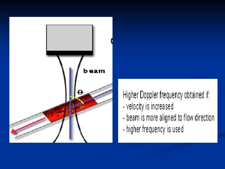
Doppler n n Doppler shift- fd= Doppler shifted frequency f 0= transducer frequency v = blood velocity θ= angle of insonification c = sound velocity in tissue
Advantage of US No radiation n Cheap n Convenience n
Comparison of imaging by X-radiation to ultrasonic imaging Advantage . 1 Excellent resolution 2. Distinguishes bone boundaries well Disadvantage. 1 Hazard(esp. to dividing cells. 2 Won’t differentiate soft tissues well . 1 Noninvasive, safe at low powers 2. Differentiates soft tissues. 1 Resolution not as good as xrays. 2. Won’t penetrate air or bone areas
A-SCAN The principle of the ultrasonic A-scanner
A-scan display
B-SCAN The principle of the ultrasonic B-scanner
Uses of Ultrasound in Medicine n Ultrasound is used for examining soft tissue inside the body. n Parts of the body that may be examined include muscles and unborn babies. n Blood flow can also be monitored using ultrasound. © 2000 ATL Ultrasound images courtesy of ATL
The Power of Ultrasound n Modern ultrasound equipment can produce n 3 D images n Colour enhancement to show blood flow n Digital files for examination on computers © 2000 ATL Ultrasound images courtesy of ATL
How Does It Work? n Medical ultrasound systems use very high frequencies - several megahertz (mega means million or 106). n A sound is a wave it has all the usual wave properties (reflection, refraction, diffraction). Ultrasound imaging makes use of the fact that sound can be reflected. n The idea is just like that used in radar and sonar.
More about how it works… l A thin layer of jelly is placed between the probe and the skin to make sure all the sound enters the body. n The probe contains a transmitter and a receiver. l A pulse of ultrasound is sent out by the transmitter. l The pulse is reflected from a surface and returns to the receiver. l The ultrasound machine measures how long it takes for the pulse to return Ultrasound probe skin Body tissue (muscle etc)
How the image is created… n Millions of sound waves are transmitted every second. n As the waves reflected at different times, the computer in the ultrasound machine calculates how far the wave travelled before being reflected (using d=vt). n Using this information the computer builds up an image of the inside of the patient. © 2000 ATL Ultrasound images courtesy of ATL