AJCC Staging Moments AJCC TNM Staging 7 th


")


- Slides: 24
AJCC Staging Moments AJCC TNM Staging 7 th Edition Lung Case #2 Contributors: Valerie W. Rusch, MD Memorial Sloan-Kettering Cancer Center, New York Peter Goldstraw, MD Royal Brompton Hospital, London, England Kelly J. Butnor, MD University of Vermont Medical Center, Burlington, Vermot Thomas W. Rice, MD Cleveland Clinic, Cleveland, Ohio
Lung Case # 2 Presentation of New Case • Newly diagnosed lung cancer patient • Presentation at Cancer Conference for treatment recommendations and clinical staging
Lung Case # 2 History & Physical • 69 yr old female who presented with an abnormal routine CXR, no symptoms • 25 pack year smoking history
Lung Case # 2 Imaging Results • Chest x-ray-5 cm right upper lobe (RUL) lung mass • CT chest-4. 5 x 5. 3 cm mass RUL lung, right paratracheal node, no hilar nodes • PET/CT-RUL lung mass, right paratracheal & right hilar lymphadenopathy • Bone scan-neg Used with permission. Swanson K, Jett J. Atlas of Cancer. Edited by Maurie Markman, David H. Johnson. © 2002 Current Medicine Inc.
Lung Case # 2 Diagnostic Procedure • Procedures – CT guided biopsy RUL lung – Mediastinoscopy with biopsy right paratracheal nodes • Pathology Reports – Poorly differentiated adenocarcinoma, bx RUL lung – Met adenocarcinoma, 2 right paratracheal nodes
Lung Case # 2 Clinical Staging • Clinical staging – Uses information from the physical exam, imaging, and diagnostic biopsy • Purpose – Select appropriate treatment – Estimate prognosis
Lung Case # 2 Clinical Staging • Synopsis- patient with 5. 3 cm adenoca lesion RUL lung, also clinically positive and biopsy proven mediastinal nodes • What is the clinical stage? – – T____ N____ M____ Stage Group______
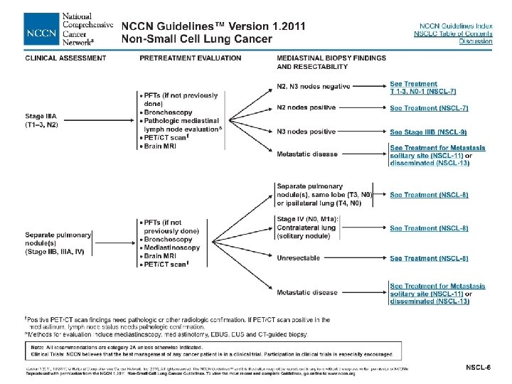
Lung Case # 2 Clinical Staging • Clinical Stage correct answer – – T 2 b N 2 M 0 Stage Group IIIA • Based on stage, treatment is selected • Review NCCN treatment guidelines for this stage
Lung Case # 2 Clinical Staging • Rationale for staging choices – T 2 b for ca >5 cm but <7 cm – N 2 because ipsilateral mediastinal nodes were clinically positive on imaging, and the diagnostic biopsy confirms the clinical category of N 2 (per new rules in AJCC 7 th) – M 0 because there was nothing to suggest distant metastases; if there was, appropriate tests would be performed before developing a treatment plan
Prognostic Factors Clinically Significant • Applicable to this case – Separate tumor nodules: none • There are no prognostic factors required for staging
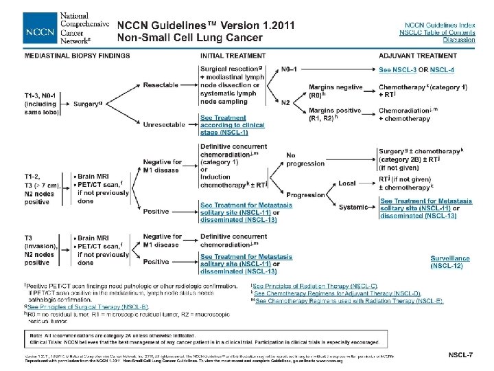
Lung Case # 2 Initial Treatment • Presentation at Cancer Conference for initial treatment recommendations – The treatment chosen based on the single lesion and clinically positive nodes in the patient, Stage IIIA, is neoadjuvant chemotherapy & radiation therapy
Lung Case # 2 Response To Therapy • Evaluation by imaging tests after neoadjuvant Rx showed no progression • Patient underwent surgical resection • Presentation at Cancer Conference for adjuvant treatment recommendations and pathologic staging
Lung Case # 2 Surgery & Findings • Procedure – RUL lung resection – Hilar and mediastinal node resection • Operative findings – No additional information
Lung Case # 2 Pathology Results • • Adenocarcinoma, RUL lung Tumor size - 3. 8 cm Grade 3 Tumor largely necrotic and inflammatory, consistent with chemo radiation effect • No pleural involvement by ca • Margins negative • 3 hilar and 3 mediastinal nodes negative
Lung Case # 2 Pathologic Staging • Pathologic staging – Uses information from the clinical staging supplemented or modified by information from surgery and the pathology report – yp is assessment at conclusion of therapy • Purpose – Additional precise data for estimating prognosis – Calculating end results (survival data) – yp – extent of response to therapy
Lung Case # 2 Pathologic Staging • Synopsis- patient with residual 3. 8 cm tumor and negative nodes after chemo & radiation therapy followed by surgery • What is the pathologic stage? (remember, clinical M may be used in pathologic staging) – – T____ N____ M____ Stage Group______
Lung Case # 2 Pathologic Staging • Pathologic Stage correct answer – – yp. T 2 a yp. N 0 c. M 0 yp. Stage Group IB • Based on pathologic stage, there is more information to estimate prognosis and adjuvant treatment recommendations
Lung Case # 2 Pathologic Staging • Rationale for staging choices – yp. T 2 a for ca >3 cm but <5 cm – yp. N 0 because hilar and mediastinal nodes were negative – c. M 0 – classified by M status prior to therapy – y prefix used to show stage during or following neoadjuvant therapy
Prognostic Factors Clinically Significant • Applicable to this case – Separate tumor nodules: none – Pleural/elastic layer invasion: PL 0 • There are no prognostic factors required for staging
AJCC Cancer Staging Atlas N 2 ipsilateral mediastinal and/or subcarinal lymph nodes
Lung Case # 2 Recap of Staging • Summary of correct answers – Clinical stage T 2 b N 2 M 0 Stage Group IIIA – Pathologic stage yp. T 2 a yp. N 0 c. M 0 yp. Stage Group IB • The staging classifications have a different purpose and therefore can be different. Do not go back and change the clinical staging based on pathologic staging information.
Staging Moments Summary • Review site-specific information if needed • Clinical Staging – Based on information before treatment – Used to select treatment options • y Pathologic Staging – Based on clinical data PLUS surgery and pathology report information – Assesses response to treatment – Used to evaluate end-results (survival)