AJCC Staging Moments AJCC TNM Staging 7 th


")


- Slides: 20
AJCC Staging Moments AJCC TNM Staging 7 th Edition Breast Case #1 Contributors: Stephen B. Edge, MD Roswell Park Cancer Institute, Buffalo, New York David R. Byrd, MD University of Washington Medical Center, Seattle, Washington David J. Winchester, MD North. Shore University Evanston Hospital, Evanston, Illinois David P. Winchester, MD North. Shore University Evanston Hospital, Evanston, Illinois
Breast Case # 1 Presentation of New Case • Newly diagnosed breast cancer patient • Presentation at Cancer Conference for treatment recommendations and clinical staging
Breast Case # 1 History & Physical • 85 yr old female who presented with an abnormal screening mammogram, no palpable breast masses, axillary nodes not palpable • No family hx, no HRT (hormone replacement therapy)
Breast Case # 1 Imaging Results • Mammogram-0. 5 cm area of microcalcifications in central left breast mid depth • Suspicious by magnification and spot compression views • Stereotactic core needle biopsy recommended • No further imaging performed ML view: magnification mammogram Used with permission. Washington University School of Medicine
Breast Case # 1 Diagnostic Procedure • Procedure – Stereotactic core needle biopsy central left breast • Pathology – – Ductal carcinoma in situ, cribriform and solid type Nuclear grade 2 Estrogen receptor positive Progesterone receptor positive
Breast Case # 1 Clinical Staging • Clinical staging – Uses information from the physical exam, imaging, and diagnostic biopsy • Purpose – Select appropriate treatment – Estimate prognosis
Breast Case # 1 Clinical Staging • Synopsis- elderly patient with 0. 5 cm DCIS lesion only visible on imaging, axilla is neg on exam and imaging • What is the clinical stage? – – T____ N____ M____ Stage Group______
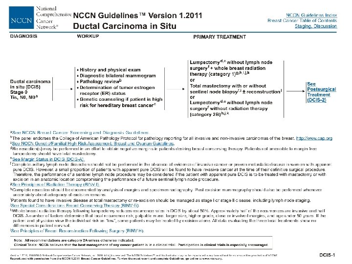
Breast Case # 1 Clinical Staging • Clinical Stage correct answer – – Tis N 0 M 0 Stage Group 0 • Based on stage, treatment is selected • Review NCCN treatment guidelines for this stage
Prognostic Factors Clinically Significant • Applicable to this case – – – Paget’s disease: no Estrogen receptor: positive Progesterone receptor: positive HER 2 status: n/a Method of node assessment: radiographic and physical examination • There are no prognostic factors required for staging
Breast Case # 1 Surgery & Findings • Procedure – Image-guided wire localized left partial mastectomy (lumpectomy) – No lymph nodes excised • Findings – Specimen radiograph reveals microcalcifications and clip in center of specimen • Final pathology deferred to permanent pathology
Breast Case # 1 Pathology Results • DCIS, cribriform and solid type, nuclear grade 2 • Invasive ductal carcinoma – 1 mm • Invasive cancer Scarff-Bloom-Richardson (SBR) Grade 1 • Margins of resection free – closest margins inferior at 5 mm • HER 2 negative
Breast Case # 1 Pathologic Staging • Pathologic staging – Uses information from the clinical staging supplemented or modified by information from surgery and the pathology report • Purpose – Additional precise data for estimating prognosis – Calculating end results (survival data)
Breast Case # 1 Pathologic Staging • Synopsis- patient with 0. 5 cm DCIS and a 1 mm infiltrating duct ca, no nodes removed • What is the pathologic stage? (remember, clinical M may be used in pathologic staging) – – T____ N____ M____ Stage Group______
Breast Case # 1 Pathologic Staging • Pathologic Stage correct answer – – p. T 1 mi (6 th Ed T 1 mic, designation changed in 7 th Ed) p. Nx c. M 0 Stage Group unknown • Based on pathologic stage, there is more information to estimate prognosis and adjuvant treatment is selected
Breast Case # 1 Pathologic Staging • Rationale for staging choices – p. T 1 mi is microinvasion <1 mm in size – p. Nx because sentinel or axillary nodes were not removed, pathologic staging cannot be completed – c. M 0 - use clinical M with pathologic staging unless there is pathologic confirmation of distant metastases
Prognostic Factors Clinically Significant • Applicable to this case – – – Paget’s disease: no SBR on invasive cancer: Grade 1 Estrogen receptor: positive Progesterone receptor: positive HER 2 status: negative Method of node assessment: radiographic and physical examination • There are no prognostic factors required for staging
AJCC Cancer Staging Atlas T 1 mi is microinvasion 0. 1 cm (1 mm) or less in greatest dimension Multiple simultaneous tumors should be indicated by (m) or the number of tumors (3)
Breast Case # 1 Recap of Staging • Summary of correct answers – Clinical stage Tis N 0 M 0 Stage Group 0 – Pathologic stage T 1 mi p. NX c. M 0 Stage Group unknown • The staging classifications have a different purpose and therefore can be different. Do not go back and change the clinical staging based on pathologic staging information.
Staging Moments Summary • Review site-specific information if needed • Clinical Staging – Based on information before treatment – Used to select treatment options • Pathologic Staging – Based on clinical data PLUS surgery and pathology report information – Used to evaluate end-results (survival)