Urological Trauma Dr Ankur Mittal Assistant Professor Urology


 • NOT")


 and")

 have gross hematuria Microhematuria does occur: usually")


 • Uncomplicated cases: conservative management with catheter drainage. • Large")
 • All penetrating and intraperitoneal injuries should be managed with")


 • Imaging – IVU – CT urogram –")


 • Suprapubic cystostomy • Primary realignment •")


")

- Slides: 54
Urological Trauma Dr Ankur Mittal Assistant Professor Urology
Trauma: 1. Given a patient with a potential urinary tract injury: 1. 2. 3. 4. To list and interpret key clinical findings To list and interpret critical investigations Construct an initial management plan To list and specify previous genitourinary anomaly. • • • Renal Bladder Urethra Ureter External Genitalia Systems:
Renal Trauma Overview • Most commonly injured GU organ • 10% of all serious injuries abdominal have associated renal injury • Mode of injury – Blunt renal truma • MVA, fall from height, assaults – Penetrating renal injuries • Gunshots and stab wounds.
Hematuria and Renal Injury • Best indicator of significant injury(microscopic or gross) • NOT related to the degree of injury • Gross Hematuria is Variable and absent in : – 7 % of grade IV renal injury – 36% of renal vascular injury – 50% of UPJ injuries
Whom to work up • Penetrating trauma: EVERYONE • Pediatric patients with microscopic hematuria. • Blunt trauma: Image with CT if: • gross hematuria • microhematuria plus shock • microhematuria plus acceleration/deceleration Mee et al. (1989) Hardeman et al (1987
Imaging of trauma patient with hematuria • CT preferred – With contrast – With “delayed” films (mandatory) – Why not get CT cystogram too? • Standard intravenous pyelogram (IVP): Forget it • “One Shot” intraoperative IVP – 2 cc/kg intravenous contrast – Single film at 10 minutes
Intraoperative One Shot IVP • Allows safe avoidance of renal exploration in 32% (Morey et al, 1999) • Highly specific for urinary extravasation • Confirms existence of the other kidney
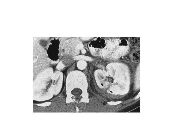
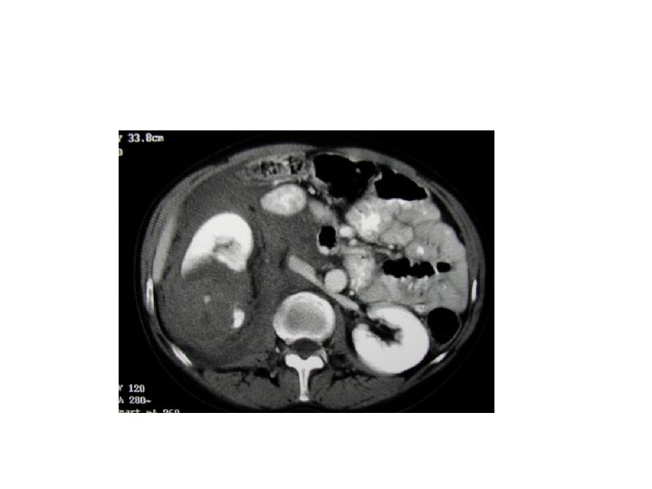
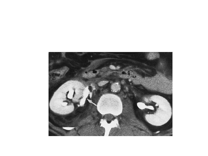
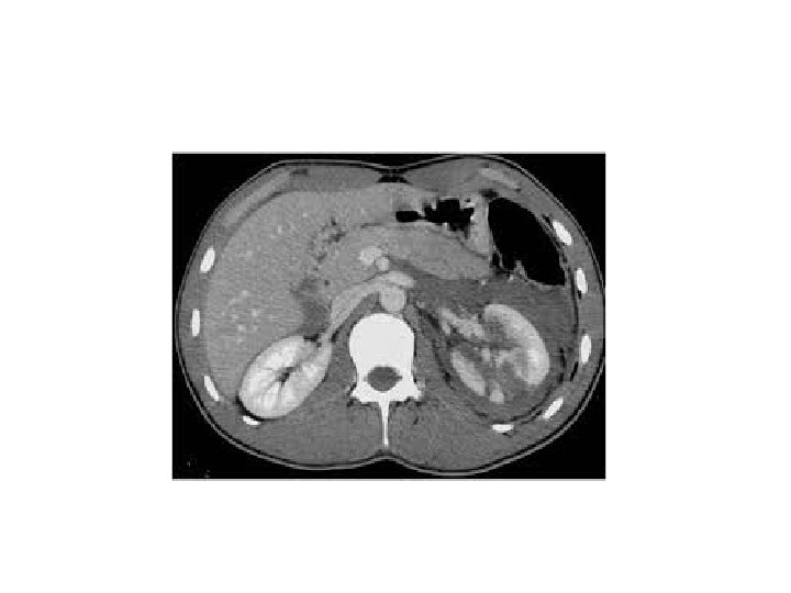
AAST Organ Injury Severity Scale for the Kidney
AAST Organ Injury Severity Scale for the Kidney
Indications for renal trauma surgery • Absolute – Hemodynamic instablity with shock – Expanding /pulsatile renal hematoma – Suspected renal pedicle avulsion (grade V) – UPJ disruption • Relative (now rare) – Urinary extravasation with non viable tissue – Renal injury together with colon /pancreatic injury – Delayed diagnosis of arterial injury
Indications for angiography with embolisation • • • Bleeding from renal segmental artery Unstable condition with grade III or IV AV fistula or pseudoaneurysm Persistent gross hematuria Blood loss extending 2 units in 24 hrs.
Management Options For Renal Trauma • Close observation – Bed rest – Serial Hemoglobins – Antibiotics if urinary extravasation • Radiographic Embolization • Urinary Diversion – Ureteral Stenting – Nephrostomy Drainage • Surgery – Renal Preservation / Reconstruction – Nephrectomy
Surgical considerations • Midline transabdominal approach • Early vascular control before opening gerotas fascia • Landmark is IMA or in presense of large hematoma , IMV.
Renal trauma in pediatric population • • Kidneys propotinally larger and less protected Less retroperitoneal and peritoneal fat Less musculature Higher sympathetic tone ie hypotention less reliable predictor of severity of renal injury.
Follow up • Repeat CECT within 72 hrs • Once pt is off hematuria and ambulatory , discharged • Adviced to avoid strenous activity for 4 -6 weeks • Follow up in opd after 3 weeks with USG and Hgm.
Bladder Trauma
Bladder: BLUNT: Overview • • Rarely isolated 80 -90 % have severe associated injuries Often high-energy injuries Associated with urethral rupture 10 -29% and pelvic fracture 6 -10%
Bladder: PENETRATING: Overview • • • Incidence 2% Associated major abdominal injuries (35%) and shock (22%) Mortality high: 12%
Bladder: Diagnosis: Physical Signs • • Suspicion: required in cases of penetrating trauma, based on trajectory Physical signs: – – – Abdominal pain Abdominal tenderness Abdominal bruising Urethral catheter does not return urine(gross hematuria in almost all cases) Delayed? • • Fever No urine output Peritoneal signs BUN / Creatinine
Bladder: Diagnosis: Hematuria • • Most (95%) have gross hematuria Microhematuria does occur: usually with minimal injury
Indications of imaging • Absolute – Gross hematuria with pelvic fracture(30 % with bladder rupture) – Penetrating injury of lower abdomen with any degree of hematuria • Relative – Gross hematuria without pelvic fracture – Microscopic hematuria with pelvic fracture
Bladder: Diagnosis Plain Cystography • Nearly 100% accurate when done properly: – – • • Adequate filling with 350 cc Drainage films Use 30% contrast Underfilling (250 cc) associated with false negatives
Bladder: Diagnosis CT Cystography • • • Preferred, especially if already getting other CTs Antegrade filling by “clamping the Foley” is not OK! Must dilute contrast (6: 1 with saline, or to about 2 -4%)
Bladder: Diagnosis CT Cystography Extraperitoneal Intraperitoneal
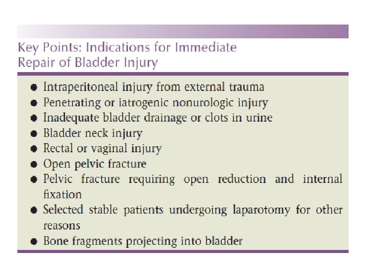
Management (extraperitoneal bladder rupture) • Uncomplicated cases: conservative management with catheter drainage. • Large bore (22 fr ) should be used. • Catheter removal 2 weeks after cystogram • Complications reported with conservative management (12% vs 5% with open repair) like fistula , clot retntion and sepsis.
Management (intraperitoneal bladder rupture) • All penetrating and intraperitoneal injuries should be managed with immediate open repair. • Catheter removal 1 week after cystogram.
Ureteral Injury • No reliable Physical findings! Usually a retrograde diagnosis • Non specific symptoms – Flank pain (36%-90%) – Fever – Ileus – Abdominal distension – fistula
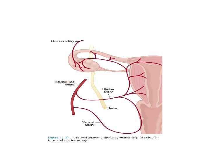
Etiology • External trauma – High speed blunt injuries – Penetrating trauma • Surgical injury – Gynecological – Obstetric – General surgery( colorectal sx) – Urologic procedures • Ureteroscopic injury
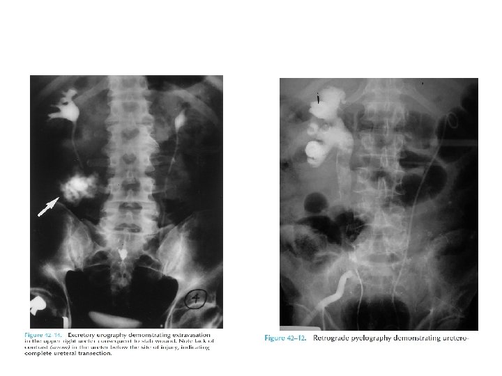
Diagnosis • Presense of hematuria(non specific) • Imaging – IVU – CT urogram – RGP – Antegrade ureterography • Intraoperative recognisation
Hematuria and ureteral injury • Nonspecific indicator • 25 – 45% patients donot demonstrate even microscopic hematuria. • Being suspicious for it is the only way you will catch it.
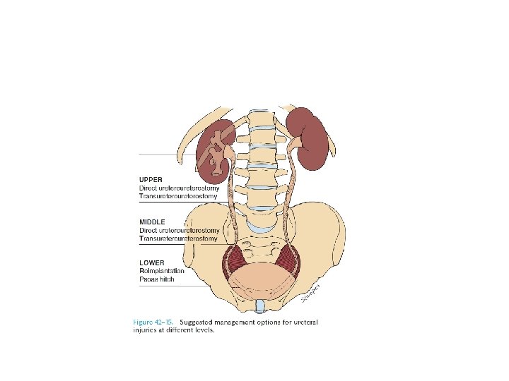
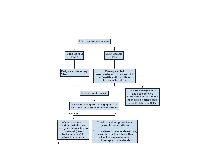
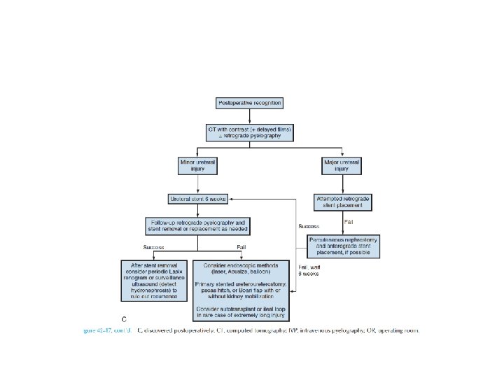
Management
Posterior Urethral Injuries
Posterior Urethra Trauma: Etiology • • • 4 -14% of pelvic fractures Bilateral pubic rami fractures (straddle fracture) and sacroiliac diasthasis Mostly males, but can happen in females Associated bladder rupture in 10 -17% Rectal injury can lead to urethral-rectal fistula in 8%
Posterior Urethra Trauma: Diagnosis • • • Blood at meatus: 50% Inability to urinate Palpable full bladder Inability to place urethral catheter High riding prostate : 34% Rarely, perineal hematoma (late finding)
• Rarely females develop proximal urethral injury • Presents with vulvar edema and blood at vaginal introitus
Retrograde Urethrogram
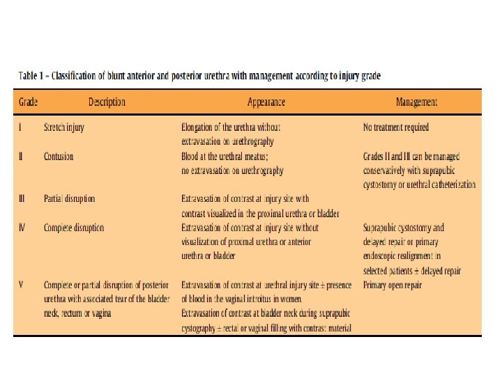
Management • Immediate open reconstruction(curently no role) • Suprapubic cystostomy • Primary realignment • Delayed reconstruction – Endoscopic treatment – Surgical reconstruction
Anterior urethral injury • • Are often isolated Majority after stradle injury Involve bulbar urethra Presents with blood at meatus, perenial hematoma, gross hematuria n urinary retention
Complications • Erectile dysfunction – 50% – Cavernosal nerve injury – Arterial insufficiency – Venous leak – Direct corporal injury • Recurent stenosis(5 -15%) • Incontinence after reconstruction <4%
QUIZ (Grades of Renal Injuries)
Thanks