Urinary tract infection and anemia in pregnancy LATEEFA

 Bacteriuria Bacteria in the")


 Pathphysiology: ascending")
 Pathphysiology: ascending")

; Rt")


 Symptomatic (1 -2%) Anatomical: Lower tract dis: asymptomatic")
 Incidence in pregnancy: 2 -10% similar to sexually active women Consequences:")
 Clinical presentation: Diagnosis:")


 dilution because the plasma volume expands more than the erythrocyte")
 Hct decreases from between")


 masurement of")
 increases risk of neural tube Deficiency occurs in")
- Slides: 26
Urinary tract infection and anemia in pregnancy LATEEFA ALDAKHYEL
Urinary Tract Infections in Pregnancy Urinary Tract Infections (terminology ) Bacteriuria Bacteria in the urine Significant bacteriureia = or > 105 CFU/m. L of urine Asymptomatic bacteriuria Lower UTI /cystitis Upper UTI / pyelonephritis
Types of UTI Recurrences 1. Relapse: same organism within 2 -3 wks 2 ndry to perineal colonization or inadequate Rx 2. Reinfection: 2 ndry to recurrent new organism within 12 wks bladder bacteriuria 3. Superinfection: new organism while on Rx 4. recurrent UTI : 2 in 6 months or = >3 in 1 year
Types of UTI Recurrences 1. Relapse: same organism within 2 -3 wks 2 ndry to perineal colonization or inadequate Rx 2. Reinfection: new organism within 12 wks 2 ndry to recurrent bladder bacteriuria 3. Superinfection: new organism while on Rx Prevention: Prenatal screening for ASB in pregnant women
Urinary Tract Infections in Pregnancy Common medical complication of pregnancy (2 -10%) Pathphysiology: ascending infection from vagina and rectum Most common causative organisms: gram –ve enteric bacteria (e. g: E. Coli 60 -80%, Proteus, K. Pnemoniae, Pseudomonas, and GBS) Lactobacilli cause no UTI
Urinary Tract Infections in Pregnancy Common medical complication of pregnancy (2 -10%) Pathphysiology: ascending infection from vagina and rectum Most common causative organisms: gram –ve enteric bacteria (e. g: E. Coli 60 -80%, Proteus, K. Pnemoniae, Pseudomonas, and GBS) Lactobacilli cause no UTI
Anatomic Changes in Pregnancy Kidneys: in length, weight, and pelves size (physiologic hydronephrosis); Rt > Lt Ureters: dilated or hydroureter (Rt > Lt), urinary stasis Mechanism: hormonal or mechanical Consequences: risk of urinary tract infections
Physiologic Changes in Pregnancy 40 -50% in renal blood flow and glomerular filtration rate (GFR) creatinine clearance serum level of creatinine, urea, uric acid by 25% Fluid volumes: extracellular volume (intravascular 50% & interstitial component) Na & Ka levels maintained Chronic loss of renal HCO 3 risk of metabolic acidosis
Risk Factors for UTI’s in Pregnancy 1. Mechanical obstruction: ureteropelvic junction, urethral or ureteric stenosis, & calculi 2. Functional obstruction: pregnancy & vesicoureteral reflux 3. Systemic diseases: DM, sickle cell trait/disease, gout, cystic renal disease
Classification of UTI’s Clinical: Asymptomatic (8%) Symptomatic (1 -2%) Anatomical: Lower tract dis: asymptomatic bacteriuria and acute cystitis Upper tract dis: acute pyelonephritis
Asymptomatic Bacteriuria (ABU) Incidence in pregnancy: 2 -10% similar to sexually active women Consequences: acute pyelonephritis (30%) Clinical presentation: ? ? Diagnosis: ? Management: outpatient Abx ( amoxil, 1 st generation cephalosporin, nitrofurantoin) length: 3 -10 days
Acute Cystitis Incidence in pregnancy: 1 -2% Consequences: acute pyelonephritis (30%) Clinical presentation: Diagnosis: Management: outpatient Abx , analgesics Length: 7 -10 days Re culture
Acute Pyelonephritis Incidence in pregnancy: 2 -4% The leading cause of ARDS and septic shock in pregnancy Most commonly in second Tx Consequences: sepsis, adult respiratory syndrome, anemia, renal failure, preterm labor Clinical presentation: fever/chills, CVA tenderness, nausea and vomiting
Acute Pyelonephritis Diagnosis: S&S Leukocytosis Urine culture Blood culture +ve in 10% Management: Inpatient - Admission - Antipyretic agents - Abx ( i. v. ampicillin or cephalosporin then p. o) Length: 10 -14 days Re culture 10 -25% recurrent
Prevention: Prenatal screening for ASB in pregnant women
Anemia in pregnancy
Physiologic anemia (dilutional anemia) dilution because the plasma volume expands more than the erythrocyte volume (The hematocrit in pregnancy normally drops several points below its pregnancy level) the oxygen-carrying capacity of the blood is not deficient
The total blood volume increase by 40%(10 -24 w) Hct decreases from between 38 and 45% in healthy women who are not pregnant to about 34% during late single pregnancy and to 30% during late multifetal pregnancy Red cell mass (driven by an increase in maternal erythropoietin production) also increases, but relatively less, compared with the increase in plasma volume Thus during pregnancy, anemia is defined as Hb < 10 g/d. L (Hct < 30%)
Women after middle age: 11. 7 to 13. 8 gm/dl
Thus during pregnancy, anemia is defined as Hb < 10 g/d. L (Hct < 30%) Women who take iron supplements have less pronounced changes in hemoglobin, as they increase their red cell mass in a more proportionate manner than those not on hematinic supplements.
Pathological anemia the oxygen-carrying capacity of the blood is deficient because of disordered erythrocyte production or excessive loss of erythrocytes through destruction or bleeding Anemia occurs in up to one third of women during the 3 rd trimester
Anemia in pregnancy Causes Iron deficiency Folate deficiency HEMOGLOBINOPATHIES
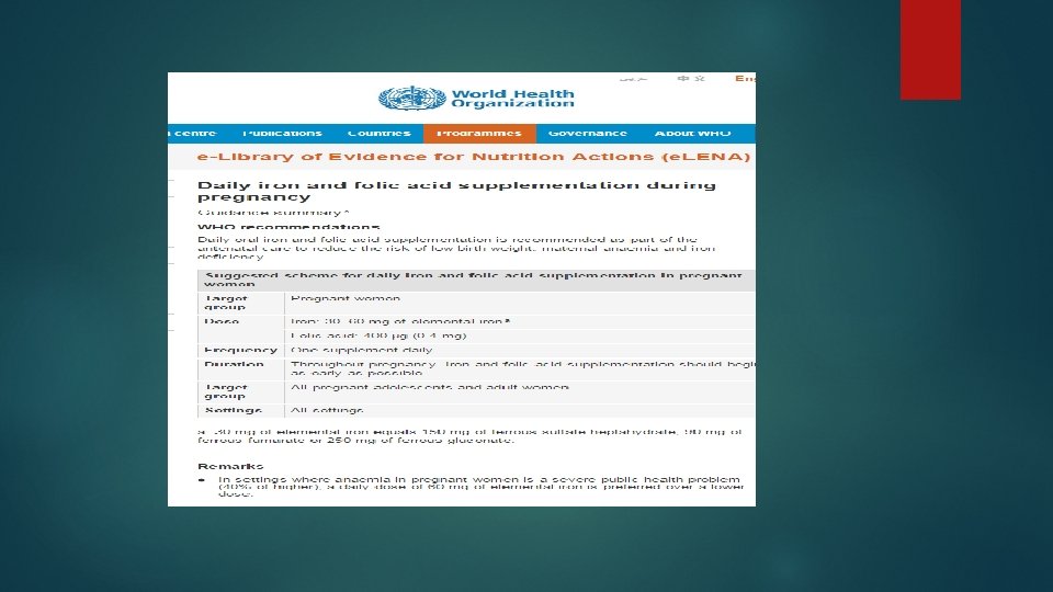
Iron deficiency anemia CBC, MCV value MCV is low (<79 f. L) masurement of serum iron, ferritin, and transferrin Typically, Hct is ≤ 30%, and MCV is < 79 f. L. Decreased serum iron and ferritin and increased serum transferrin levels confirm the diagnosis. Usually ferrous sulfate 325 mg po once/day parenteral therapy IM: 20% of pregnant women do not absorb enough supplemental oral iron absolute non-compliance IV: faster increases in Hb and better replenishment of iron stores in comparison with oral therapy,
Folate deficiency ( Megaloblastic Macrocytic Anemia) increases risk of neural tube Deficiency occurs in 0. 5 to 1. 5% of pregnant women Diagnosis Measurement of serum folate Severe megaloblastic anemia may warrant bone marrow examination and further treatment in a hospital Treatment is folate 1 mg po bid