Thoracic Empyema Pleural empyema also known as pyothorax


, forming a film")


 <")


 with pleural infection who were")


- Slides: 41
Thoracic Empyema
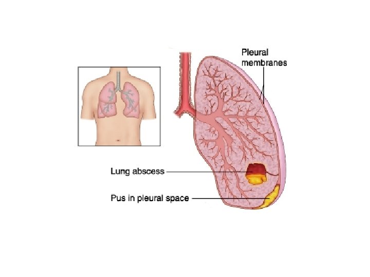
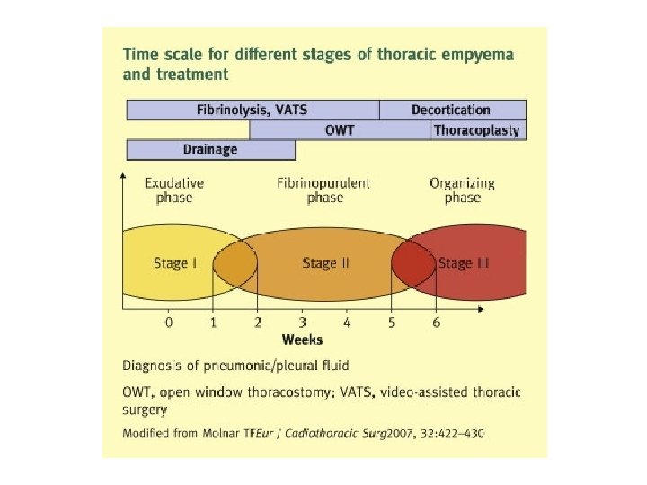
Pleural empyema, also known as pyothorax or purulent pleuritis, is an accumulation of pus in the pleural cavity that can develop when bacteria invade the pleural space. Causes of empyema Pneumonia (bacterial or TB) Chest Surgery Broncho-pleural fistula Lung Abscess Trauma Chest tube Three stages: Exudative, when there is an increase in pleural fluid with or without the presence of pus Fibrinopurulent, when fibrous septa form localized pus pockets Organizing stage, when there is scarring of the pleura membranes with possible inability of the lung to expand. Simple pleural effusions occur in up to 40% of bacterial pneumonias.
The incidence of pleural empyema and the prevalence of specific causative microorganisms varies depending on the source of infection (community acquired vs. . hospital acquired pneumonia), the age of the patient and host immune status. Independent risk factors for developing empyema include: Diabetes mellitus, Immunosuppression including corticosteroid use and HIV infection, Neoplasm, Pre existent pulmonary disease, Gastro-esophageal reflux, Alcohol misuse Intravenous drug abuse, Aspiration or poor oral hygiene (anaerobic infection) The associated mortality and morbidity is high; 20% of patients with empyema die and approximately 20% require surgery to recover.
Historical Perspective: The Egyptian physician Imhotep initially described pleural infection around 3000 BC (““An ailment which I will treat, ” “An ailment with which I will contend, ” or “An ailment not to be treated”). Hippocrates has been more famously credited with its recognition in 500 BC. Until the 19 th century open thoracic drainage was the recommended treatment but carried an associated mortality of up to 70%. This high mortality was probably due to respiratory failure produced by the large open pneumothorax left by drainage. Closed tube drainage was first described in 1876 but was not widely adopted until the influenza epidemic of 1917. An Empyema Commission, advocated adequate pus drainage with a closed chest tube, avoidance of early open drainage, obliteration of the pleural space and proper nutritional support. These changes reduced mortality to 4. 3% during the later stages of this epidemic.
Historical Perspective: The introduction of antibiotics both reduced the incidence of empyema and changed its bacteriology. Before antibiotics, 60 to 70% of cases were due to Streptococcus pneumonia which now only accounts for approximately 10% of culturepositive cases. The prevalence of Staphylococcus aureus rose and the development of staphylococcal resistance in the 1950’s increased complications and mortality. More recently, the reported prevalence of anaerobic infections and Gramnegative organisms has risen.
Pleural Fluid: The volume of pleural fluid is small (<1 ml), forming a film about 10 mm thick between the visceral and parietal pleural surfaces. Pleural fluid contains protein at concentrations similar to the interstitial fluid, a small number of cells (predominantly mesothelial cells, macrophages and lymphocytes) and some large molecular weight proteins such as lactate dehydrogenase (LDH). Compared with serum, healthy pleural fluid contains more bicarbonate, lower levels of sodium, and similar glucose. The p. H of normal pleural fluid is around 7. 6. These parameters change when disease processes affecting the adjacent lung or vascular tissue activate an immune response. Water and small molecules pass freely between mesothelial cells, while larger particles may be transported by cytoplasmic transport mechanisms or via pleurolymphatic communications.
Early Stage Empyema: In the early exudative stage there is fluid movement into the pleural space due to increased capillary vascular permeability. This is accompanied by the production of proinflammatory cytokines such as interleukin 8 (IL-8) and tumor necrosis factor a (TNFa). These produce active changes in the pleural mesothelial cells to facilitate fluid entry into the pleural cavity. The fluid is a free-flowing exudate characterized by a low white cell count, an LDH level less than half that in the serum, normal p. H and glucose levels and does not contain bacterial organisms. This is “simple para-pneumonic effusion”. Treatment with antibiotics at this stage is likely to be adequate and most do not require drainage.
Fibrinopurulent Stage Increasing fluid accumulation and bacterial invasion across the damaged endothelium. Bacterial invasion accelerates the immune response, promoting further migration of neutrophils and activation of the coagulation cascade leading to increased procoagulant and depressed fibrinolytic activity. Increased levels of plasminogen activator inhibitors and decreased tissue type plasminogen activator (t. PA) , favoring fibrin deposition and promoting septations. Neutrophil phagocytosis and bacterial death fuel the inflammatory process by the release of more bacteria cell wall derived fragments, increased lactic acid, a fall in pleural fluid p. H, increased glucose metabolism, and a rise in LDH. Characteristic biochemical features of a fibrinopurulent collection low p. H (<7. 20), low glucose, and high LDH. Also called “complicated para-pneumonic” effusion.
Organizing phase Fibroblasts proliferate. A solid fibrous pleural peel begins to form which occasionally encases the lung preventing re-expansion, impairing lung function and creating a persistent pleural space with continuing potential for infection.
Bacteriology of community-acquired and hospital-acquired pleural infection Common organisms Community-acquired Streptococcus spp. (52%) < S milleri < S pneumoniae < S intermedius Staphylococcus aureus (11%) Gram-negative aerobes (9%) < Enterobacteriaceae < Escherichia coli Anaerobes (20%) < Fusobacterium spp. < Bacteroides spp. < Peptostreptococcus spp. < Mixed Hospital-acquired Staphylococci < Meticillin-resistant S aureus (MRSA) (25%) < S aureus (10%) Gram-negative aerobes (17%) < E coli < Pseudomonas aeruginosa < Klebsiella spp. Anaerobes (8%)
Microbiology: Surgical Management of Thoracic Empyema in HIV Infected Patients Failing Conventional Anti-Microbial and Closed Drainage Treatment Total CD 4<200 Surgery MRSA Streptococcus 3 3 1 2 MSSA Enterococcus Staph. Epi. 1 1 Gram Negative Enterobacter 2 2 Anaerobes Peptostreptococcus 2 Propionibacterium A. 2 Other Mycobacterium T. 3 3 1 Fungus Candida A. PCP Cryptococcus Alternaia Aspergillus Nocardiosis Penicillium 4 3 1 1 1 4 3 1 1 Gram Positive 1 1 1 2 1 3 1 1 1
Specialists: A chest physician or thoracic surgeon should be involved in the care of all patients requiring chest tube drainage for pleural infection. Delay to pleural drainage is probably associated with increased morbidity, duration of hospital stay, and may lead to increased mortality. Misdiagnosis, inappropriate antibiotics and chest tube malpositioning have been cited as important factors contributing to the inadequate management of pleural infection. A surgical opinion is appropriate at any stage if a patient is not settling with drainage and antibiotics. Management of pleural infection in adults: British Thoracic Society pleural disease guideline 2010 Thorax 2010; 65(Suppl 2): ii 41 eii 53.
Nutrition: Clinicians should ensure adequate nutrition in patients with pleural infection. Poor nutrition was identified during the First World War as an adverse determinant of outcome from pleural empyema but is frequently overlooked. Patients with pleural infection suffer catabolic consequences which may lead to further immunodeficiency and slow recovery. Hypoalbuminemia is associated with a poor outcome from pleural infection and clinicians should provide adequate nutritional support and consider supplemental feeding. Management of pleural infection in adults: British Thoracic Society pleural disease guideline 2010 Thorax 2010; 65(Suppl 2): ii 41 eii 53.
DVT Risk All patients with pleural infection are at high risk for the development of venous thromboembolism and should receive adequate prophylaxis with heparin unless contraindicated. All acutely ill patients with pneumonia and/or pleural infection who have been admitted to hospital should receive prophylactic dose low molecular weight heparin treatment unless contraindicated (bleeding, thrombocytopenia, significant renal impairment, allergy to low molecular weight heparins). Inpatients with renal impairment, unfractionated heparin should be used (5000 units subcutaneously twice daily). Mechanical prophylaxis and thromboembolic deterrent stockings should be used in those with contraindications to anticoagulant treatment. Management of pleural infection in adults: British Thoracic Society pleural disease guideline 2010 Thorax 2010; 65(Suppl 2): ii 41 eii 53.
Progression to Empyema Features of ongoing sepsis and raised C-reactive protein in patients with pneumonia after 3 days may indicate progression to pleural infection. All patients with suspected pleural infection should have blood cultures for aerobic and anaerobic bacteria performed. For patients in hospital with community-acquired pneumonia the median time to improvement in heart rate and blood pressure is 2 days, and 3 days for temperature, respiratory rate, and oxygen saturation. A failure to respond to initial management may indicate the presence of a para-pneumonic effusion or empyema as a complication of pneumonia. Management of pleural infection in adults: British Thoracic Society pleural disease guideline 2010 Thorax 2010; 65(Suppl 2): ii 41 eii 53.
Progression to Empyema Ongoing fever and symptoms or signs of sepsis or example, elevated white cell count and/or inflammatory markers such as C reactive protein (CRP). CRP is a sensitive marker of progress in pneumonia. Failure of the CRP level to fall by 50% is associated with an adverse outcome and increased incidence of empyema and a repeat chest x-ray. The following variables were positively predictive: (1) albumin <3. 0; (2) CRP >100; (3) platelet count >400, 000; (4) sodium <130 ; (5) intravenous drug abuse; and (6) chronic alcohol abuse. Using two or more points as the cut-off, the sensitivity was 87%, specificity 68. 3%, positive predictive value 17. 7% and negative predictive value 98. 5%. Management of pleural infection in adults: British Thoracic Society pleural disease guideline 2010 Thorax 2010; 65(Suppl 2): ii 41 eii 53.
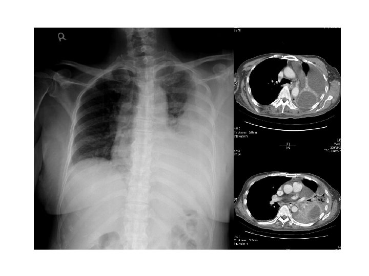
Contrast-enhanced CT scanning may be of value in patients when the diagnosis is in doubt or an underlying abnormality is thought either to be associated with the empyema or potentially its cause, such as an esophageal perforation or bronchogenic carcinoma. CT scanning can help to differentiate pleural empyema from a parenchymal lung abscess and may also help to formulate management decisions about drainage. Empyemas are usually lenticular in shape and compress the lung parenchyma, while lung abscesses often have an indistinct boundary between the lung parenchyma and collection. The “split pleura” sign caused by enhancement of both parietal and visceral pleural surfaces and their separation in empyema is characteristic of a pleural collection. Pleural thickening is seen in 100% of empyemas and 56% of exudative parapneumonic effusions. Moderate (<2 cm) mediastinal lymphadenopathy is seen in over one-third of patients with pleural infection. Management of pleural infection in adults: British Thoracic Society pleural disease guideline 2010 Thorax 2010; 65(Suppl 2): ii 41 eii 53.
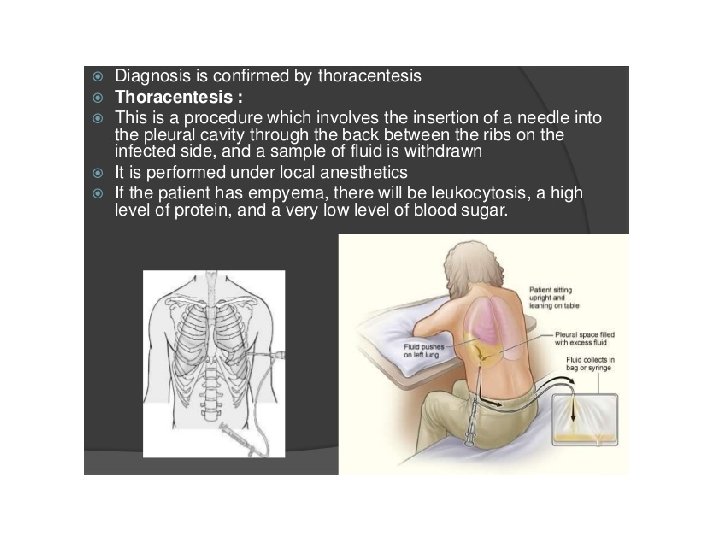
Fluid Sampling All patients with a pleural effusion in association with sepsis or a pneumonic illness require diagnostic pleural fluid sampling. Pleural fluid characteristics remain the most reliable diagnostic test to guide management in association with a pneumonic illness or recent chest trauma or surgery and who have features of ongoing sepsis. Imaging guidance should be used since this minimizes risks of lung puncture and improves the recovery rate of pleural fluid. Sampling using thoracic ultrasound is simple, safer, and reduces patient discomfort. Small effusions (ie, <10 mm thickness) will usually resolve with antibiotics alone. Observation may be appropriate for these patients, but an increase in the size of the effusion or ongoing sepsis should warrant re-evaluation and diagnostic pleural fluid sampling. Management of pleural infection in adults: British Thoracic Society pleural disease guideline 2010 Thorax 2010; 65(Suppl 2): ii 41 eii 53.
Fluid Sampling Pleural fluid p. H should be assessed in all non-purulent effusions when pleural infection is suspected. If pleural fluid p. H measurement is not available, pleural fluid glucose assessment should be performed where pleural infection is possible. pleural fluid p. H of <7. 2 is also the single most powerful indicator to predict a need for chest tube drainage, and that pleural fluid LDH (>1000) and glucose <34 did not improve diagnostic accuracy. Where pleural fluid p. H measurement is not available glucose and LDH should be measured, a pleural fluid glucose level <34 may be used as an alternative marker to indicate a need for chest drain insertion. Pleural fluid glucose may be lowered in situations other than pleural infection, such as rheumatoid effusions. Occasionally a pleural fluid p. H of >7. 6 will be obtained in a complicated para-pneumonic effusion as a result of infection with Proteus spp. Its ability to produce ammonia by urea splitting can produce alkalotic fluid. Management of pleural infection in adults: British Thoracic Society pleural disease guideline 2010 Thorax 2010; 65(Suppl 2): ii 41 eii 53.
Antibiotics All patients should receive antibiotics targeted to treat the bacterial profile of pleural infection and based on local antibiotic policies and resistance patterns. Antibiotics to cover anaerobic infection should be used in all patients except those with culture proven pneumococcal infection. Macrolide antibiotics are not indicated unless there is objective evidence for or a high clinical index of suspicion of ‘atypical’ pathogens. Where possible, antibiotic choice should be guided by bacterial culture results. Management of pleural infection in adults: British Thoracic Society pleural disease guideline 2010 Thorax 2010; 65(Suppl 2): ii 41 eii 53.
Drainage Patients with frankly purulent or turbid/cloudy pleural fluid on sampling should receive prompt pleural space chest tube drainage. The presence of organisms identified by Gram stain and/or culture from a nonpurulent pleural fluid sample should lead to prompt chest tube drainage. Pleural fluid p. H <7. 2 in patients with suspected pleural infection indicates a need for chest tube drainage. Poor clinical progress during treatment with antibiotics alone should lead to prompt patient review, repeat pleural fluid sampling, and probably chest tube drainage. Patients with a loculated pleural collection should receive early chest tube drainage. A small-bore catheter 10 to 14 will be adequate for most cases of pleural infection. However, there is no consensus on the size of the optimal chest tube for drainage. Chest tube insertion should be performed under imaging guidance wherever possible. Management of pleural infection in adults: British Thoracic Society pleural disease guideline 2010 Thorax 2010; 65(Suppl 2): ii 41 eii 53.
Intrapleural Fibrinolytics There is no indication for the routine use of intrapleural fibrinolytics in patients for pleural infection. There is currently interest in other intrapleural agents including combination therapy with fibrinolytics and fluid viscosity and biofilm-disrupting agents such as streptodornase and deoxyribonuclease (DNase). In experimental/translational studies, this combination reduced infected pus viscosity when compared with fibrinolytics (streptokinase) alone and can disrupt infected biofilms. Short term drainage benefits are not associated with reduced mortality, the frequency of surgery, the length of hospital stay, or long-term radiological and lung function outcome. Streptokinase that was associated with an excess of immunological adverse reactions such as fever, leucocytosis and malaise, but no excess of systemic or intrapleural bleeding and no systemic activation of the fibrinolytic cascade Management of pleural infection in adults: British Thoracic Society pleural disease guideline 2010 Thorax 2010; 65(Suppl 2): ii 41 eii 53.
VATS vs. Intrapleural Fibrinolytics UT Southwestern Study Patients (20) with pleural infection who were suitable for general anesthesia were randomized to receive either immediate VATS or chest tube insertion with additional instillation of intrapleural streptokinase for 3 days. The surgical group had higher primary treatment success (10/11 patients) and all streptokinase medical failures (5/9 patients) were salvaged by surgery without requiring thoracotomy. Surgical patients also had a shorter drainage period (5. 8 vs. 9. 8 days), hospital stay (8. 7 vs. 12. 8 days), and cost savings. Wait MA, Sharma S, Hohn J, et al. A randomized trial of empyema therapy. Chest 1997; 111: 1548 e 51. (1 L).
Prognosis: All patients with empyema require outpatient follow-up with a repeat chest X-ray and inflammatory biochemistry analysis within 4 weeks following discharge. Chest radiograph returns to normal in the majority of patients by 6 months. Patients be advised to return sooner if symptoms redevelop. Long-term result of pleural empyema are rare but include bronchopleural fistula formation, recurrent empyema and pleural thickening, which may lead to functional lung impairment. Approximately 15% of adult patients with pleural infection die within 1 year of the event, although deaths are usually due to co-morbid conditions and not directly due to sepsis from the empyema. Mortality in children is generally reported to be less than 3%. No reliable clinical, radiological or pleural fluid characteristics accurately determine patients’ prognosis at initial presentation. Management of pleural infection in adults: British Thoracic Society pleural disease guideline 2010 Thorax 2010; 65(Suppl 2): ii 41 eii 53.
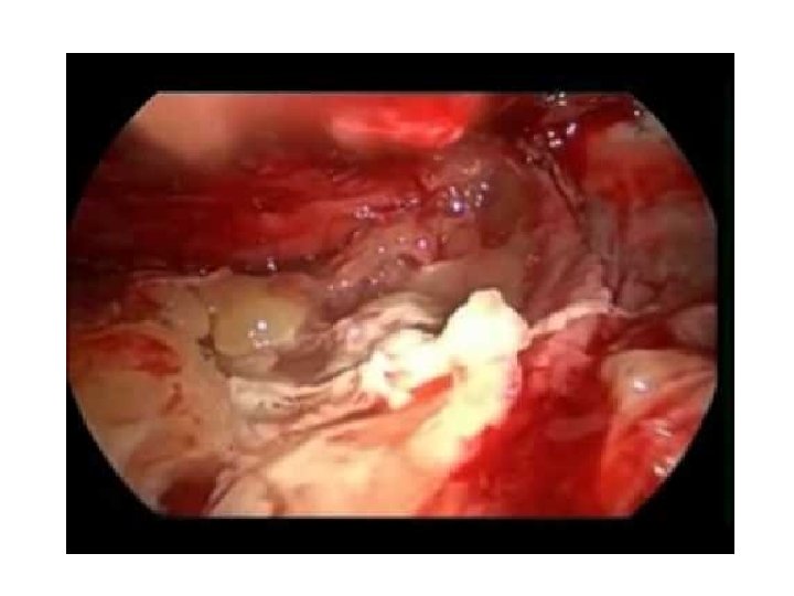
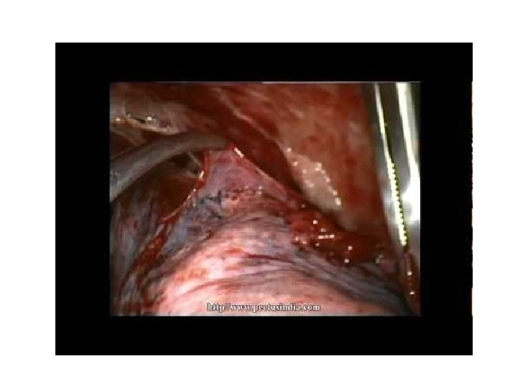
Empyema Surgery Drainage of fluid and pus – free up the pleural space Decortication – free up the trapped lung, re-expansion, obliterate any residual pleural space Chest tube Insertions – post-operative drainage
Thoracoplasty is removal of the skeletal support of a portion of the chest, done by removal of varying number of rib segments to approximate the chest wall to the underlying lung or mediastinum or pleural space obliteration. Alexander procedure collapsed the apical cavities of pulmonary tuberculosis Schede procedure which aims at reducing intrathoracic spaces or filling them with living tissue implants
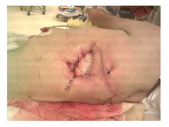
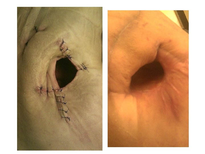
An Eloesser flap is a single stage procedure for the treatment of severe pleural empyema, and involves a U-shaped incision and the resection of a number of subjacent posterolateral ribs. The U-shaped flap is then folded into the pleural space creating a permanent communication. Described by Dr. Leo Eloesser in 1935 while treating TB empyema in San Francisco. Dr Leo Eloesser (1881 - 1976) American thoracic surgeon.
A Clagett thoracotomy is a three stage procedure performed for treatment severe empyema 1. the resection of a posterolateral lower rib and the formation of an open window in the lateral aspect of the chest 2. continuous drainage and irrigation of the cavity with antibiotic solution 3. once the healthy granulation tissue has lined the cavity and the patient has clinically recovered, the space is filled with antibiotic solution and the thoracotomy wound closed The technique was first described at the Mayo clinic in 1963 by Dr O. Theron Clagett.
Chest Tubes There are two types of chest drainage collection: 1. Dry Suction Water Seal a. Collection chamber: Chest fluid drains into here b. Water Seal: 2 cm water to prevent entry of air c. Suction regulator: No sound or water evaporation 2. Water Seal a. Collection chamber: Chest fluid drains into here b. Water Seal: 2 cm water to prevent entry of air c. Suction: Fill to 20 cm, need to keep filled with water
Dry Suction Chest Drainage
Water Seal Chest Drainage
Care of Chest Tubes • How much wall suction? It doesn’t matter as long as suction is set on the dry suction. • Do not turn suction valve off. This is regulated to keep bubbles low. You need to keep the system open so air can escape. • What if the chest tube comes out? Do not put anything in wound. Cover with a non occlusive gauze dressing. Air needs to be able to get out. • What if chest Tube is disconnected? Plug it back in. • How do we transport chest tube patient? Disconnect from suction, the tube is to water seal. Do not tip over the water seal container. Water seal is OK for short term and so patient can walk. • Securing the Tube: A lot of tape is bad psychologically for the patient and irritates the skin. No petroleum gauze only a small amount of tape and a gauze sponge.