Esophageal Motility Pathophysiology of Reflux Disease Maha Saja


 • What")
 • Teeth perform cutting and grinding action. • Chewing is largely a")


 and subsequently")


 • Propels food from mouth to stomach. • Complicated process since pharynx")

")
 Voluntary stage Pharyngeal stage Esophageal stage Changes Nervous control Food is “squeezed”")


- Slides: 40
Esophageal Motility & Pathophysiology of Reflux Disease Maha Saja Msaja@ksu. edu. sa
Objectives • Mastication & chewing • Salivary glands • Secretion of saliva • Contents of saliva • Functions of saliva • Control of salivary secretion • Swallowing • Types of esophageal peristalsis • Function of lower esophageal sphincter
The Early Response to a Meal
What is the Early Response of the GIT to a Meal It can be divided into phases; 1. The Cephalic Phase → Before food is ingested. 2. The Oral Phase → When ingested food is in the mouth. 3. The Esophageal Phase → When food is transferred from mouth to esophagus.
The Cephalic Phase • Occurs before ingesting food. • Thinking or anticipating food, smelling or seeing food. • Aim to prepare the GIT for the meal. • Stimuli cause an increase in parasympathetic neural outflow to gut leading to enhanced GI secretions (salivary, gastric, pancreatic. . Etc).
The Oral Phase • Occurs when food is in the mouth. • Same features as cephalic phase but here there is contact between food and GI surface. • Adding more stimuli from mouth → taste. • The response is similar to the cephalic phase → an increase in parasympathetic neural outflow to gut leading to enhanced GI secretions (salivary, gastric, pancreatic. . Etc).
The Mouth
Where Does Food Digestion Start From? • At the mouth (oral cavity) • What is meant by digestion? • What are the types of digestion? ü Mechanical. ü Chemical. • What is the role of the mouth in digestion?
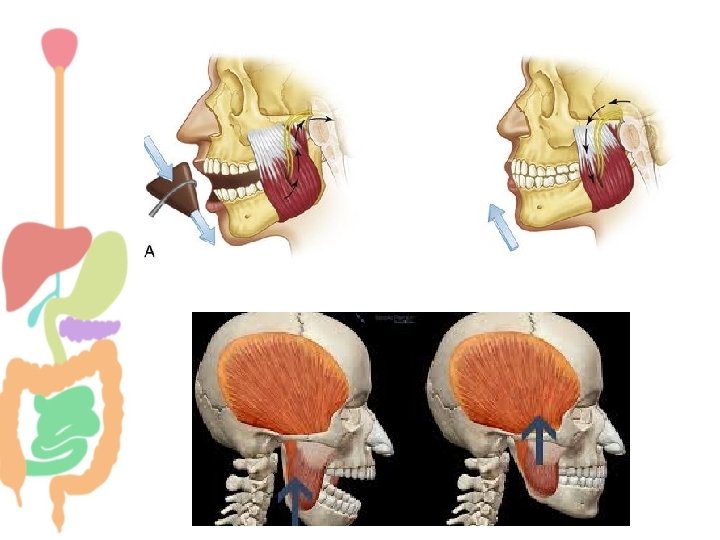
Chewing (Mastication) • Teeth perform cutting and grinding action. • Chewing is largely a reflex (5 th CN). Food in mouth Reflex inhibition to muscles of mastication Jaw drops • What is the importance of mastication? The drop initiates a stretch reflex in jaw muscles Rebound contraction
The Salivary Glands
Salivary Glands Are exocrine glands. The secretory unit is made up of acini and ducts.
THE SECRETORY UNIT The basic building block of all salivary glands ACINI - water and ions derived from plasma Saliva formed in acini flows down DUCTS to empty into the oral cavity.
Salivary Glands + Many tiny buccal glands
Salivary Glands Based on type of secretion they are classified into Serous Mucinous Clear, salty, watery containing ptyalin (αamylase). Thick slimy secretion of mucus (mucin). e. g. Parotid gland e. g. buccal glands Mixed A mixture of both e. g. Submandibular & sublingual
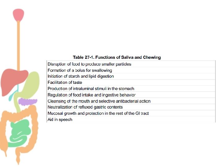
Salivary Secretions • We secrete ≈ 800 – 1500 ml of saliva per day (average 1 L). • p. H of saliva = 6. 0 -7. 0 • What are the functions of saliva? ü Digestion. ü Lubrication. ü Cleansing. ü Protection ü Proteolytic enzymes. ü Antibodies.
Two stage process Mechanism of Salivary Secretion 1 st stage → Acini Isotonic concentration of major ions similar to that in plasma 2 nd stage → Ducts hypotonic Secondary secretion
TWO STAGE HYPOTHESIS OF SALIVA FORMATION Most proteins Water & electrolytes Na+ Cl- resorbed Some proteins Isotonic primary saliva electrolytes K+ secreted & HCO 3 Hypotonic final saliva into mouth
Ions in Saliva • What is the normal composition of saliva? • How does the level of ions compare to their level in plasma? • What happens to the composition of saliva in cases of increased flow of secretion?
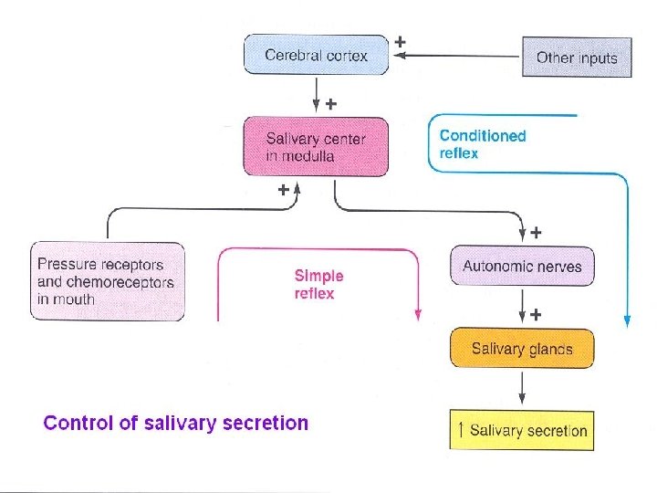
Regulation of Salivary Secretion • Exclusively neural. • Increased secretion of salivary glands in response to a meal is a reflex, two types of salivary reflexes; • Unconditioned (born with it). • Conditioned reflex (learnt by experience). • Salivation may occur in response to reflexes originating in the stomach and upper small intestine. E. g. Nauses, presence of irritating food.
Mechanism of salivary secretion. Initial saliva is produced by acinar cells (1) and subsequently modified by ductal epithelial cells (2). ATP, Adenosine triphosphate
• Nerve supply of salivary glands • I- Sympathetic nerves • Originate in the superior cervical ganglion and reach the 3 pairs of salivary glands through blood vessels • Functions: • Act on mucous cells and produce small amount of viscous secretion. • Cause vasoconstriction.
II- Parasympathetic nerves Originate in the superior & inferior salivary nuclei in brain stem. Fibers from the superior salivary nucleus leave in VII cranial nerve supply both submandibular and sublingual glands. Fibers from the inferior salivary nucleus leave the medulla in IX cranial nerve supply the parotid gland.
Stimulation of both sympathetic and parasympathetic nerves stimulates salivary secretion. Regulation of salivary secretion by the autonomic nervous system. ACh, Acetylcholine; β, β receptor; c. AMP, cyclic adenosine monophosphate; CN, cranial nerve; M, muscarinic receptor; NE, norepinephrine; T 1 -T 3, thoracic segments.
The Esophageal Phase
Esophagus • Collapsible muscular tube that conveys food from pharynx to stomach (10 inches long). • Food passes through quickly because of peristalsis
Esophageal Sphincters 1. Upper esophageal sphincter. 2. Lower esophageal sphincter. • Propel food from mouth to stomach. • Protect airway during swallowing. • Protect esophagus from acidic gastric contents.
Swallowing (Deglutition) • Propels food from mouth to stomach. • Complicated process since pharynx is a shared space between respiration & swallowing. • Food should move without compromising respiration.
Stages of Swallowing 1. Voluntary stage. 2. Pharyngeal stage. 3. Esophageal stage. Involuntary • Voluntary moves bolus of food from mouth to pharynx. • Pharyngeal stage moves bolus of food from pharynx to esophagus. • Esophageal stage move bolus from esophagus to stomach.
Swallowing (Deglutition)
Swallowing (Deglutition) Voluntary stage Pharyngeal stage Esophageal stage Changes Nervous control Food is “squeezed” posteriorly into the pharynx by the tongue Soft palate-up Larynx – anterior & up. Epiglottis –down Esopahgus opens UES –relaxes Respiration inhibited A wave of peristalsis starts Primary peristalsis Secondary peristalsis LES- relax Swallowing reflex Stage Food stimulates touch receptors in pharynx Afferent via CN V & IX Swallowing center Motor efferent via CN V, IX, X, & XII Myenteric plexus Vagus
Esophageal Stage • Primary peristalsis → continuation of the peristaltic wave that started in the pharynx. • Secondary peristalsis → starts at the point of esophageal distention by retained food. • “Receptive relaxation” of LES & stomach → a wave of relaxation that travels along the myenteric plexus ahead of peristaltic wave. Allows LES & stomach to prepare to receive the food bolus.
Primary Secondary
Esophageal stage
Lower Esophageal Sphincter • Also known as gastroesophageal sphincter • Extends 3 cm above its junction with stomach • Formed by circular muscles • Normally remains tonically constricted • Relaxes ahead of esophageal peristaltic wave • Helps to prevent reflux of gastric juice • Valvelike mechanism of short portion of esophagus that extend slightly into the stomach also helps in preventing reflux
Thank you