
Thank you for viewing this presentation We would

- Slides: 153
Thank you for viewing this presentation. We would like to remind you that this material is the property of the author. It is provided to you by the ERS for your personal use only, as submitted by the author. Ó 2011 by the author
Non CPAP therapies in OSA An ERS Task Force Report 2011
Members ERS Task Force Group • Chairs: – Johan Verbraecken (Antwerp, B) – Winfried Randerath (Solingen, G) • Collaborators: – – – – Stefan Andreas (Immenhausen, G) Jean-Louis Pepin (Grenoble, F) Ingo Fietze (Berlin, G) Ian E Smith (Cambridge, UK) Marie Marklund (Umea, Sw) An Boudewyns (Antwerp, B) Joachim Maurer (Mannheim, G) Arschang Valipour (Vienna, A) Thomas Verse (Hamburg, G) Boris A. Stuck (Mannheim, G) Evert Hamans (Antwerp, B) Bernd Sanner (Wuppertal, G) Jean Roch Paoli (Bastia, F) Laurent Lacassagne (Saint-Jean, F) Floriant Jalbert (Toulouse, F) Georges Bettega (Grenoble, F)
ERS Task Force Study Group Objectives • Many patients with obstructive sleep apnoea (OSA) look for alternative conservative or surgical therapies to avoid treatment with continuous positive airway pressure. In view of the high prevalence and the relevant impairment of these patients, lots of methods are offered that promise a definite cure or relevant improvement of OSA. The Sleep and control of breathing Group of the European Respiratory Society established a task force to evaluate the scientific literature on non-CPAP therapies in the treatment of OSA according to the standards of evidence-based medicine. A literature search was performed (Medline and hand search of relevant papers). The studies were evaluated according the recommendations of the “Oxford Centre for Evidence-based Medicine Levels of Evidence”. • The following treatment options were evaluated: – Part I. Conservative treatment – Part II. Oral devices – Part III: Nasal and velopharyngeal surgery – Part IV: Hypopharyngeal and maxillofacial surgery
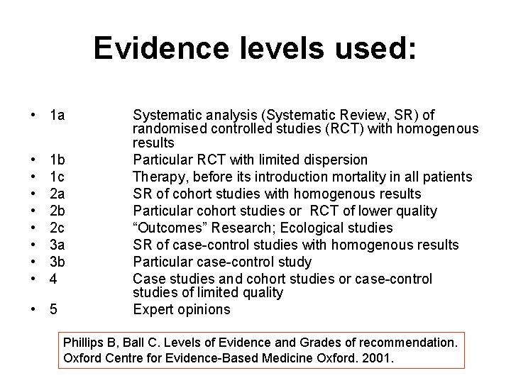
Evidence levels used: • 1 a • • 1 b 1 c 2 a 2 b 2 c 3 a 3 b 4 • 5 Systematic analysis (Systematic Review, SR) of randomised controlled studies (RCT) with homogenous results Particular RCT with limited dispersion Therapy, before its introduction mortality in all patients SR of cohort studies with homogenous results Particular cohort studies or RCT of lower quality “Outcomes” Research; Ecological studies SR of case-control studies with homogenous results Particular case-control study Case studies and cohort studies or case-control studies of limited quality Expert opinions Phillips B, Ball C. Levels of Evidence and Grades of recommendation. Oxford Centre for Evidence-Based Medicine Oxford. 2001.
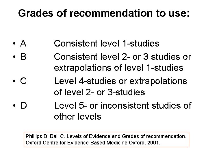
Grades of recommendation to use: • A • B • C • D Consistent level 1 -studies Consistent level 2 - or 3 studies or extrapolations of level 1 -studies Level 4 -studies or extrapolations of level 2 - or 3 -studies Level 5 - or inconsistent studies of other levels Phillips B, Ball C. Levels of Evidence and Grades of recommendation. Oxford Centre for Evidence-Based Medicine Oxford. 2001.
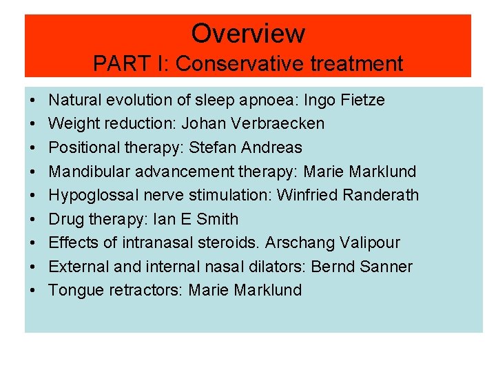
Overview PART I: Conservative treatment • • • Natural evolution of sleep apnoea: Ingo Fietze Weight reduction: Johan Verbraecken Positional therapy: Stefan Andreas Mandibular advancement therapy: Marie Marklund Hypoglossal nerve stimulation: Winfried Randerath Drug therapy: Ian E Smith Effects of intranasal steroids. Arschang Valipour External and internal nasal dilators: Bernd Sanner Tongue retractors: Marie Marklund
Non-CPAP Therapies in Obstructive Sleep Apnoea Natural evolution of sleep apnoea Ingo Fietze ERS Task Force “Non-CPAP therapies in obstructive sleep apnoea“
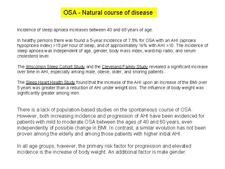
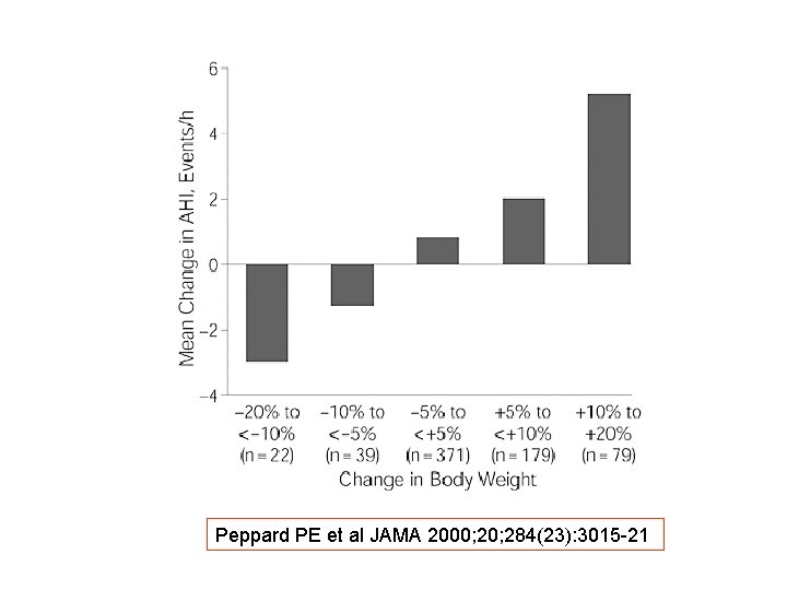
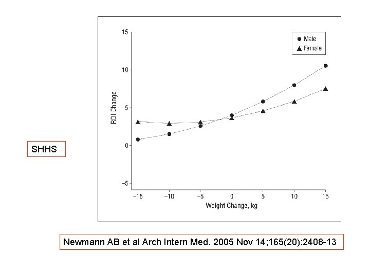
OSA - Natural course of disease Incidence of sleep apnoea increases between 40 and 65 years of age. In healthy persons there was found a 5 -year incidence of 7. 5% for OSA with an AHI (apnoea hypopnoea index) >15 per hour of sleep, and of approximately 16% with AHI >10. The incidence of sleep apnoea was independent of age, gender, body mass index, waist-hip radio, and serum cholesterol level. The Wisconsin Sleep Cohort Study and the Cleveland Family Study revealed a significant increase over time in AHI, especially among male, obese, older, and snoring patients. The Sleep Heart Health Study found that the increase of the AHI upon an increase of the BMI over 5 years was greater than a reduction of AHI under weight loss. The influence of body weight was significantly greater among men. There is a lack of population-based studies on the spontaneous course of OSA. However, both increasing incidence and progression of AHI have been evidenced for patients with mild to moderate OSA between the ages of 40 and 60 years, even independently of possible change in BMI. In contrast, a similar evolution has not been proven among the elderly and among those patients with higher initial AHI. In all age groups, however, the primary risk factor for progression and elevated incidence is the increase of body weight. An additional factor is male gender.
OSA - Natural course of disease Recommendation level • Presented data show a trend to worsening of sleep disordered breathing over time. Most studies are retrospective and observational (case series). Based on these data a spontaneous improvement cannot be expected. Grade of recommendation C
Non-CPAP Therapies in Obstructive Sleep Apnoea Weight reduction Johan Verbraecken ERS Task Force “Non-CPAP therapies in obstructive sleep apnoea“
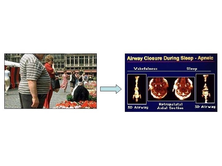
OSA – Weight reduction Rationale • OSA is caused by pharyngeal and/or laryngeal collapse. Pharyngeal fat deposits lead to a decrease in pharyngeal patency and underline the risk factor of obesity. Weight reduction will lead to a decrease in critical closing pressure (Pcrit), and consequently decreases the severity of OSA.
Peppard PE et al JAMA 2000; 284(23): 3015 -21
SHHS Newmann AB et al Arch Intern Med. 2005 Nov 14; 165(20): 2408 -13
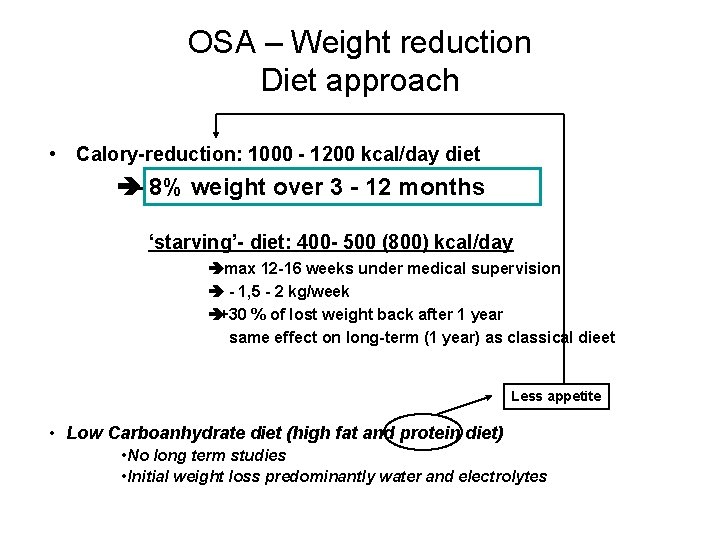
OSA – Weight reduction Diet approach • Calory-reduction: 1000 - 1200 kcal/day diet è- 8% weight over 3 - 12 months ‘starving’- diet: 400 - 500 (800) kcal/day èmax 12 -16 weeks under medical supervision è - 1, 5 - 2 kg/week è+30 % of lost weight back after 1 year same effect on long-term (1 year) as classical dieet Less appetite • Low Carboanhydrate diet (high fat and protein diet) • No long term studies • Initial weight loss predominantly water and electrolytes
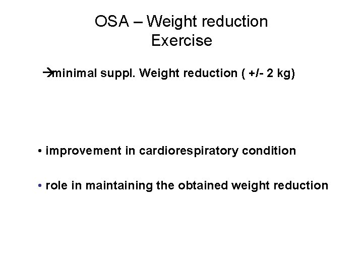
OSA – Weight reduction Exercise àminimal suppl. Weight reduction ( +/- 2 kg) • improvement in cardiorespiratory condition • role in maintaining the obtained weight reduction
OSA – Weight reduction ‘Behavior’ therapy àminimal effect (cfr. exercise) • + diet has better result than diet allone • role in maintaining the obtained weight reduction
OSA – Weight reduction Diet approach: results • In the diet intervention studies: – mean BMI reduction of 4. 7 ± 2. 5 kg/m 2 – accompanied by a decrease in AHI of 21 ± 13/h (44%). • 246 patients (39%) showed a partial improvement in AHI • 145 patients (23%) were cured from OSA.
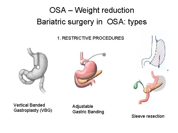
OSA – Weight reduction Bariatric surgery in OSA: types 1. RESTRICTIVE PROCEDURES Vertical Banded Gastroplasty (VBG) Adjustable Gastric Banding Sleeve resection
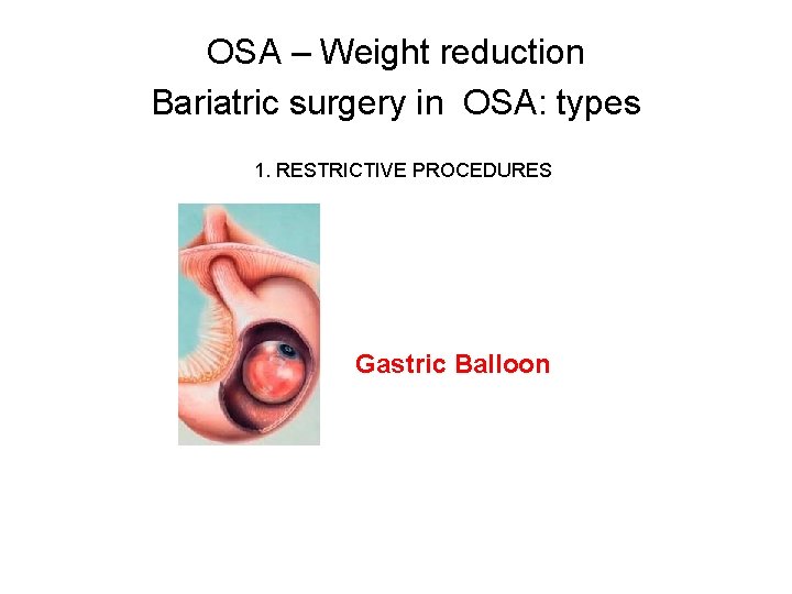
OSA – Weight reduction Bariatric surgery in OSA: types 1. RESTRICTIVE PROCEDURES Gastric Balloon
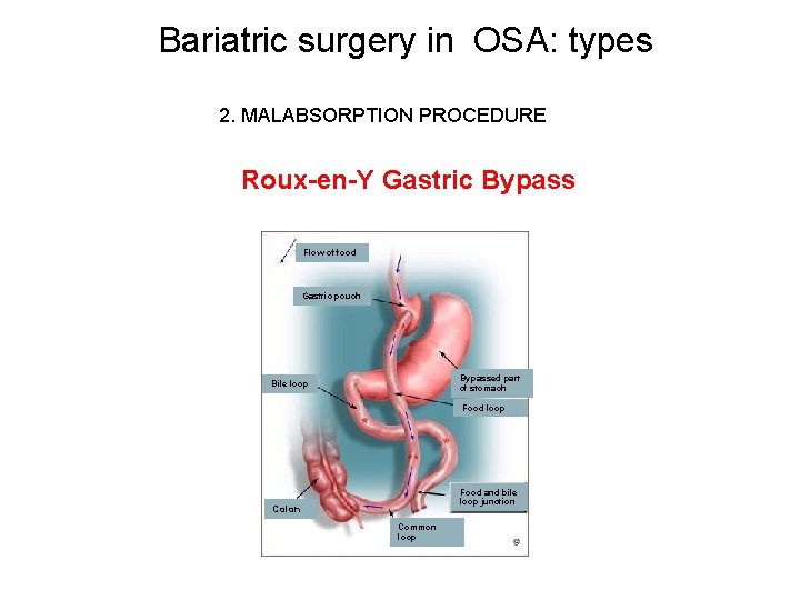
Bariatric surgery in OSA: types 2. MALABSORPTION PROCEDURE Roux-en-Y Gastric Bypass Flow of food Gastric pouch Bypassed part of stomach Bile loop Food and bile loop junction Colon Common loop
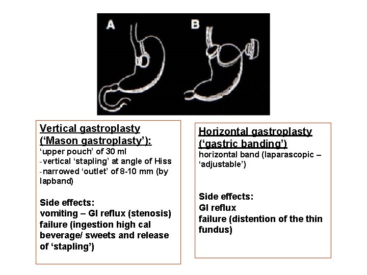
Vertical gastroplasty (‘Mason gastroplasty’): ‘upper pouch’ of 30 ml -vertical ‘stapling’ at angle of Hiss -narrowed ‘outlet’ of 8 -10 mm (by lapband) Side effects: vomiting – GI reflux (stenosis) failure (ingestion high cal beverage/ sweets and release of ‘stapling’) Horizontal gastroplasty (‘gastric banding’) horizontal band (laparascopic – ‘adjustable’) Side effects: GI reflux failure (distention of the thin fundus)
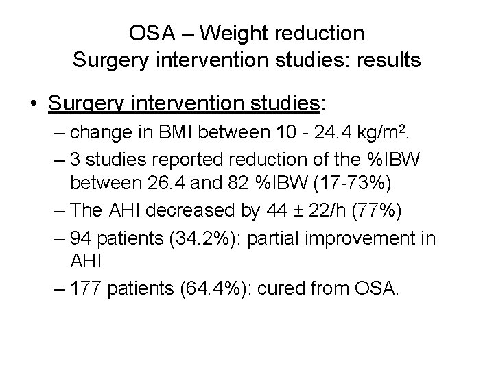
OSA – Weight reduction Surgery intervention studies: results • Surgery intervention studies: – change in BMI between 10 - 24. 4 kg/m 2. – 3 studies reported reduction of the %IBW between 26. 4 and 82 %IBW (17 -73%) – The AHI decreased by 44 ± 22/h (77%) – 94 patients (34. 2%): partial improvement in AHI – 177 patients (64. 4%): cured from OSA.
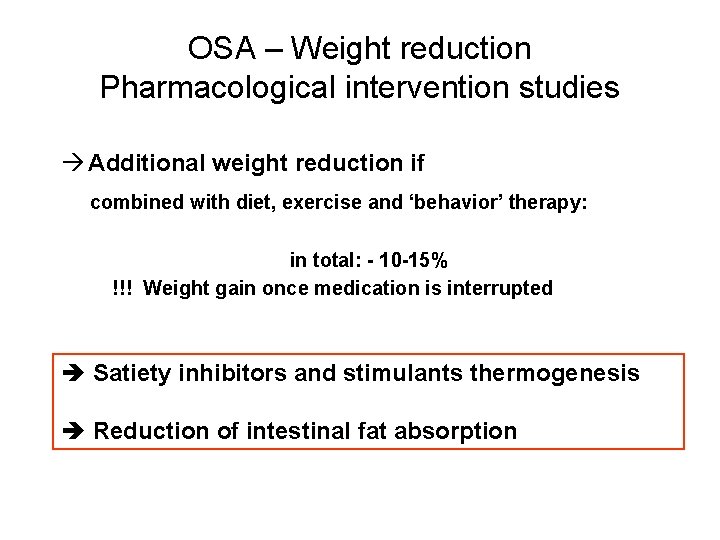
OSA – Weight reduction Pharmacological intervention studies à Additional weight reduction if combined with diet, exercise and ‘behavior’ therapy: in total: - 10 -15% !!! Weight gain once medication is interrupted è Satiety inhibitors and stimulants thermogenesis è Reduction of intestinal fat absorption
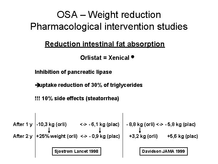
OSA – Weight reduction Pharmacological intervention studies Reduction intestinal fat absorption Orlistat = Xenical â Inhibition of pancreatic lipase èuptake reduction of 30% of triglycerides !!! 10% side effects (steatorrhea) After 1 y -10, 3 kg (orli) <-> - 6, 1 kg (plac) After 2 y +25% weight (orli) <-> - 0, 9 kg (plac) Sjostrom Lancet 1998 - 8, 8 kg (orli) <-> - 5, 8 kg (plac) +3, 2 kg (orli) +5, 6 kg (plac) Davidson JAMA 1999
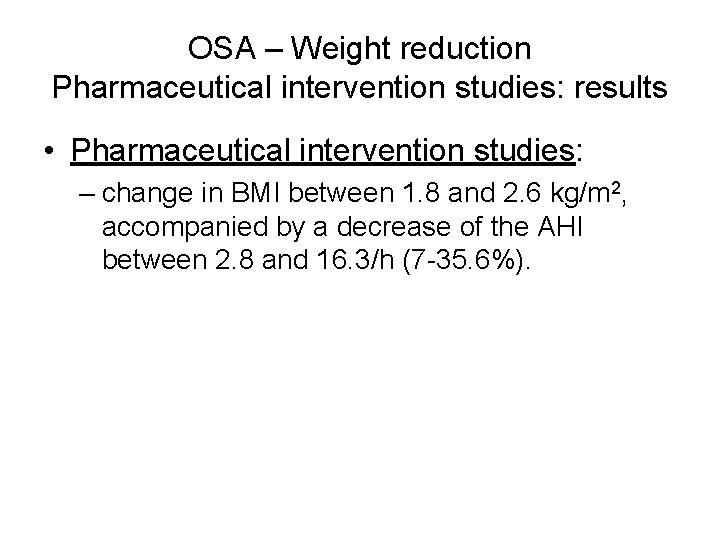
OSA – Weight reduction Pharmaceutical intervention studies: results • Pharmaceutical intervention studies: – change in BMI between 1. 8 and 2. 6 kg/m 2, accompanied by a decrease of the AHI between 2. 8 and 16. 3/h (7 -35. 6%).
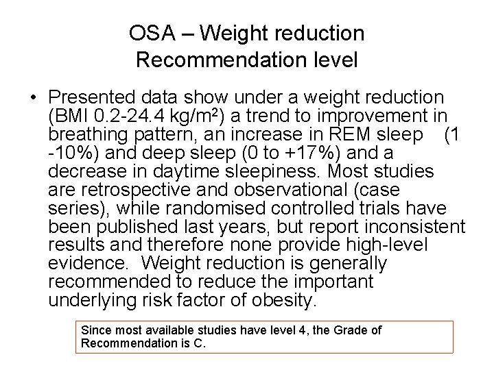
OSA – Weight reduction Recommendation level • Presented data show under a weight reduction (BMI 0. 2 -24. 4 kg/m 2) a trend to improvement in breathing pattern, an increase in REM sleep (1 -10%) and deep sleep (0 to +17%) and a decrease in daytime sleepiness. Most studies are retrospective and observational (case series), while randomised controlled trials have been published last years, but report inconsistent results and therefore none provide high-level evidence. Weight reduction is generally recommended to reduce the important underlying risk factor of obesity. Since most available studies have level 4, the Grade of Recommendation is C.
Non-CPAP Therapies in Obstructive Sleep Apnoea Positional therapy Stefan Andreas ERS Task Force “Non-CPAP therapies in obstructive sleep apnoea“
OSA – Body position Rationale Body position effects: • Pharyngeal cross-sectional area • closing pressures of the pharynx • Ventilatory drive • And thus nocturnal respiratory disturbances Thus, there is evidence suggesting effects of position during sleep
Definition positional OSA A supine AHI of at least twice that in the lateral position Using this definition a prevalence of approximately 50% is reported
Patients responding to positional therapy are an easy to treat subset • patients with positional OSA tend to have a lower AHI, to be younger and to be less obese • Accordingly, patients with a clear improvement of the AHI with positional therapy tended to be younger, to have a lower AHI and to be less obese
Clinical studies on positional therapy • Different devices such as a tennis ball, vests, positional alarm, verbal instruction and pillows are used • Significant but moderate effects on AHI • Limitations: – most studies were uncontrolled – small studies – long term compliance poor or not evaluated
OSA – Positional therapy Recommendation level • Positional therapy is not recommended for the treatment of OSA. • If positional therapy is used, follow up sleep studies have to be performed to document individual success. • Long term compliance has to be secured by follow up studies. Grade of Recommendation C
Non-CPAP Therapies in Obstructive Sleep Apnoea Muscle and nerve stimulation Winfried J. Randerath ERS Task Force “Non-CPAP therapies in obstructive sleep apnoea“
OSA - Upper airway muscle and nerve stimulation Background Obstructive sleep apnoea • Diminished activity of dilating muscles during sleep (Remmers 78). • Insufficient muscle response against hypercapnia, hypoxia or negative pharyngeal pressure (Block 84). • Muscles contract more intensively to counterbalance collapsible forces (Deegan 98). Genioglossus muscle • One of the most important dilators • Pushes forward tongue; enlarges cross-sectional area of upper airways • Activity increased in OSAS during wakefulness (compensation) (Mezzanotte 92, Wiegand 90).
OSA – muscle and nerve stimulation Point of interest Does stimulation or training of the upper airway muscles • optimize the dilating forces ? • increase the width of the upper airways ? • allow for effective therapy of OSAS ?
OSA – muscle and nerve stimulation Literature search Search in PUBMED • Keyword combinations: Neurostimulation OR electrical stimulation OR upper airway muscles OR genioglossus stimulation OR hypoglossus nerve stimulation AND sleep apnoea syndrome. • Limit: Clinical Trial; no other limitations. Manual search through references Results • 28 studies were found, 13 included in further evaluation. • Excluded studies: not on the electrical stimulation, but on pathophysiology, effects of mandibular advancement, drugs, surgery. • 3 studies level of evidence 1 b, the others level 4.
OSA – muscle and nerve stimulation Topics • Acute efficacy of electrical stimulation on upper airway patency. • Efficacy of apnoea-triggered neurostimulation in clinical use. • Tongue muscle training by electrical stimulation. • Oropharyngeal exercise
OSA – muscle and nerve stimulation Acute efficacy of electrical stimulation on the upper airway patency • Stimulation with surface and intraneural electrodes. • Effective reduction of upper airways resistance in healthy persons and in OSAS (Schwartz 1996, 1991, Mann 2002, Oliven 2001, 2003, 2007, Isono 1999, Schnall 1995). • Highest efficacy by stimulation of the genioglossus muscle (Mann 02, Oliven 01, 07).
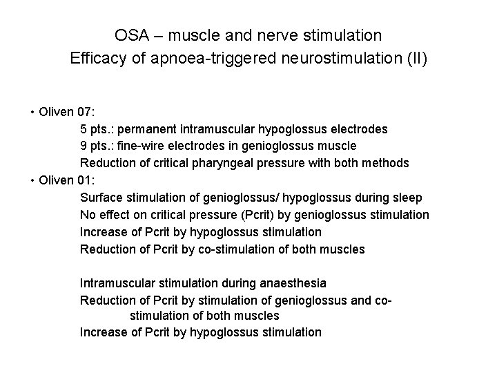
OSA – muscle and nerve stimulation Efficacy of apnoea-triggered neurostimulation (I) • Conflicting results. • Significant improvement of respiratory disturbances under intraneural stimulation of hypoglossus nerve (Schwartz 96). • Transcutaneous electrical stimulation of the genioglossus muscle Miki 89: 6 OSAS patients. Stimulation with 15 -40 V when apnoeas for >5 sec. Apnoea index: before 39 ± 19, under stimulation 12 ± 5/h Slow wave sleep: before 14 ± 4, under stimulation 22 ± 5% Guilleminault 95: No enlargement of the upper airways Undesirable contractions of the platysma and arousals • Intraoral stimulation Motions of tongue muscle without positive effects on upper airway patency (Guilleminault 95, Oliven 03)
OSA – muscle and nerve stimulation Efficacy of apnoea-triggered neurostimulation (II) • Oliven 07: 5 pts. : permanent intramuscular hypoglossus electrodes 9 pts. : fine-wire electrodes in genioglossus muscle Reduction of critical pharyngeal pressure with both methods • Oliven 01: Surface stimulation of genioglossus/ hypoglossus during sleep No effect on critical pressure (Pcrit) by genioglossus stimulation Increase of Pcrit by hypoglossus stimulation Reduction of Pcrit by co-stimulation of both muscles Intramuscular stimulation during anaesthesia Reduction of Pcrit by stimulation of genioglossus and costimulation of both muscles Increase of Pcrit by hypoglossus stimulation
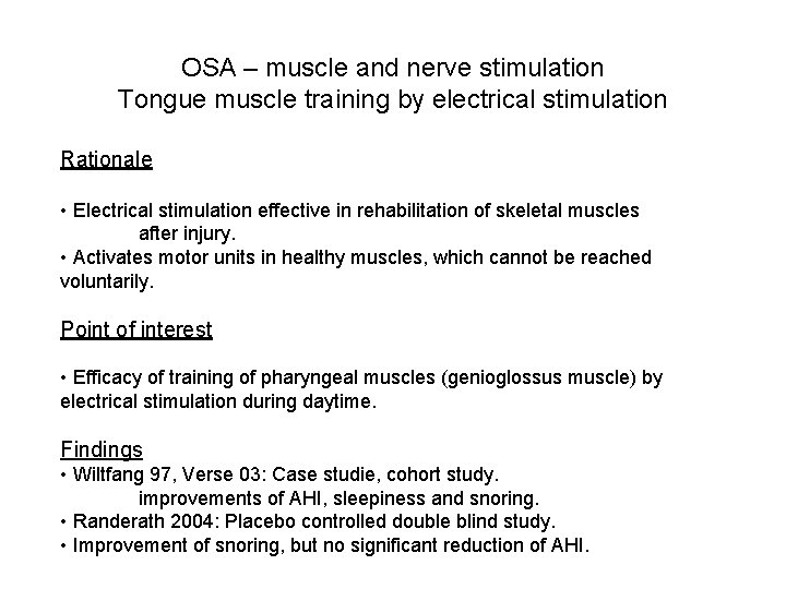
OSA – muscle and nerve stimulation Tongue muscle training by electrical stimulation Rationale • Electrical stimulation effective in rehabilitation of skeletal muscles after injury. • Activates motor units in healthy muscles, which cannot be reached voluntarily. Point of interest • Efficacy of training of pharyngeal muscles (genioglossus muscle) by electrical stimulation during daytime. Findings • Wiltfang 97, Verse 03: Case studie, cohort study. improvements of AHI, sleepiness and snoring. • Randerath 2004: Placebo controlled double blind study. • Improvement of snoring, but no significant reduction of AHI.
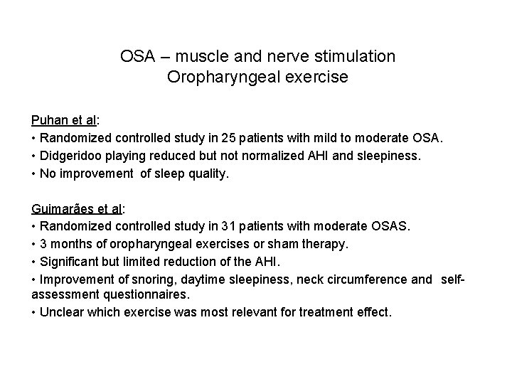
OSA – muscle and nerve stimulation Oropharyngeal exercise Puhan et al: • Randomized controlled study in 25 patients with mild to moderate OSA. • Didgeridoo playing reduced but normalized AHI and sleepiness. • No improvement of sleep quality. Guimarães et al: • Randomized controlled study in 31 patients with moderate OSAS. • 3 months of oropharyngeal exercises or sham therapy. • Significant but limited reduction of the AHI. • Improvement of snoring, daytime sleepiness, neck circumference and selfassessment questionnaires. • Unclear which exercise was most relevant for treatment effect.
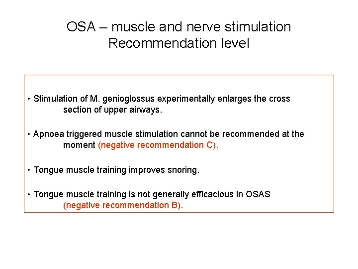
OSA – muscle and nerve stimulation Recommendation level • Stimulation of M. genioglossus experimentally enlarges the cross section of upper airways. • Apnoea triggered muscle stimulation cannot be recommended at the moment (negative recommendation C). • Tongue muscle training improves snoring. • Tongue muscle training is not generally efficacious in OSAS (negative recommendation B).
Non-CPAP Therapies in Obstructive Sleep Apnoea Drug therapy Ian E. Smith ERS Task Force “Non-CPAP therapies in obstructive sleep apnoea“
OSA – Drug therapy Rationale • Although there may be a predisposing airway abnormality, it is changes in respiratory drive, airway tone or surface forces that cause airway closure during sleep. • Pharmaceutical agents might reduce sleep apnoea by increasing respiratory drive, changing sleep structure (in particular suppressing REM sleep), increasing upper airway muscle tone, changing respiratory and cardiovascular reflexes that may perpetuate apnoeas and reducing surface forces that encourage closure of the upper airway.
OSA – Drug therapy Search strategy • Data bases individually searched: Pub. Med and Medline. Keyword combinations: Sleep apnea/ apnoea (drug* or pharmacological and (treatment*)) or progesterone or progestogen or medroxy* or "tricyclic anti depressant*" or protriptyline or amitriptyline or imipramine or ssri or fluoxetine or clonidine or modafinil or stimulant* or Buspirone or doxapram or dopram or naloxone or narcan or "opiod antagonist*" or nicotin* or "ACE inhibitor*" or ACE-inhibitor* or cilazapril or captopril or enalapril or fosinopril or imidapril or lisinopril or perindopril or quinapril or ramipril or trandolapril or "anti hypertensive*" or anti-hypertensive* or antihypertensive* or baclofen or mirtazapine or steroid* or *steroid or fluticasone • Reports on snorers or subjects with predominantly central apnoeas were excluded. Even with these filters a large number of low quality studies were identified. For nearly all of the drugs that have been trialled in the last 20 years there is at least one randomised controlled trial (RCT) and so uncontrolled studies and review articles were excluded.
OSA – Drug therapy Conclusions and recommendations • At the present time there is no evidence that any drug is likely to benefit an unselected patient with OSA. Small studies have reported positive effects of certain agents on short-term outcomes. Longer studies with measures of symptomatic responses of cholinergic agents and upper airway lubricants are supported by the results of single night studies already published. It is likely that better characterisation of the predominant mechanisms of OSA in individual patients will lead to better results and this also needs further study. It is recommended that none of the drugs investigated to date are used to treat OSA (Grade of Recommendation C for most drugs trialled, Recommendation B for mirtazapine and protriptyline).
Non-CPAP Therapies in Obstructive Sleep Apnoea Effect of intranasal steroids Arschang Valipour ERS Task Force “Non-CPAP therapies in obstructive sleep apnoea“
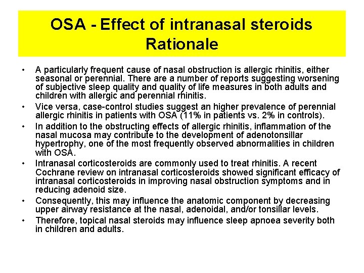
OSA - Effect of intranasal steroids Rationale • • • A particularly frequent cause of nasal obstruction is allergic rhinitis, either seasonal or perennial. There a number of reports suggesting worsening of subjective sleep quality and quality of life measures in both adults and children with allergic and perennial rhinitis. Vice versa, case-control studies suggest an higher prevalence of perennial allergic rhinitis in patients with OSA (11% in patients vs. 2% in controls). In addition to the obstructing effects of allergic rhinitis, inflammation of the nasal mucosa may contribute to the development of adenotonsillar hypertrophy, one of the most frequently observed abnormalities in children with OSA. Intranasal corticosteroids are commonly used to treat rhinitis. A recent Cochrane review on intranasal corticosteroids showed significant efficacy of intranasal corticosteroids in improving nasal obstruction symptoms and in reducing adenoid size. Consequently, this may influence the anatomic component by decreasing upper airway resistance at the nasal, adenoidal, and/or tonsillar levels. Therefore, topical nasal steroids may influence sleep apnoea severity both in children and adults.
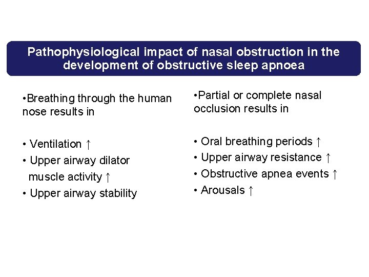
Pathophysiological impact of nasal obstruction in the development of obstructive sleep apnoea • Breathing through the human nose results in • Partial or complete nasal occlusion results in • Ventilation ↑ • Upper airway dilator muscle activity ↑ • Upper airway stability • Oral breathing periods ↑ • Upper airway resistance ↑ • Obstructive apnea events ↑ • Arousals ↑
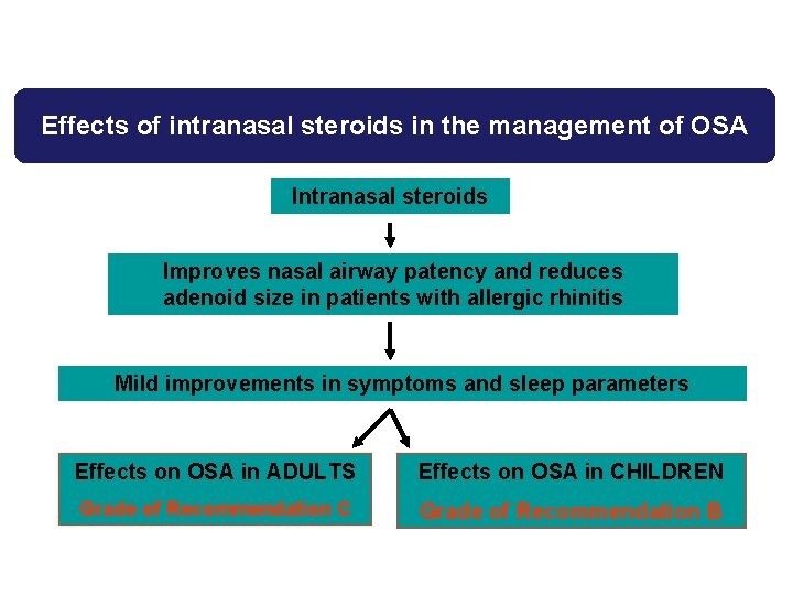
Effects of intranasal steroids in the management of OSA Intranasal steroids Improves nasal airway patency and reduces adenoid size in patients with allergic rhinitis Mild improvements in symptoms and sleep parameters Effects on OSA in ADULTS Effects on OSA in CHILDREN Grade of Recommendation C Grade of Recommendation B
Non-CPAP Therapies in Obstructive Sleep Apnoea External and internal nasal dilators Bernd Sanner ERS Task Force “Non-CPAP therapies in obstructive sleep apnoea“
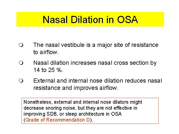
Nasal Dilation in OSA m The nasal vestibule is a major site of resistance to airflow. m Nasal dilation increases nasal cross section by 14 to 25 %. m External and internal nose dilation reduces nasal resistance and improves airflow. Nonetheless, external and internal nose dilators might decrease snoring noise, but they are not effective in improving SDB, or sleep architecture in OSA (Grade of Recommendation D).
Overview PART II: oral devices • Mandibular advancement therapy: Marie Marklund • Tongue retractors: Marie Marklund
Non-CPAP Therapies in Obstructive Sleep Apnoea Mandibular advancement therapy Marie Marklund ERS Task Force “Non-CPAP therapies in obstructive sleep apnoea“
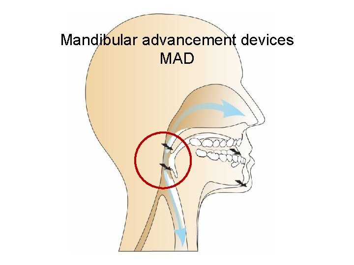
Mandibular advancement devices MAD
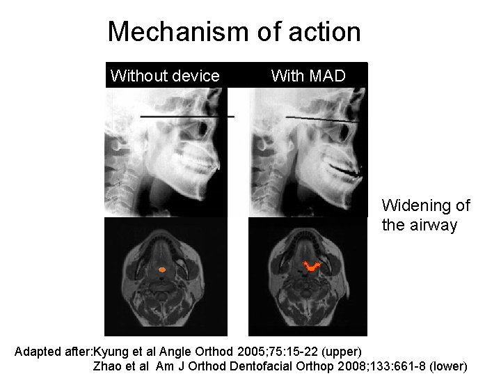
Mechanism of action Without device With MAD Widening of the airway Adapted after: Kyung et al Angle Orthod 2005; 75: 15 -22 (upper) Zhao et al Am J Orthod Dentofacial Orthop 2008; 133: 661 -8 (lower)
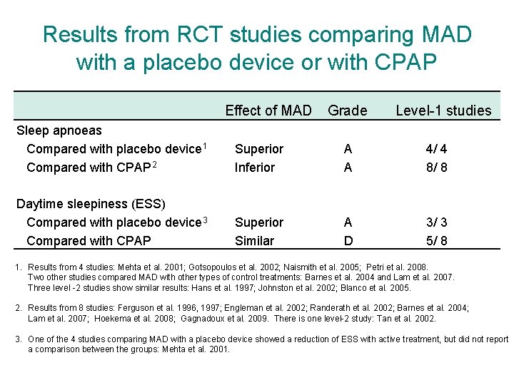
Results from RCT studies comparing MAD with a placebo device or with CPAP Effect of MAD Grade Level-1 studies Sleep apnoeas Compared with placebo device 1 Compared with CPAP 2 Superior Inferior A A 4/ 4 8/ 8 Daytime sleepiness (ESS) Compared with placebo device 3 Compared with CPAP Superior Similar A D 3/ 3 5/ 8 1. Results from 4 studies: Mehta et al. 2001; Gotsopoulos et al. 2002; Naismith et al. 2005; Petri et al. 2008. Two other studies compared MAD with other types of control treatments: Barnes et al. 2004 and Lam et al. 2007. Three level -2 studies show similar results: Hans et al. 1997; Johnston et al. 2002; Blanco et al. 2005. 2. Results from 8 studies: Ferguson et al. 1996, 1997; Engleman et al. 2002; Randerath et al. 2002; Barnes et al. 2004; Lam et al. 2007; Hoekema et al. 2008; Gagnadoux et al. 2009. There is one level-2 study: Tan et al. 2002. 3. One of the 4 studies comparing MAD with a placebo device showed a reduction of ESS with active treatment, but did not report a comparison between the groups: Mehta et al. 2001.
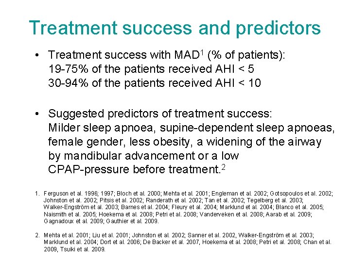
Treatment success and predictors • Treatment success with MAD 1 (% of patients): 19 -75% of the patients received AHI < 5 30 -94% of the patients received AHI < 10 • Suggested predictors of treatment success: Milder sleep apnoea, supine-dependent sleep apnoeas, female gender, less obesity, a widening of the airway by mandibular advancement or a low CPAP-pressure before treatment. 2 1. Ferguson et al. 1996; 1997; Bloch et al. 2000; Mehta et al. 2001; Engleman et al. 2002; Gotsopoulos et al. 2002; Johnston et al. 2002; Pitsis et al. 2002; Randerath et al. 2002; Tan et al. 2002; Tegelberg et al. 2003; Walker-Engström et al. 2003; Barnes et al. 2004; Fleury et al. 2004; Marklund et al. 2004; Blanco et al. 2005; Naismith et al. 2005; Hoekema et al. 2008; Petri et al. 2008; Vanderveken et al. 2008; Aarab et al. 2009; Gagnadoux et al. 2009; Gauthier et al. 2009. 2. Mehta et al. 2001; Liu et al. 2001; Johnston et al. 2002; Sanner et al. 2002, Walker-Engström et al. 2003; Marklund et al. 2004; Dort et al. 2006; De Backer et al. 2007, Hoekema et al. 2008; Petri et al. 2008; Chan et al. 2009, Tsuiki et al. 2009.
Other treatment effects from MAD • Lowered blood pressure 1 • Beneficial effects on cardiac and endothelial function 2 • Reduced lapses of attention in simulated driving 3 1. 2. 3. Barnes et al. 2004; Gotsopoulos et al. 2004; Lam et al. 2007; Gauthier et al. 2009. Coruzzi et al. 2006; Itzhaki et al. 2007; Hoekema et al. 2008; Trzepizur et al. 2009. Hoekema et al. 2008.
Device design • Clinically insignificant differences in effects on symptoms and sleep apnoeas from various designs of custom-made MAD´s. 1 • Monoblock custom-made MAD had a better effect on sleep apnoeas and snoring than a monoblock thermoplastic device. 2 • Larger advancement was related to higher efficacy, smaller vertical opening was preferred by the patients. 3 1. Results from 5 studies: Bloch et al. 2000; Rose et al. 2002; Lawton et al. 2005; Gauthier et al. 2007; Gahzal et al. 2009. 2. Vanderveken et al. 2008. 3. Pitsis et al. 2002: Tegelberg et al. 2003; Walker-Engström et al. 2003; Fleury et al. 2004; Aarab et al. 2009.
Side-effects and compliance • Mild side-effects such as: jaw discomfort, tooth tenderness, excessive salivation and temporary occlusal changes were common in the shorter term. • The side-effects occurred more frequently from MAD than from a control plate. 1 • Compliance with MAD was reported to be higher than with CPAP 2 and MAD was often preferred by the patients. 3 • After one year 76% of the patients continued treatment and 65% were still using their devices after 4 years. 1. Gotsopoulos et al. 2002. 2. Randerath et al. 2002; Barnes et al. 2004; Gagnadoux et al. 2009. 3. Ferguson, 1996, 1997; Randerath, 2002; Tan, 2002; Gagnadoux, 2009.
MAD Recommendations • MAD is indicated in patients with mild to moderate OSA (Grade of Recommendation A) and in patients who do not tolerate CPAP. • CPAP more effectively reduces sleep apnoeas than MAD, while the positive effects on symptoms and health are more similar between these treatments. • Evidence on beneficial cardiovascular effects from MAD is emerging. • Evaluated, custom-made MAD that advance the mandible at least 50% of maximum protrusion is recommended. No golden standard design of MAD is identified. • Mild side-effects are common. • Follow-up should be performed regularly long term.
Non-CPAP Therapies in Obstructive Sleep Apnoea Tongue retaining devices Marie Marklund ERS Task Force “Non-CPAP therapies in obstructive sleep apnoea“
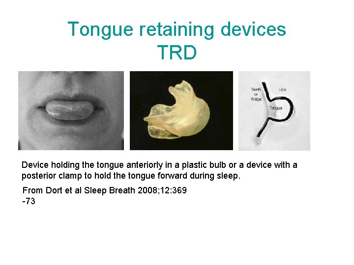
Tongue retaining devices TRD Device holding the tongue anteriorly in a plastic bulb or a device with a posterior clamp to hold the tongue forward during sleep. From Dort et al Sleep Breath 2008; 12: 369 -73
TRD Results • TRD reduced sleep apnoeas compared with a control device according to one RCT study. 1 • The effect was similar compared with the effect of MAD, according to another RCT-study. 2 91% of the patients preferred MAD. • Daytime sleepiness (ESS) decreased from TRD. 1, 2 The effect was similar as control treatment. 1 1. 2. Dort et al. 2008. Deane et al. 2009.
TRD Recommendations • TRD is indicated in the treatment of patients with mild to moderate OSA (Grade of Recommendation C), when other treatments have failed or are not possible. • There are few RCT-studies of effects from TRD. Two studies show reductions in sleep apnoeas from TRD, although the symptomatic effects are mainly unknown and compliance might be a limitation. • The treatment effect should be monitored and the patients should be strictly followed-up.
Overview PART III: nasal and velopharyngeal surgery • Nasal surgery. Arschang Valipour • Tonsils: – Tonsillectomy and tonsillotomy. Arschang Valipour – Radiofrequency surgery tonsils. Evert Hamans • Palate: – – Uvulopalatopharyngoplasty and LAUP. Jean-Louis Pepin Radiofrequency surgery. Boris A. Stuck Uvulopalatal flap. Thomas Verse Pillar method. Maurer Joachim
Non-CPAP Therapies in Obstructive Sleep Apnoea Nasal surgery Arschang Valipour ERS Task Force “Non-CPAP therapies in obstructive sleep apnoea“
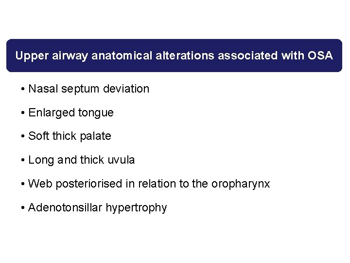
Upper airway anatomical alterations associated with OSA • Nasal septum deviation • Enlarged tongue • Soft thick palate • Long and thick uvula • Web posteriorised in relation to the oropharynx • Adenotonsillar hypertrophy
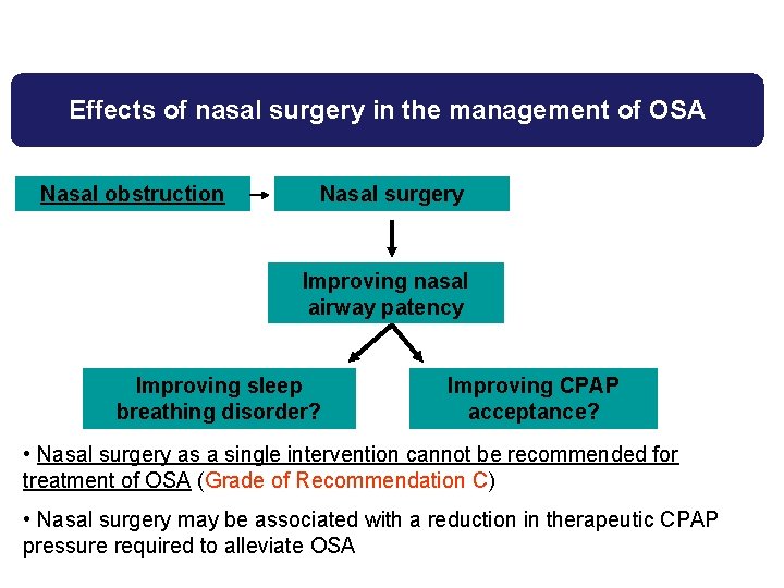
Effects of nasal surgery in the management of OSA Nasal obstruction Nasal surgery Improving nasal airway patency Improving sleep breathing disorder? Improving CPAP acceptance? • Nasal surgery as a single intervention cannot be recommended for treatment of OSA (Grade of Recommendation C) • Nasal surgery may be associated with a reduction in therapeutic CPAP pressure required to alleviate OSA
Non-CPAP Therapies in Obstructive Sleep Apnoea Tonsillectomy and tonsillotomy Arschang Valipour ERS Task Force “Non-CPAP therapies in obstructive sleep apnoea“
Adenotonsillar hypertrophy and OSA • Adenotonsillar hypertrophy is a rare cause of OSA in adults • Adenotonsillar hypertophy is the most common aetiology of OSA in children • Degree of adenotonsillar hypertrophy in children correlates with disease severity • Pediatric OSA associated with adenotonsillar hypertrophy has detrimental effects on growth, metabolism, blood pressure, and cognitive function
Effects of adenotonsillectomy for treatment of OSA in children • Adenotonsillectomy can be recommended in the presence of adenotonsillar hypertrophy associated with pediatric OSA (Grade of Recommendation C) • Reported clinical benefits: • Sleep respiratory disturbances and architecture • Qo. L scores • Growth, metabolism • Cognitive function and behaviour
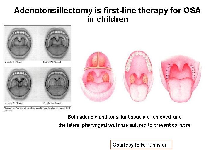
Adenotonsillectomy is first-line therapy for OSA in children Both adenoid and tonsillar tissue are removed, and the lateral pharyngeal walls are sutured to prevent collapse Courtesy to R Tamisier
Effects of adenotonsillectomy for treatment of OSA in adults • Tonsillectomy can be recommended in the presence of tonsillar hypertrophy associated with OSA in adults (Grade of Recommendation C) • Reported clinical benefits: • Sleep respiratory disturbances and architecture • Residual sleep disordered breathing is common
Non-CPAP Therapies in Obstructive Sleep Apnoea Radiofrequency surgery tonsils Evert Hamans ERS Task Force “Non-CPAP therapies in obstructive sleep apnoea“
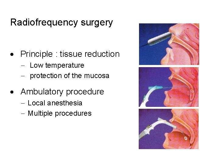
Radiofrequency surgery · Principle : tissue reduction - Low temperature - protection of the mucosa · Ambulatory procedure - Local anesthesia - Multiple procedures
Radiofrequency surgery tonsils Rationale • Radiofrequency (RF) surgery of the tonsils is developed to reduce post operative pain and bleeding risk • The key innovation of RF surgery is the concept of subtotal intracapsular tonsil reduction that avoids injury to the pharyngeal constrictor muscles • Radiofrequency can be used to create small channels in the tonsil with dissipation of ionising energy to surrounding tissue, with subsequent tissue death days or weeks later that leads to shrinkage and volume reduction (1) 1. Nelson L. Radiofrequency treatment for obstructive tonsilar hypertrophy. Arch Otolaryngol Head Neck Surg 2000; 126: 736 -740.
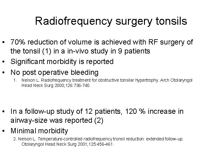
Radiofrequency surgery tonsils • 70% reduction of volume is achieved with RF surgery of the tonsil (1) in a in-vivo study in 9 patients • Significant morbidity is reported • No post operative bleeding 1. Nelson L. Radiofrequency treatment for obstructive tonsilar hypertrophy. Arch Otolaryngol Head Neck Surg 2000; 126: 736 -740. • In a follow-up study of 12 patients, 120 % increase in airway-size was reported (2) • Minimal morbidity 2. Nelson L. Temperature-controlled radiofrequency tronsil reduction: extended follow-up. Otolaryngol Head Neck Surg 2001; 125: 456 -461.
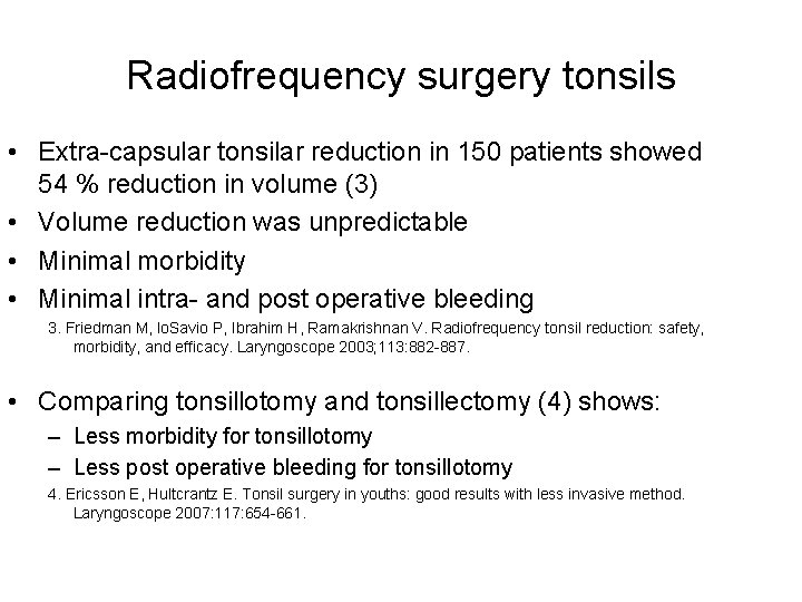
Radiofrequency surgery tonsils • Extra-capsular tonsilar reduction in 150 patients showed 54 % reduction in volume (3) • Volume reduction was unpredictable • Minimal morbidity • Minimal intra- and post operative bleeding 3. Friedman M, lo. Savio P, Ibrahim H, Ramakrishnan V. Radiofrequency tonsil reduction: safety, morbidity, and efficacy. Laryngoscope 2003; 113: 882 -887. • Comparing tonsillotomy and tonsillectomy (4) shows: – Less morbidity for tonsillotomy – Less post operative bleeding for tonsillotomy 4. Ericsson E, Hultcrantz E. Tonsil surgery in youths: good results with less invasive method. Laryngoscope 2007: 117: 654 -661.
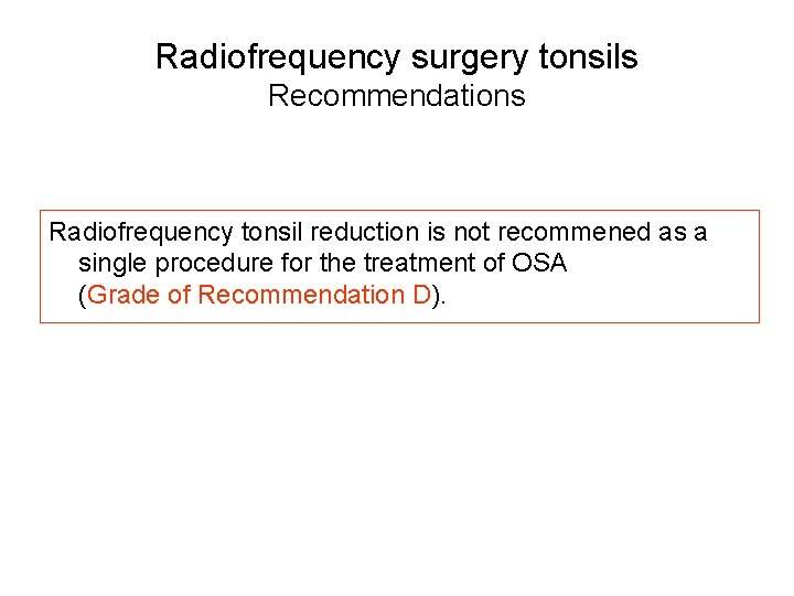
Radiofrequency surgery tonsils Recommendations Radiofrequency tonsil reduction is not recommened as a single procedure for the treatment of OSA (Grade of Recommendation D).
Non-CPAP Therapies in Obstructive Sleep Apnoea Uvulopalatopharyngoplasty (UPPP) Jean-Louis Pépin ERS Task Force “Non-CPAP therapies in obstructive sleep apnoea“
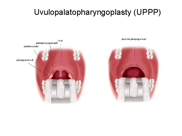
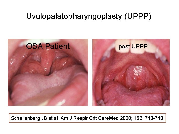
Uvulopalatopharyngoplasty (UPPP) Rationale • • • Uvulopalatopharyngoplasty (UPPP) and laser-assisted uvulopalatoplasty (LAUP) aim to diminish anatomical upper airway obstruction at the oropharyngeal level by reducing soft palate redundancy. UPPP enlarges the retro- palatal airway by trimming and reorienting the posterior and anterior lateral pharyngeal pillars, and by excising the uvula and the posterior soft palate. LAUP is an office-based surgical procedure that progressively shortens and tightens the uvula and palate through a series of carbon dioxide laser incisions and vaporizations. In the large majority of apnoeic patients, upper airway collapses occur at multiple levels i. e. both at the retropalatal level and behind the tongue base. Therefore, a surgical success with UPPP or LAUP can only be anticipated when pharyngeal collapse is limited to the retropalatal area which is rarely the case in obese or severe sleep apnoea. For these reasons surgical procedures dedicated to soft palate have been essentially studied in selected mild to moderate OSA populations with predominant oropharyngeal narrowing.
Uvulopalatopharyngoplasty (UPPP)
Uvulopalatopharyngoplasty (UPPP) OSA Patient post UPPP Schellenberg JB et al Am J Respir Crit Care. Med 2000; 162: 740 -748
Uvulopalatopharyngoplasty Recommendations • UPPP is a single-level surgical procedure working only in selected patients with obstruction limited to the oropharyngeal area. • Therefore, it is generally not recommended and may only be performed in selected cases. • When proposing UPPP, potential benefits should be weighed against the risk of frequent long-term side effects among them velopharyngeal insufficiency, dry throat, and abnormal swallowing are the most common. • Analysed studies were prospective but with selection biases, and therefore do not provide high-level evidence. Grade of Recommendation C
Non-CPAP Therapies in Obstructive Sleep Apnoea Laser assisted uvuloplasty (LAUP) Jean-Louis Pépin ERS Task Force “Non-CPAP therapies in obstructive sleep apnoea“
Laser assisted uvuloplasty (LAUP) Results • Two randomised controlled trials are available: – Ferguson et al Am J Respir Crit Care Med 2003; 167(1): 15 -9 • compared LAUP to conservative treatment • 46 mild OSA patients with a mean follow-up of 7 months. • There was a statistically significant but not clinically relevant 21% reduction in AHI after LAUP (from 19 to 15/h) • No improvement was found for subjective sleepiness and quality of life. Surgery reduced snoring intensity and frequency. – Larrosa et al Eur Respir J 2004; 24(1): 66 -70 • failed to find a difference between LAUP and a Sham surgery in sleepiness, quality of life or respiratory disturbances. • After LAUP surgery, airway is further compromised by edema in the early postoperative period with a potential risk of OSA exacerbation
Laser assisted uvuloplasty (LAUP) Recommendations • In mild OSA, LAUP has not demonstrated any significant effect neither on OSA severity nor in symptoms or quality of life domains and is therefore not recommended. Grade of Recommendation B
Non-CPAP Therapies in Obstructive Sleep Apnoea Palate: Radiofrequency surgery Boris A. Stuck ERS Task Force “Non-CPAP therapies in obstructive sleep apnoea“
Palate: Radiofrequency surgery Rationale • The term “radiofrequency surgery” in the context of palatal interventions is usually focusing on the interstitial electrosurgical treatment with stiffening of the tissue. • Furthermore, cutting devices based on radiofrequency energy have also been developed to excise palatal tissue (radiofrequency assisted uvulopalatoplasty: RAUP). • Finally, various combinations have been proposed, for example the combination of interstitial radiofrequency surgery and RAUP (RF-UPP). Tissue excision is usually performed as a single step procedure, whereas the interstitial application routinely requires repeated treatment sessions.
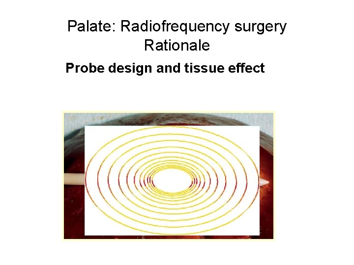
Palate: Radiofrequency surgery Rationale Probe design and tissue effect
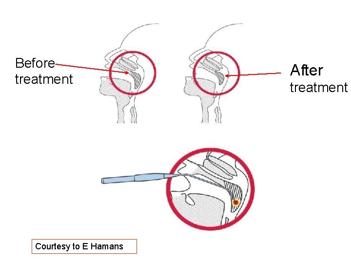
Before treatment Courtesy to E Hamans After treatment
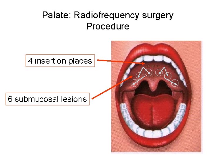
Palate: Radiofrequency surgery Procedure 4 insertion places 6 submucosal lesions
Palate: Radiofrequency surgery Results • Improvements in respiratory parameters have been described in groups of mild to moderate and more severely affected patients under interstitial radiofrequency at the soft palate. • Moreover, radiofrequency assisted uvulopalatoplasty has been shown to be superior to interstitial radiofrequency surgery, but was associated with higher postoperative morbidity. • However, there are inconsistent findings in daytime sleepiness or other sleep parameters. • A most recent, placebo-controlled study has shown that a single step RF-intervention at the soft palate is not effective to treat OSA. Back LJ et al Laryngoscope 2009; 119(8): 1621 -7
Palate: Radiofrequency surgery Recommendations • No controlled trials are available in sleep apnea. • All case series demonstrate a significant reduction in AHI after radiofrequency surgery. • Radiofrequency surgery is not generally recommended and may only be considered in mild disease refusing or not requiring CPAP as long as the individual anatomy appears suitable. Grade of Recommendation C
Non-CPAP Therapies in Obstructive Sleep Apnoea Uvulopalatal flap Thomas Verse ERS Task Force “Non-CPAP therapies in obstructive sleep apnoea“ Verse T. Dept. ORL HNS, Asklepios Klinik Harburg, Hamburg, Germany
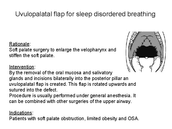
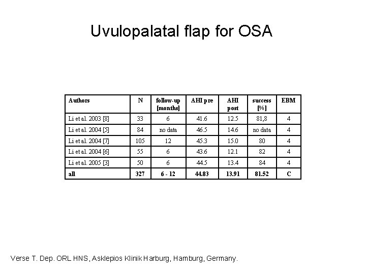
Uvulopalatal flap for sleep disordered breathing Rationale: Soft palate surgery to enlarge the velopharynx and stiffen the soft palate. Intervention: By the removal of the oral mucosa and salivatory glands and incisions bilaterally into the posterior pillar an uvulopalatal flap is created. This flap is rotated upwards and sutured into the defect. Procedure is usually performed under general anesthesia. It can be combined with other surgeries of the upper airway. Indications: Patients with soft palate obstruction, limited obesity and OSA.
Uvulopalatal flap for OSA Authors N follow-up [months] AHI pre AHI post success [%] EBM Li et al. 2003 [8] 33 6 41. 6 12. 5 81, 8 4 Li et al. 2004 [5] 84 no data 46. 5 14. 6 no data 4 Li et al. 2004 [7] 105 12 45. 3 15. 0 80 4 Li et al. 2004 [6] 55 6 43. 6 12. 1 82 4 Li et al. 2005 [3] 50 6 44. 5 13. 4 84 4 all 327 6 - 12 44. 83 13. 91 81. 52 C Verse T. Dep. ORL HNS, Asklepios Klinik Harburg, Hamburg, Germany.
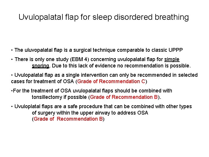
Uvulopalatal flap for sleep disordered breathing • The uluvopalatal flap is a surgical technique comparable to classic UPPP • There is only one study (EBM 4) concerning uvulopalatal flap for simple snoring. Due to this lack of evidence no recommendation is possible. • Uvulopalatal flap as a single intervention can only be recommended in selected cases for treatment of OSA (Grade of Recommendation C) • For the treatment of OSA uvulopalatal flaps should be combined with tonsillectomy if possible (Grade of Recommendation B). • Uvuloplatal flaps are a safe procedure that can be combined with other types of surgery within the upper airway to address OSA (Grade of Recommendation B)
Non-CPAP Therapies in Obstructive Sleep Apnoea Pillar Method Joachim Maurer ERS Task Force “Non-CPAP therapies in obstructive sleep apnoea“
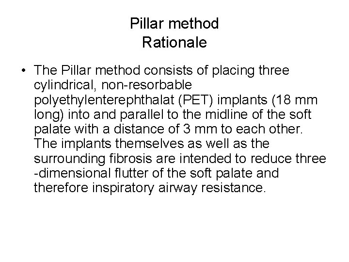
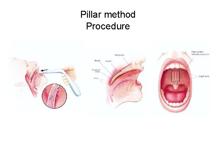
Pillar method Rationale • The Pillar method consists of placing three cylindrical, non-resorbable polyethylenterephthalat (PET) implants (18 mm long) into and parallel to the midline of the soft palate with a distance of 3 mm to each other. The implants themselves as well as the surrounding fibrosis are intended to reduce three -dimensional flutter of the soft palate and therefore inspiratory airway resistance.
Pillar method Procedure
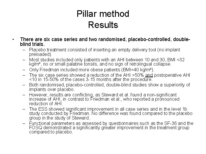
Pillar method Results • There are six case series and two randomised, placebo-controlled, doubleblind trials. – Placebo treatment consisted of inserting an empty delivery tool (no implant preloaded). – Most studies included only patients with an AHI between 10 and 30, BMI <32 kg/m², no or small palatine tonsils, and no sign of retrolingual collapse. – Only Friedman included more obese patients (BMI<40 kg/m²). – The six case series showed a reduction of the AHI >50% and postoperative AHI <10 in 15 -50% of the cases 3 -15 months after the procedure. – Both randomised, placebo-controlled, double-blind studies show a superiority of implants over placebo. – However, results are conflicting, as Steward et al. found a non-significant increase of AHI, in contrast to Friedman et al. , who reported a pronounced reduction of AHI. – The ESS showed significant improvement in all case series and in the level 1 b study conducted by Friedman. No difference was found compared to the placebo group in the study of Steward. – Functional parameters as assessed by questionnaires such as the SF-36 and the FOSQ demonstrated a significantly greater improvement in the treatment group compared to placebo.
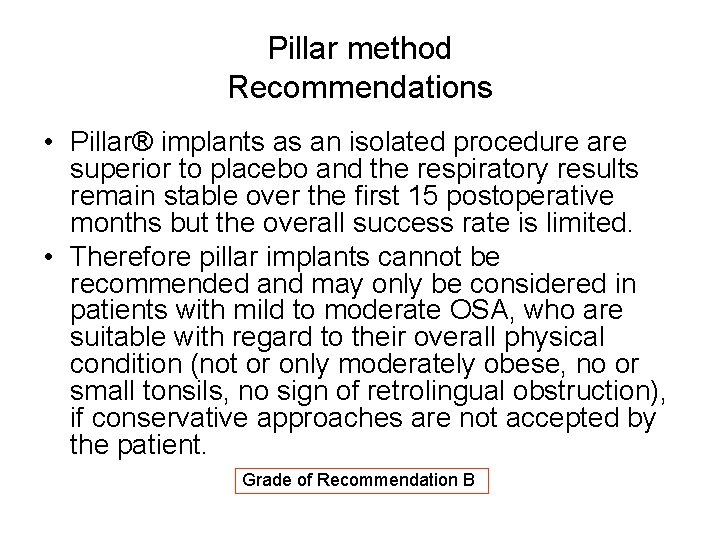
Pillar method Recommendations • Pillar® implants as an isolated procedure are superior to placebo and the respiratory results remain stable over the first 15 postoperative months but the overall success rate is limited. • Therefore pillar implants cannot be recommended and may only be considered in patients with mild to moderate OSA, who are suitable with regard to their overall physical condition (not or only moderately obese, no or small tonsils, no sign of retrolingual obstruction), if conservative approaches are not accepted by the patient. Grade of Recommendation B
Overview Part IV: hypopharyngeal and maxillofacial surgery – Tongue base and hypopharynx • • • Radiofrequency ablation therapy of the tongue base. Boris A. Stuck Hyoid suspension. Thomas Verse Other treatments. An Boudewyns – – Partial resection tongue base. An Boudewyns Maxillo-facial surgery • • Genioglossal advancement. Laurent Lacassagne, Floriant Jalbert, Jean Roch Paoli, Georges Bettega Maxillo-mandibular advancement. Jean Roch Paoli, Laurent Lacassagne Distraction osteogenesis. Floriant Jalbert, Laurent Lacassagne, Jean Roch Paoli, Georges Bettega Multilevel surgery. Laurent Lacassagne, Floriant Jalbert, Jean Roch Paoli
Non-CPAP Therapies in Obstructive Sleep Apnoea Radiofrequency tongue base Boris A. Stuck ERS Task Force “Non-CPAP therapies in obstructive sleep apnoea“
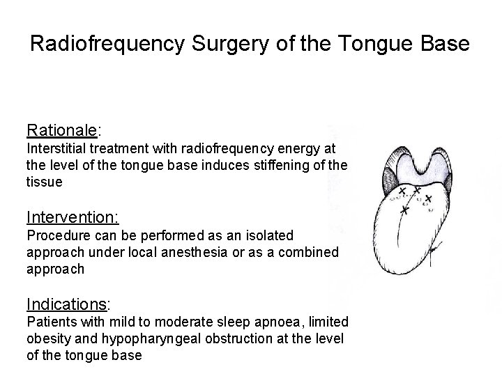
Radiofrequency Surgery of the Tongue Base Rationale: Indikationen: Interstitial treatment with radiofrequency energy at • the level of the tongue base induces stiffening of the Zungengrundhyperplasie tissue • Intervention: OSA Procedure can be performed as an isolated Chirurgie: approach under local anesthesia or as a combined • approach Lokalanästhesie • Indications: 1 -2 Sitzungen Patients with mild to moderate sleep apnoea, limited obesity and hypopharyngeal obstruction at the level of the tongue base
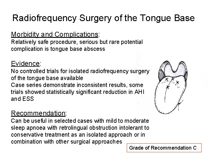
Radiofrequency Surgery of the Tongue Base Morbidity and Complications: Relatively safe procedure, serious but rare potential complication is tongue base abscess Indikationen: Evidence: • No controlled trials for isolated radiofrequency surgery Zungengrundhyperplasie of the tongue base available • Case series demonstrate inconsistent results, some OSA trials showed statistically significant reduction in AHI and ESS Chirurgie: • Recommendation: Lokalanästhesie Can be useful in selected cases with mild to moderate • sleep apnoea with retrolingual obstruction intolerant to 1 -2 Sitzungen conservative treatment as an isolated approach or in combination with other surgical approaches Grade of Recommendation C
Non-CPAP Therapies in Obstructive Sleep Apnoea Hyoid suspension Thomas Verse ERS Task Force “Non-CPAP therapies in obstructive sleep apnoea“ Verse T. Dept. ORL HNS, Asklepios Klinik Harburg, Hamburg, Germany
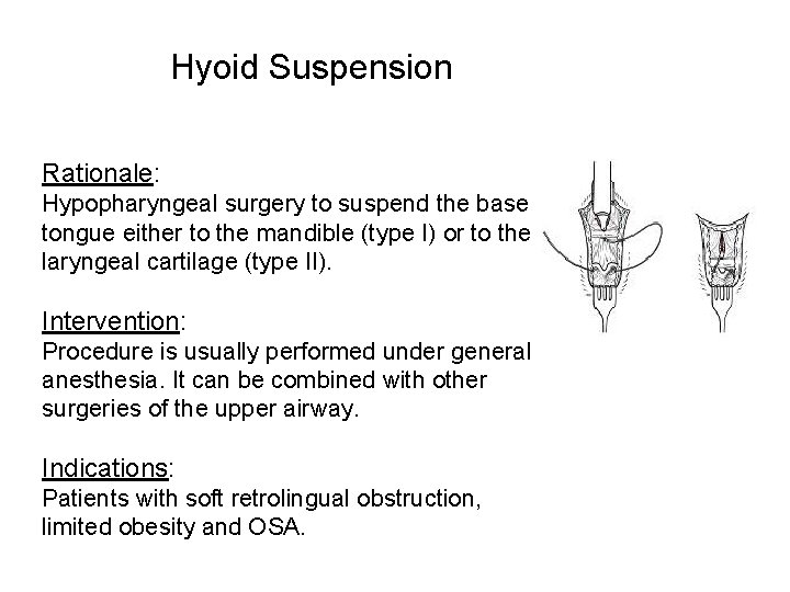
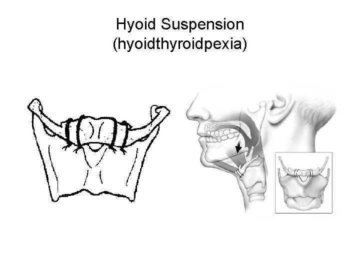
Hyoid Suspension Rationale: Hypopharyngeal surgery to suspend the base of tongue either to the mandible (type I) or to the laryngeal cartilage (type II). Intervention: Procedure is usually performed under general anesthesia. It can be combined with other surgeries of the upper airway. Indications: Patients with soft retrolingual obstruction, limited obesity and OSA.
Hyoid Suspension (hyoidthyroidpexia)
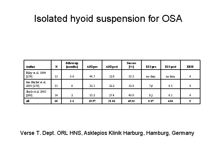
Isolated hyoid suspension for OSA Author N follow-up [months] AHI pre AHI post Success [%] ESS pre ESS post EBM Riley et al. 1994 [279] 15 3 -6 44. 7 12. 8 53. 3 no data 4 den Herder et al. 2005 [278] 31 6 32. 1 22. 2 52. 0 7, 6 4. 3 4 Stuck et al. 2005 [280] 14 2 35. 2 27. 4 40. 0 9, 1 6. 1 4 all 60 2 -6 35. 97 21. 06 49. 53 8. 07 4. 86 C Verse T. Dept. ORL HNS, Asklepios Klinik Harburg, Hamburg, Germany
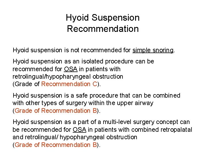
Hyoid Suspension Recommendation Hyoid suspension is not recommended for simple snoring. Hyoid suspension as an isolated procedure can be recommended for OSA in patients with retrolingual/hypopharyngeal obstruction (Grade of Recommendation C). Hyoid suspension is a safe procedure that can be combined with other types of surgery within the upper airway (Grade of Recommendation B). Hyoid suspension as a part of a multi-level surgery concept can be recommended for OSA in patients with combined retropalatal and retrolingual/ hypopharyngeal obstruction (Grade of Recommendation B).
Non-CPAP Therapies in Obstructive Sleep Apnoea Tongue base surgery An Boudewyns ERS Task Force “Non-CPAP therapies in obstructive sleep apnoea“
Tongue base soft tissue surgery Rationale: • Reduction of the tongue volume by resection and removal (partial glossectomy -tongue base reduction-lingualplasty) • Tongue suspension (Repose R) Intervention: To be performed under general anesthesia Can be combined with other surgical interventions of the upper airway Populations studied: Severe OSA with concomittant obesity Previous upper airway surgery CPAP intolerance
Tongue base soft tissue surgery Results: Variable success rates (> 50% reduction in AHI and postop AHI <20) ranging from 20 to 100% Improvement in snoring and daytime sleepiness Evidence levels: 90% level 4 studies , 2 level 2 b studies Recommendation: Tongue base reduction and suspension techniques have little role as a single treatment option for patients with moderate to severe OSAS. Grade of Recommendation C. Potential value: limited morbidity, lack of cosmetic changes, possibility to combine with other surgical techniques. No data for patients with milder disease.
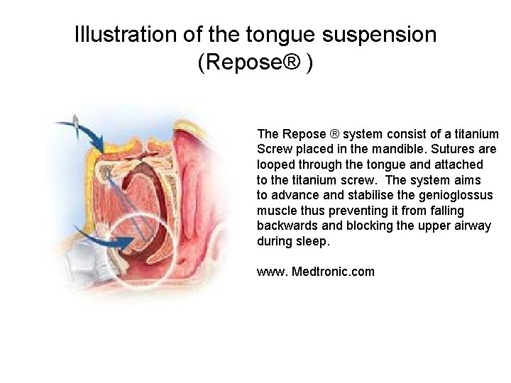
Illustration of the tongue suspension (Repose® ) The Repose ® system consist of a titanium Screw placed in the mandible. Sutures are looped through the tongue and attached to the titanium screw. The system aims to advance and stabilise the genioglossus muscle thus preventing it from falling backwards and blocking the upper airway during sleep. www. Medtronic. com
Non-CPAP Therapies in Obstructive Sleep Apnoea Genioglossal advancement Laurent Lacassagne, Floriant Jalbert, Jean Roch Paoli, Georges Bettega ERS Task Force “Non-CPAP therapies in obstructive sleep apnoea“
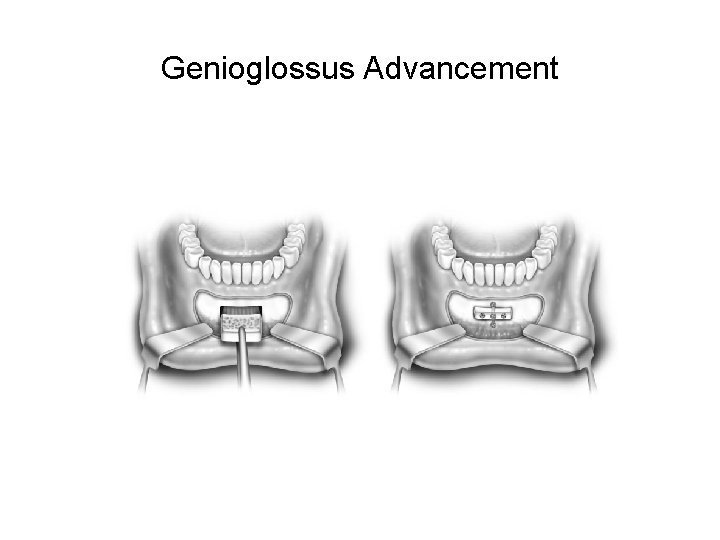
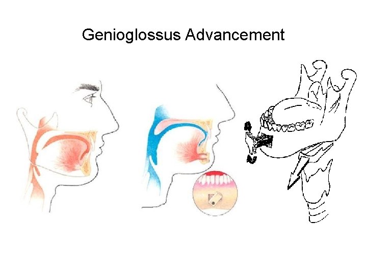
Genioglossus Advancement Rationale: Genioglossus advancement (GA) consists in : an advancement of the genial tubercle and genioglossus muscle. sliding genioplasty with a complete manipulation of the chin position that is moved forward geniotomy with a rectangular bicortical osteotomy centered over the genial tubercle Overview of evidence : No study investigates the effect of isolated genioglossus advancement In multilevel surgery, when genioglossus advancement is the only hypopharyngeal procedure associated with velar surgery, GA demonstrated an improvement of postoperative AHI with a success rate of 60% in 5 series including 117 patients.
Genioglossus Advancement
Genioglossus Advancement
Genioglossus Advancement Recommendation: GA is efficient used in multilevel surgery for treatment of hypopharyngeal airway impairment, but should not be recommended as a single procedure for the surgical treatment of OSA. Grade of Recommendation C
Non-CPAP Therapies in Obstructive Sleep Apnoea Distraction Osteogenesis Floriant Jalbert, Laurent Lacassagne, Jean Roch Paoli, Georges Bettega ERS Task Force “Non-CPAP therapies in obstructive sleep apnoea“
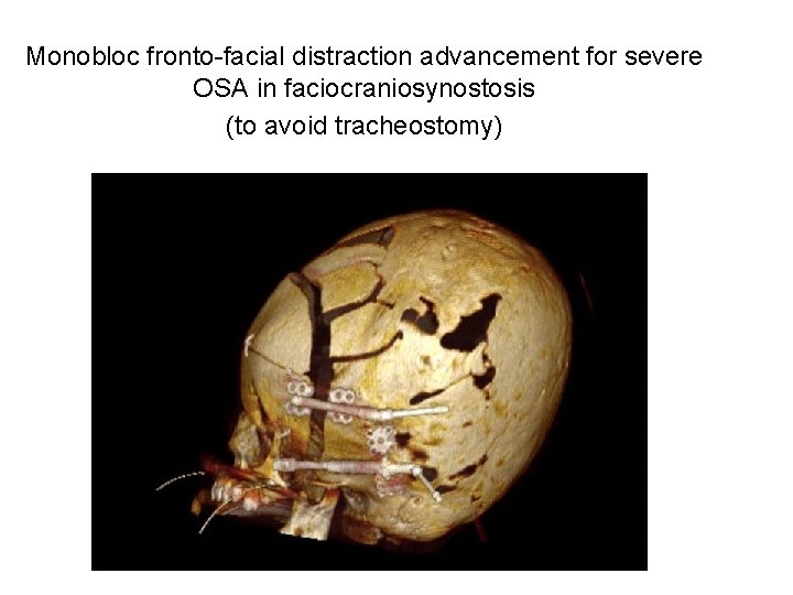
Distraction osteogenesis Rationale: Distraction osteogenesis (DOG) provides progressive bone lengthening associated with stretching of the surrounding soft tissues. Overview of evidence: Most publications deal with the use the DOG in mandibular lengthening in pediatric micrognathia (such Pierre Robin sequence) in order to avoid tracheostomy or to decanulate children. But DOG remains controversial in mild obstruction. In severe midfacial hypoplasia (such faciocraniosynostosis) DOG is also used for maxillo-zygomatic advancement to improve airway impairment Recommendation: DOG is efficiently applied and can be recommended in severe congenital micrognathia (mandibular lengthening: Grade of Recommendation B) or midface hypoplasia (midface advancement: Grade of Recommendation C)
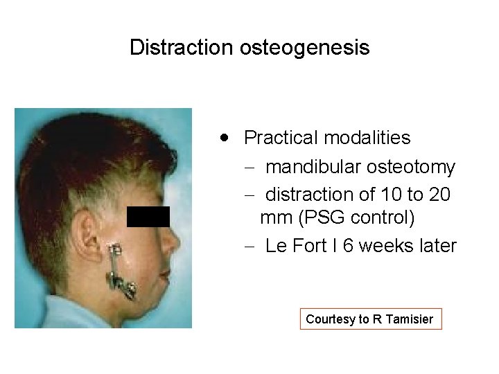
Distraction osteogenesis · Practical modalities - mandibular osteotomy - distraction of 10 to 20 mm (PSG control) - Le Fort I 6 weeks later Courtesy to R Tamisier
Monobloc fronto-facial distraction advancement for severe OSA in faciocraniosynostosis (to avoid tracheostomy)
Non-CPAP Therapies in Obstructive Sleep Apnoea Maxillo-mandibular advancement Jean Roch Paoli, Laurent Lacassagne ERS Task Force “Non-CPAP therapies in obstructive sleep apnoea“
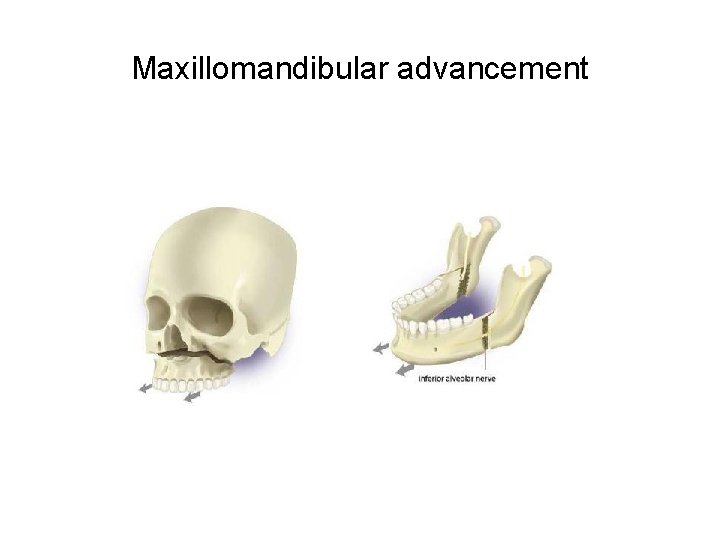
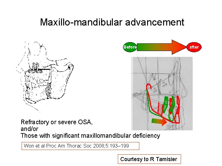
Maxillomandibular advancement Rationale: Advancement of the maxilla and the mandible (10 to 15 mm) can provide improvement of airway impairment Overview of evidence : Success rate ranges from 67 to 100% with postoperative AHI <10 Transient anaesthesia or sometimes residual neurosensitive deficit (hypoesthesia of the lower lip) was the most common complication. Changes in patient profile remain acceptable. MMA has been shown to be effective over the long term.
Maxillomandibular advancement
Maxillo-mandibular advancement Before Refractory or severe OSA, and/or Those with significant maxillomandibular deficiency Won et al Proc Am Thorac Soc 2008; 5: 193– 199 Courtesy to R Tamisier after
Maxillomandibular advancement Recommendation: MMA seems to be as efficient as CPAP in patients with OSA who refuse conservative treatment, particularly in a young OSA population without excessive BMI or other comorbidities. Grade of Recommendation B
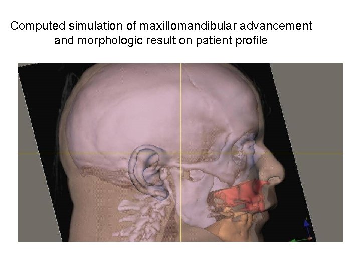
Computed simulation of maxillomandibular advancement and morphologic result on patient profile
Non-CPAP Therapies in Obstructive Sleep Apnoea Multilevel surgery Laurent Lacassagne, Floriant Jalbert, Jean Roch Paoli ERS Task Force “Non-CPAP therapies in obstructive sleep apnoea“
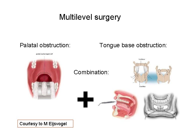
Multilevel surgery Palatal obstruction: Tongue base obstruction: Combination: + Courtesy to M Eijsvogel
Multilevel surgery Rationale : The goal of multilevel surgery (MLS) is to improve nasal airway blockage and both to enlarge the retropalatal airway and the retrolingual airway (Septoplasty, alar valve or alar rim reconstruction and turbinectomy, UPPP, often combined with adenotonsillectomy, mandibular osteotomy with genioglossus advancement (GA), hyoid myotomy suspension (HS), laser midline glossectomy with lingualplasty, and radiofrequency tongue base reduction (RTBR)…) Overview of evidence : MLS may be considered in younger OSA patients in whome CPAP is unsuccessful or has been rejected for a long term period The success rate of MLS on AHI is about 53. 6% at 6 months Recommendation: MLS cannot be recommended as a substitute for CPAP but as a salvage procedure for OSA patients in whom CPAP and other conservative therapies failed. Surgical success of MLS for OSA is often unpredictable and less effective than CPAP. Grade of Recommendation C
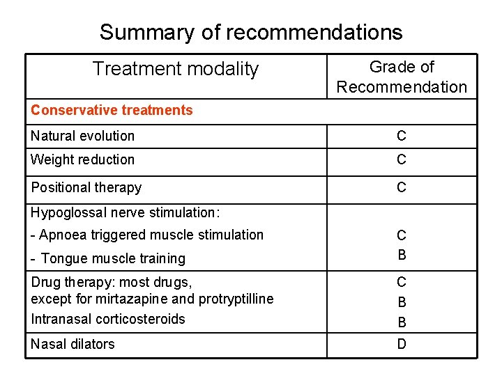
Summary of recommendations Treatment modality Grade of Recommendation Conservative treatments Natural evolution C Weight reduction C Positional therapy C Hypoglossal nerve stimulation: - Apnoea triggered muscle stimulation - Tongue muscle training C B Drug therapy: most drugs, except for mirtazapine and protryptilline Intranasal corticosteroids C B B Nasal dilators D
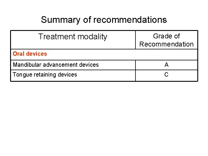
Summary of recommendations Treatment modality Grade of Recommendation Oral devices Mandibular advancement devices A Tongue retaining devices C
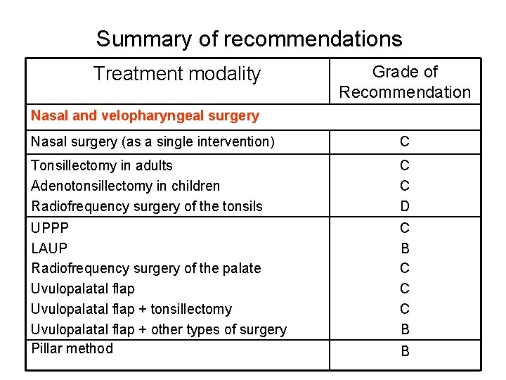
Summary of recommendations Treatment modality Grade of Recommendation Nasal and velopharyngeal surgery Nasal surgery (as a single intervention) C Tonsillectomy in adults Adenotonsillectomy in children Radiofrequency surgery of the tonsils C C D UPPP LAUP Radiofrequency surgery of the palate Uvulopalatal flap + tonsillectomy Uvulopalatal flap + other types of surgery Pillar method C B C C C B B
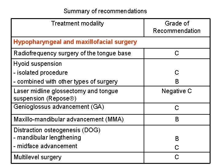
Summary of recommendations Treatment modality Grade of Recommendation Hypopharyngeal and maxillofacial surgery Radiofrequency surgery of the tongue base C Hyoid suspension - isolated procedure - combined with other types of surgery C B Laser midline glossectomy and tongue suspension (Repose ) Genioglossus advancement (GA) Negative C C Maxillo-mandibular advancement (MMA) B Distraction osteogenesis (DOG) - mandibular lengthening - midface advancement B C Multilevel surgery C
MC QUESTIONS ERS Task Force “Non-CPAP therapies in obstructive sleep apnoea“
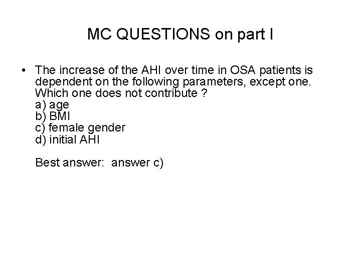
MC QUESTIONS on part I • The increase of the AHI over time in OSA patients is dependent on the following parameters, except one. Which one does not contribute ? a) age b) BMI c) female gender d) initial AHI Best answer: answer c)
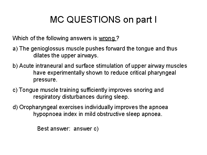
MC QUESTIONS on part I Which of the following answers is wrong ? a) The genioglossus muscle pushes forward the tongue and thus dilates the upper airways. b) Acute intraneural and surface stimulation of upper airway muscles have experimentally shown to reduce critical pharyngeal pressure. c) Tongue muscle training sufficiently improves snoring and respiratory disturbances during sleep. d) Oropharyngeal exercises individually improves the apnoea hypopnoea index in mild obstructive sleep apnoea. Best answer: answer c)
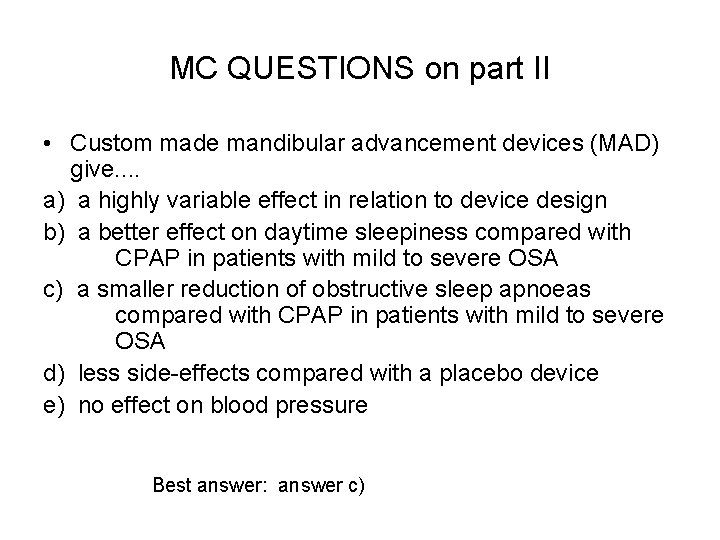
MC QUESTIONS on part II • Custom made mandibular advancement devices (MAD) give. . a) a highly variable effect in relation to device design b) a better effect on daytime sleepiness compared with CPAP in patients with mild to severe OSA c) a smaller reduction of obstructive sleep apnoeas compared with CPAP in patients with mild to severe OSA d) less side-effects compared with a placebo device e) no effect on blood pressure Best answer: answer c)
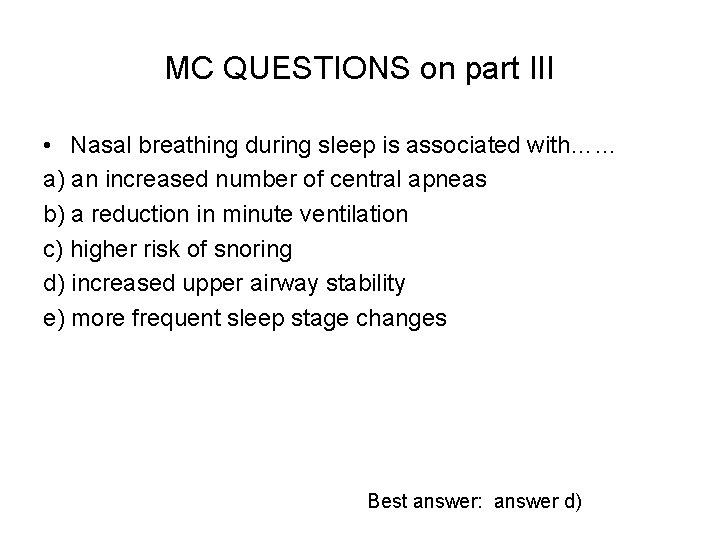
MC QUESTIONS on part III • Nasal breathing during sleep is associated with…… a) an increased number of central apneas b) a reduction in minute ventilation c) higher risk of snoring d) increased upper airway stability e) more frequent sleep stage changes Best answer: answer d)
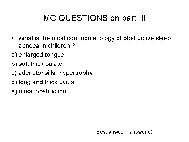
MC QUESTIONS on part III • What is the most common etiology of obstructive sleep apnoea in children ? a) enlarged tongue b) soft thick palate c) adenotonsillar hypertrophy d) long and thick uvula e) nasal obstruction Best answer: answer c)
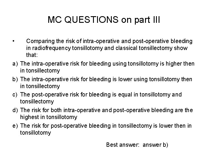
MC QUESTIONS on part III • a) b) c) d) e) Comparing the risk of intra-operative and post-operative bleeding in radiofrequency tonsillotomy and classical tonsillectomy show that: The intra-operative risk for bleeding using tonsillotomy is higher then in tonsillectomy The intra-operative risk for bleeding is lower using tonsillotomy then in tonsillectomy The post-operative risk for bleeding is equal in tonsillotomy and tonsillectomy The risk for both intra-operative and post-operative bleeding are the highest in tonsillotomy The risk for post-operative bleeding in tonsillectomy is lower then in tonsillotomy Best answer: answer b)
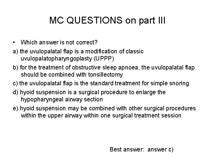
MC QUESTIONS on part III • Which answer is not correct? a) the uvulopalatal flap is a modification of classic uvulopalatopharyngoplasty (UPPP) b) for the treatment of obstructive sleep apnoea, the uvulopalatal flap should be combined with tonsillectomy c) the uvulopalatal flap is the standard treatment for simple snoring d) hyoid suspension is a surgical procedure to enlarge the hypopharyngeal airway section e) hyoid suspension may be combined with other surgical procedures within the upper airway within one surgical treatment session Best answer: answer c)
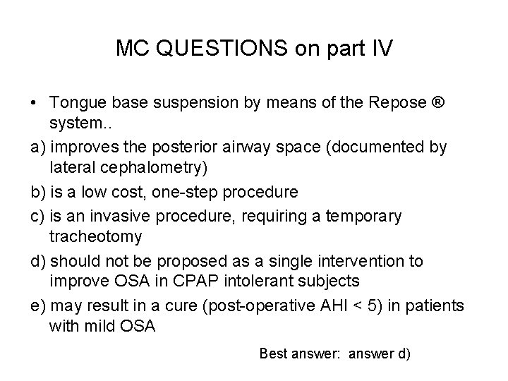
MC QUESTIONS on part IV • Tongue base suspension by means of the Repose ® system. . a) improves the posterior airway space (documented by lateral cephalometry) b) is a low cost, one-step procedure c) is an invasive procedure, requiring a temporary tracheotomy d) should not be proposed as a single intervention to improve OSA in CPAP intolerant subjects e) may result in a cure (post-operative AHI < 5) in patients with mild OSA Best answer: answer d)
Affiliations • • • • • Winfried Randerath, Bethanien Hospital, Institute of Pneumology, University Witten/Herdecke, Solingen, Germany Johan Verbraecken, Antwerp University Hospital, Dept of Pulmonary Medicine, and Multidisciplinary Sleep Disorders Centre, Edegem, Belgium Stefan Andreas, Lungenfachklinik Immenhausen, Germany Georges Bettega, Service de Chirurgie Plastique et Maxillo-Faciale, CHU Grenoble Cedex, France An Boudewyns, Antwerp University Hospital , Dept of Otorhinolaryngology Head and Neck Surgery, and Multidisciplinary Sleep Disorders Centre, Edegem, Belgium Florian Jalbert, Department of Maxillofacial Surgery, CHU Toulouse Purpan, France Jean Roch Paoli, CH Bastia, Plastic and maxillofacial department, Bastia, France Bernd Sanner, Bethesda Krankenhaus, Wuppertal, Germany Ian E Smith, Respiratory Support and Sleep Centre, Papworth Hospital, University of Cambridge, United Kingdom Boris A Stuck, Sleep Disorders Center, Department of Otorhinolaryngology, Head and Neck Surgery, University Hospital Mannheim, Germany Laurent Lacassagne, Service de Pneumologie, Clinique de l’Union, Saint Jean, France Marie Marklund, Department of Othodontics, Umea University, Umea, Sweden Jean-Louis Pepin, EFCR Sleep and Respiration Unit CHU Grenoble Cedex, France Arschang Valipour, Otto-Wagner-Hospital, Dept Respir and Crit Care Med, Ludwig-Boltzmann-Institute for COPD and Respiratory Epidemiology, Vienna, Austria Thomas Verse, Department of Otorhinolaryngology, Head and Neck Surgery Asklepios Clinic Harburg, Germany Ingo Fietze, Center of Interdisciplinary Sleep Medicine, Charité - Universitätsmedizin Berlin, CCM/CVK, Germany Joachim T. Maurer, Universitäts-HNO-Klinik Mannheim, Schlafmedizinisches Zentrum, Mannheim, Germany Evert Hamans, Antwerp University Hospital, Dept of Otorhinolaryngology Head and Neck Surgery, and Multidisciplinary Sleep Disorders Centre, Edegem, Belgium