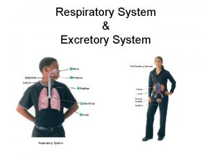
The Upper Respiratory System Nose Pharynx throat Middle


- Slides: 45

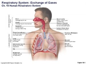
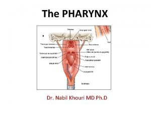
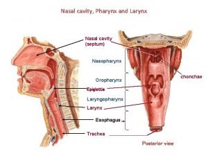
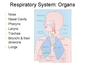
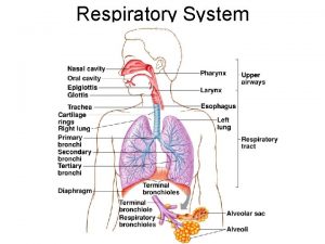
The Upper Respiratory System § § Nose Pharynx (throat) Middle ear Eustachian tubes

Structures of Upper Respiratory System Figure 24. 1

The Lower Respiratory System § § § Larynx Trachea Bronchial tubes Alveoli Pleura

Structures of Lower Respiratory System Figure 24. 2

Normal Microbiota of Respiratory System § Suppress pathogens by competitive inhibition in upper respiratory system § Lower respiratory system is sterile

Upper Respiratory System Diseases § § § Pharyngitis Laryngitis Tonsillitis Sinusitis Epiglottitis: H. influenzae type b

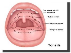
Streptococcal Pharyngitis – Is it? ? § Also called strep throat § Streptococcus pyogenes § Sore throat, usually starting quickly § Severe pain when swallowing § A fever (101° F or above) § Red and swollen tonsils, sometimes with white patches or streaks of pus § Nausea and/or vomiting § Swollen lymph nodes in the neck § Diagnosis by enzyme immunoassay (EIA) tests Figure 24. 3

Diphtheria § Corynebacterium diphtheriae: Gram-positive rod § Diphtheria toxin produced by lysogenized C. diphtheriae § Sore throat, fever, and adherent membrane on throat.

Diphtheria § Diphtheria membrane: Fibrin, tissue, bacterial cells Figure 24. 5

Diphtheria § Prevented by DTa. P vaccine § Diphtheria toxoid § Cutaneous diphtheria § Infected skin wound leads to slow-healing ulcer

Otitis Media § § § S. pneumoniae (35%) H. influenzae (20– 30%) M. catarrhalis (10– 15%) S. pyogenes (8– 10%) S. aureus (1– 2%) Incidence of S. pneumoniae reduced by vaccine Figure 24. 6

The Common Cold § Rhinoviruses (50%) § Coronaviruses (15– 20%) § Runny nose, coughing, sneezing, headache, fever, nasal congestion, sore throat. § OTC treatments § NO ANTIBIOTICS!!!!!

Lower Respiratory System Diseases § Bacteria, viruses, and fungi cause § Bronchitis § Bronchiolitis § Pneumonia

Pertussis (Whooping Cough) § Bordetella pertussis § Gram-negative coccobacillus § Capsule § Tracheal cytotoxin of cell wall damaged ciliated cells § Pertussis toxin § Prevented by DTa. P vaccine (acellular Pertussis cell fragments) Figure 24. 7

Pertussis (Whooping Cough) § Stage 1: Catarrhal stage, like common cold § Stage 2: Paroxysmal stage—violent coughing sieges (10 -12 days later) § Stage 3: Convalescence stage § Can last upwards of 6 weeks.

Tuberculosis § Mycobacterium tuberculosis § Acid-fast rod; transmitted from human to human Figure 24. 8

Tuberculosis § M. bovis: <1% U. S. cases; not transmitted from human to human § M. avium-intracellulare complex infects people with late-stage HIV infection

Tuberculosis Figure 14. 10 c

Worldwide Distribution of Tuberculosis Figure 24. 11 a

U. S. Distribution of Tuberculosis Figure 24. 11 b

Treatment of Tuberculosis § Treatment: Prolonged treatment with multiple antibiotics § Vaccines: BCG, live, avirulent M. bovis; not widely used in United States § There is MDR-TB and XDR-TB

A Positive Tuberculin Skin Test Figure 24. 10

Diagnosis of Tuberculosis § Tuberculin skin test screening § Positive reaction means current or previous infection § Followed by X-ray or CT exam, acid-fast staining of sputum, culturing of bacteria

Pneumococcal Pneumonia § Streptococcus pneumoniae § Gram-positive encapsulated diplococci Figure 24. 12

Pneumococcal Pneumonia § Symptoms: Infected alveoli of lung fill with fluids; interferes with oxygen uptake § Diagnosis: Optochin-inhibition (drug-test; disk assay) test or bile solubility test; serological typing of bacteria § Treatment: Penicillin, fluoroquinolones § Prevention: Pneumococcal vaccine

Haemophilus influenzae Pneumonia § Gram-negative coccobacillus § Predisposing factors: Alcoholism, poor nutrition, cancer, or diabetes § Symptoms: Resemble those of pneumococcal pneumonia § Diagnosis: Isolation; special media for nutritional requirements § Treatment: Cephalosporins

Mycoplasmal Pneumonia § Primary atypical pneumonia; walking pneumonia § Mycoplasma pneumoniae § Pleomorphic, -less bacteria wall § Common in children and young adults Figure 24. 13

Mycoplasma pneumoniae Figure 11. 20

Mycoplasmal Pneumonia § Symptoms: Mild but persistent respiratory symptoms; low fever, cough, headache § Diagnosis: PCR and serological testing § Treatment: Tetracyclines

Legionellosis § Legionella pneumophila § Gram-negative rod § Found in water § Transmitted by inhaling aerosols; not transmitted from human to human

Legionellosis § Symptoms: Potentially fatal pneumonia that tends to affect older men who drink or smoke heavily § Diagnosis: Culture on selective media, DNA probe § Treatment: Erythromycin

Chlamydial Pneumonia § Chlamydophila pneumoniae § Transmitted from human to human Figure 11. 24 b

Chlamydial Pneumonia § Symptoms: Mild respiratory illness common in young people; resembles mycoplasmal pneumonia § Diagnosis: Serological tests § Treatment: Tetracyclines

Q Fever § § Causative agent: Coxiella burnetii Reservoir: Large mammals Tick vector Can be transmitted via unpasteurized milk

Coxiella burnetii, the Cause of Q Fever Figure 24. 14

Q Fever § Symptoms: Mild respiratory disease lasting 1– 2 weeks; occasional complications such as endocarditis occur § Diagnosis: Growth in cell culture § Treatment: Doxycycline and chloroquine

Viral Pneumonia § Viral pneumonia occurs as a complication of influenza, measles, or chickenpox § Viral etiology suspected if no other cause is determined

Respiratory Syncytial Virus (RSV) § § Common in infants; 4500 deaths annually Causes cell fusion (syncytium) in cell culture Symptoms: Pneumonia in infants Diagnosis: Serological test for viruses and antibodies § Treatment: Ribavirin, palivizumab

Influenza (Flu) § Symptoms: Chills, fever, headache, and muscle aches § No intestinal symptoms § 1% mortality, very young and very old § Treatment: Zanamivir and oseltamivir inhibit neuraminidase § Prophylaxis: Multivalent vaccine

The Influenza Virus § Hemagglutinin (HA) spikes used for attachment to host cells § Neuraminidase (NA) spikes used to release virus from cell Figure 24. 15

Influenza Serotypes Type Antigenic Subtype Year Severity A H 3 N 2 H 1 N 1 H 2 N 2 H 3 N 2 H 1 N 1 1889 1918 1957 1968 1977 Moderate Severe Moderate Low B None 1940 Moderate C None 1947 Very mild

Histoplasmosis § Histoplasma capsulatum, dimorphic fungus Figure 24. 16

Histoplasmosis Distribution Figure 24. 17

Coccidioidomycosis § § § Causative agent: Coccidioides immitis Reservoir: Desert soils of Southwest U. S. Symptoms: Fever, coughing, weight loss Diagnosis: Serological tests Treatment: Amphotericin B

U. S. Endemic Area for Coccidioidomycosis Figure 24. 19
 Ccmc ear nose and throat
 Nikolai gogol 1836
 Elements of morphology
 Conducting zone and respiratory zone
 Inspiration anatomy and physiology
 Upper and lower respiratory system
 Respiratory zone
 Site:slidetodoc.com
 Classification of upper respiratory tract infection
 Anatomy of the upper respiratory tract
 Rsv sympyoms
 Upper respiratory tract
 What is the major function of the respiratory system
 Acute upper respiratory infection unspecified คือ
 Inferior meatus
 Digestive system cavity
 Upper adams middle school
 Montgomery upper middle school
 Tiny air sacs at the end of the bronchioles
 Circulatory system and respiratory system work together
 Pterygopharyngeus
 4 muscles of pharynx
 Trachea is also called
 Pharynx subdivisions
 Lingual tonsil
 Pre trematic nerve meaning
 Roof of nasal cavity
 Pharynx to esophagus
 Horizontal
 Pharynx function earthworm
 Plicae circulares kerckringi
 Pharynx and larynx
 Physiology of pharynx
 Pharynx hiperemija
 Sara run isafold
 Laryngopharynx
 Taenia omentalis
 Buonator
 Development of respiratory system
 Respiratory epithelium
 Epliglottis
 Microvilli
 Muscular pharynx
 Pharynx
 Pharynx food pipe
 Pharynx
