Pharyngeal arches Primitive pharynx stomodeum Hind brain vesicle

Pharyngeal arches

Primitive pharynx stomodeum Hind brain vesicle pericardial sac Septum tranversum notochord Dorsal aorta

Primitive pharynx

- 4 th week of IUL

• Initially pharyngeal arches confined to lateral wall of the primitive pharynx. • But gradually they extend ventrally and fuse with their counterparts of the opposite side in the floor of the primitive pharynx

Lateral wall

Mesodermal bar Endoderm Ectoderm Pharyngeal cleft Closing membrane Pharyngeal pouch


Pharyngeal arches cut in cross section

The Derivatives arranged in 3 Groups - MESODERMAL -ECTODERMAL -ENDODERMAL

MESODERMAL -Arch Artery derivatives artery -Cartilagenous rod -Muscular component -Traversed by Nerves

Fate of Pharyngeal Arch Arteries Sixth Aortic Arch Fourth Aortic Arch -Second Aortic arch Fifth Aortic Arch On right side- Third Aortic Arch -On Regress -right artery right. Pulmonary side entirely Disappears -right Subclavian artery on both sides Ventral part form Common left side- On except Stapedial artery Carotid artery First Aortic Arch artery -Left Pulmonary On left side arteriosus - -Ductus part of Arch of Aorta Dorsal part persist as stem -Disappears of Internal Carotid arterya part for Except Maxillay artery

Pharyngeal Arch cartilage Derivatives of • Derived from Neural crest cells Mesoderm Perichondrium Bone Cartilagenous bar Ligament

First arch cartilage Anterior ligament of malleus Malleus, Sphenomandibular ligament -Incus,
 cartilage Styloid process Stapes Stylohyoid ligament -Lesser cornu -upper portion of body")
2 ndarch(reicherts) cartilage Styloid process Stapes Stylohyoid ligament -Lesser cornu -upper portion of body of hyoid

Third arch - Greater cornu - lower part of body of hyoid

Fourth Arch Lamina of thyroid cartilage Sixth Arch Arytenoid cartilage Cricoid cartilage

NERVES -Nerves of branchial arches are derived from HIND BRAIN VESICLE Pre trematic Post trematic

- POST TREMATIC - PRE TREMATIC

Mandibular nerve Chorda tympani Facial nerve Glosopharyngeal nerve Superior Laryngeal nerve Recurrent Laryngeal nerve

Mesodermal derivatives Muscular components - Myoblasts migrate from paraxial mesoderm to the site

First arch

Second Arch

Third Arch

4 th and 6 th arch 4 th arch – Cricothyroid , Inferior pharyngeal constrictor 6 th arch – Intrinsic laryngeal muscle (except cricothyroid)
Carotid body – 3 rd arch b)Aortic body - 4")
Other mesodermal derivatives are a)Carotid body – 3 rd arch b)Aortic body - 4 th arch c)Fibro areolar stroma of face, neck tongue

Arch Skeletal Derivatives Muscles Nerve Artery 1 Meckel cartilage, malleus, incus, anterior ligament of malleus, sphenomandibular ligament Muscles of mastication, mylohyoid, anterior digastric, tensor palatini, tensor tympani Trigeminal Maxillary 2 Reichert cartilage 1. Stapes 2. Styloid process 3. Stylohyoid ligament 4. Lesser cornu/upper portion of body of hyoid 1. Stapedius Facial Stapedial Glossopharyn geal 3 2. Posterior digastric 3. Stylohyoid 4. Muscles of facial expression Greater cornu/ lower part of Stylopharyngeus body of hyoid 4 Thyroid cartilage Cricothyroid muscle Superior laryngeal Inferior pharyngeal nerve contrictor Common carotid, portion of internal carotid Rt subclavian Part arch of aorta 6 Arytenoid cartilage Intrinsic muscle of Recurrent larynx( except laryngeal cricothyroid) nerve Rt and Lft pulmonary artery Cricoid cartilage

Ectodermal Derivatives Covering 1 st arch forms: - Skin over upper and lower jaw - Tragus of auricle - Skin of upper and lower lip - Mucosa of gums

ECTODERMAL DERIVATIVES OR CLEFTS n dorsal part - forms: epithelial lining of external acoustic meatus - cuticular layer of tympanic membrane.

Second arch grows

n The second arch grows much faster than the succeeding arches. n The space between the overhanging 2 nd arch & 3 rd, 4 th, 6 th arch is called cervical sinus. n The 2 nd, 3 rd and 4 th clefts loose contact with the outside.

ENDODERMAL DERIVATIVES • Grouped as – Lateral derivatives, arising from pharyngeal pouches – Ventral derivatives, developing in the floor of the primitive pharynx.
 • 4 pairs • Pouches develop - craniocaudal sequence. •")
PHARYNGEAL POUCH( lateral derivatives) • 4 pairs • Pouches develop - craniocaudal sequence. • 1 st pouch - between the 1 st& 2 nd pharyngeal arch. • Ventral part obliterated by developing tongue • Dorsal part except the first one divides into ventral and dorsal wings.

FIRST POUCH

• Dorsal part - tubotympanic recess – Medial part- auditory tube – Lateral part- primitive tympanic cavity from which gives rise to • Mucous lining of tympanic cavity • Mastoid antrum • Mastoid air cells • Mucous layer of tympanic membrane. • Distal end of tubotympanic recess contact with bottom of 1 st

SECOND POUCH • Endoderm proliferates as tiny solid buds and grows into mesenchyme • Central cells of these buds breaks down to form tonsillar crypts. • 20 weeks - lymphoid tissue organizes into lymphatic
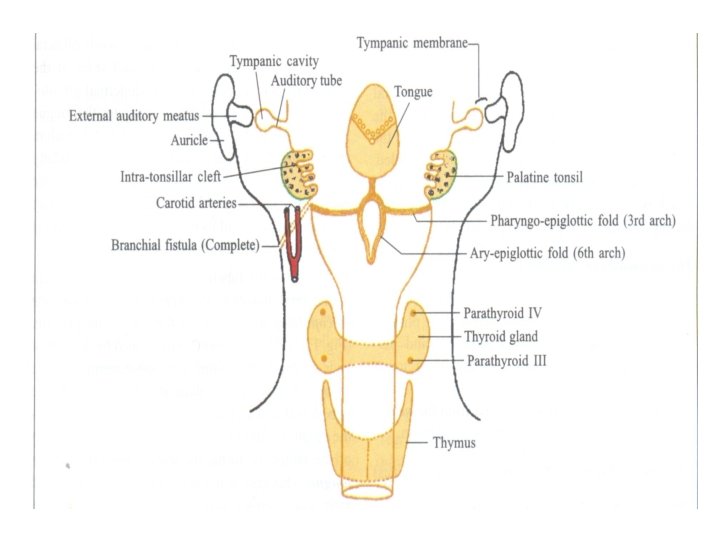

Remember • Endoderm of the second pharyngeal pouch forms stratified squamous non keratinized epithelium • Mesoderm forms the lymphoid tissue and connective tissue elements of the tonsil • Due to development of the tonsil, second pouch is mostly obliterated & in adults this pouch as intratonsillar cleft

THIRD POUCH

• Dorsal wing - inferior parathyroid gland. • Ventral wing - thymic rudiment. • Caudal migration of thymic rudiments and inferior parathyroid gland. • Inferior parathyroid gland separate from the thymus and gain permanent attachment to the lower pole of thyroid gland.

FOURTH POUCH

FOURTH POUCH • Dorsal part - superior parathyroid gland • Ventral part + fifth pouch = caudal pharyngeal complex. • Neural crest cells migrate to form Caudal pharyngeal complex exhibit 3 elements – thymic element – Lateral thyroid element – Ultimo - branchial body

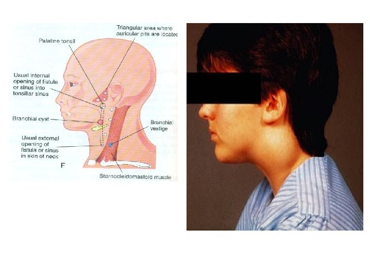
BRANCHIAL SINUSES

BRANCHIAL FISTULA

1 ST ARCH SYNDROME • Insufficient migration of neural crest cells to the 1 st arch( 4 th week ) • Associated with anomalies of ears, mandible, face, palate Macrostomia Deformed Auricle Preauricular appendages Hypoplasia of mandible Cheek defect between auricle and the mouth

Di. George anomaly -22 q deletion features Features: -Cardiac abnormality -Abnormal facial features -Thymic aplasia -Cleft palate -Hypocalcemia
- Slides: 47