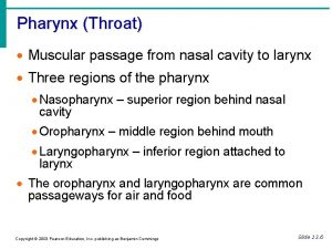
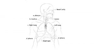
Respiratory system Respiratory system n Nasal cavity pharynx


- Slides: 30

Respiratory system

Respiratory system

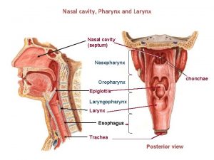
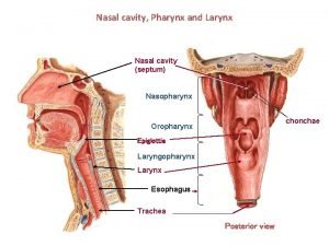
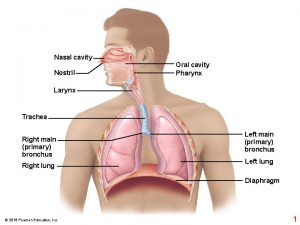
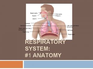
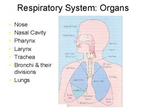
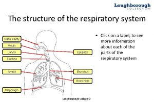
n Nasal cavity, pharynx & larynx. n Trachea & bronchi. n Lung.

一、trachea

mucosa submucosa trachea (HE) adventitia Mixed gland

EM: epithelium of trachea 3. Goblet C 1. Ciliated C 2. Brush C 5. Basal C 4. Small granule C

The wall of trachea 1. Mucosa=epithelium+lamina propria u u pseudostratiated ciliated columnar… Ciliated C, goblet C, brush C, basal C, small granular C. 2. Submucosa=CT+ mixed glands 3. Adventitia=16 -20 ‘C’ shaped hayline cartilage rings.

二、lung n atmosphere & alveoli n alveoli & blood (blood & tissue) bronchiole l ung Gas exchange lung

L ung Low power

lung n parenchyma: bronchial trees & alveoli. u Principal bronchi(Br)—lobular、 segmental Br—smaller Br—bronchiole —terminal bronchiole—respiratory bronchiole—alveolar duct、alveolar sac—alveoli. u Conduction portion : above terminal bronchioles. u Respiratory portion : respiratory bronchiole to alveoli. u Pulmonary lobule: bronchiole and it’s ramifications.

Conduction portion- lobular Br & small Br • Epithelium • Goblet C • Glands • Cartilage plate • SMC

Conduction portion- bronchiole & terminal brochiole • Simple ciliated columnar. • Goblet C (-) • Gland (-) • Cartilage plate (-) • SMC

Rules of structural variance in conducting portion Small Br bronchiol e Epi Pseudostratifie Simple d ciliated Ciliated columnar Goblet C + + +/- Terminal bronchiole Simple columnar** clara cell - Mixed gland Cartilag e plate SMC + + +/- - + + ++

Respiratory portion- Respiratory bronchiole • The wall is interrupted • Simple cuboidal Epi, clara C • SMC

Respiratory portion- Alveolar duct & alveolar sac Alveolar duct • The wall, many alveoli • SMC** • Simple cuboidal, squamous Alveolar sac • No SMC**

Respiratory portion-alveolus 肺泡管 Simple alveolar Epi.

(1) Respiratory portion n ###constituents:respiratory bronchiole,alveolar duct, alveolar sac,alveolus。 1. Respiratory bronchiole u The wall, few alveoli open into. u Simple cuboidal Epi, SMC.

(2) Respiratory portion 2. Alveolar duct & alveolar sac The wall, many alveoli open into. u Knobs in the wall of alveolar duct u Knobs is sphincter-like SMC. u

(3) Respiratory portion 3. alveolus: u type I alveolar C u type II alveolar C u alveolar septum.

macrophage Type II alveolar C Elastic fiber

type I alveolar C

Type II alveolar C Osmiophilic Multilamellar bodies

blood-air barrier

Alveoli n Type I cell u squamous, covered most surface of the alveoli. n Type II cell LM: round or cuboidal, cell body protrude into the lumen. u EM: osmiophilic multilamellar body, with abundant phospholipids. u Functions: secret surfactant, reduce surface tension, stabilize alveoli. u

n Alveolar septum: continuous Cap. , elastic fiber. macrophage dust C. @blood-air barrier includes: 1. mucus layer on surface of alveoli 2. type I alveolar C 3. BM of alveolar C 4. delicate layer of CT 5. continuous Cap. BM 6. continuous Cap. Endothelium.

1 2 3 4 Terminal bronchiole Respiratory bronchiole Alveolar duct Alveolar sac

Rules of structural variance in respiratory portion wall Epi. SMC Respiratory Not integrated bronchiole Alveoli open Alveolar Many alveoli duct open into Simple columan thin Simple cuboidal Alveolar sac alveolus Alveolar Epi. no of alveoli Irregular vesicular Simple cuboidal A few Alveolar Epi. no

Alveolar epithelial cell shape cytoplasm functions Type squamous Phagocytotic Covered 95% of the surface of alveoli to I vesicle Osmiophilic Type round cuboidal multilamellar II body ** benefit gas exchange. Secret surfactant to reduce surface tension and stabilize the alveoli. Type I cell can not regenerate. Type II cell can differentiate into Type I cell.

keypoints n The basic structure of trachea. n Histologic structure of conduction portion. n Histologic structure of respiratory portion. n Esp. the alveolar epithelium

 Dorsal cavity
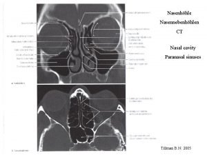
 Respiratory system nasal cavity
 Respiratory system nasal cavity
 Respiratory system nasal cavity
 Palatopharyngeal arch
 Pyloric orifice function
 Drainage
 Dr mujahid nazal
 Saddle nose pictures
 Meatus of nose
 Meatus drainage
 Nasal cavity
 Two pronged plastic device for delivering oxygen
 Chapter 5 the skeletal system figure 5-10
 Labeled
 Chapter 5 the skeletal system figure 5-10
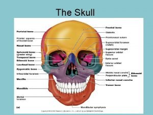
 Bones that form nasal cavity
 Internal nares
 Sinus sphenoidalis mündung
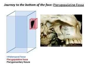
 Pterygopalatine fossa boundaries
 Drainage
 Ventral body cavity
 Basic anatomy terminology
 Left mesenteric sinus
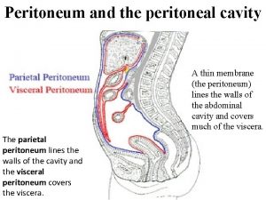
 Peritoneal cabity
 Respiratory distress nasal flaring
 Conducting zone of the respiratory system function
 Pharynx in digestive system
 Digestive respiratory and circulatory system
 Pharyngis
 Dr nabil khouri
