Respiratory system Upper respiratory system 1 Nose pharynx


- Slides: 38
Respiratory system
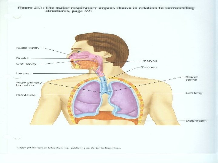
• Upper respiratory system 1. Nose, pharynx, and associated structures • Lower respiratory system 1. Larynx, trachea, bronchi, lungs • Conducting portion 1. structures that warm, moisten, and bring air into lungs 2. consists of upper and lower systems
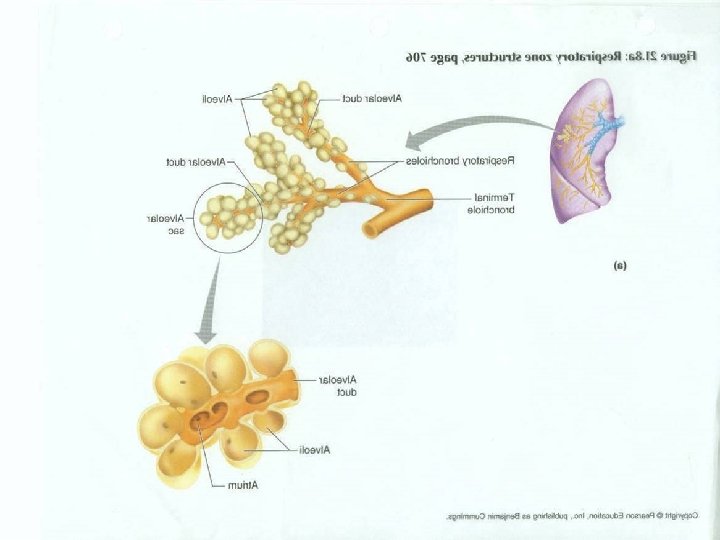
• Tissues in lungs where gas is exchanged • Consists of respiratory bronchioles, alveolar ducts, alveolar sacs, and alveoli
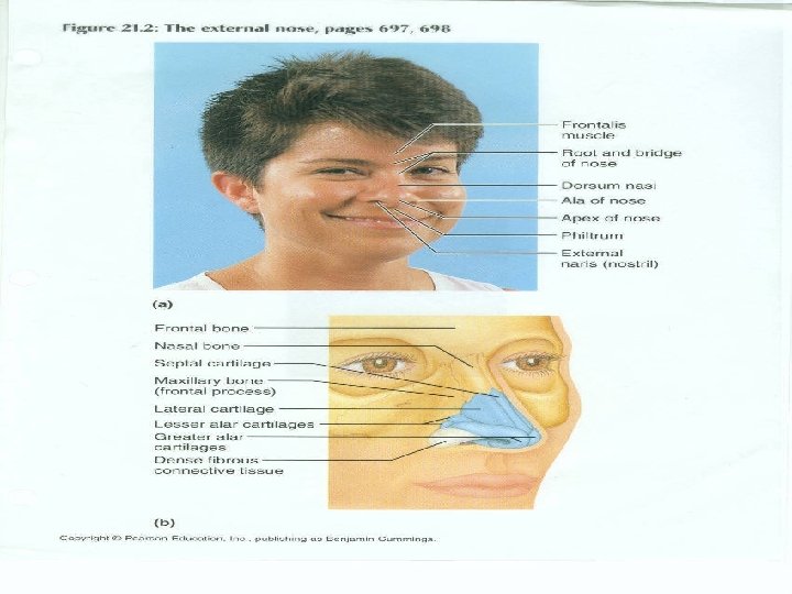
• External portion 1. Hyaline cartilage 2. nostrils 3. functions a. warming, moistening, and filtering air b. detecting olfactory stimuli c. modify speech vibrations as they pass through resonating chambers
• Internal portions 1. large cavity in anterior portion of skull 2. includes muscle and mucous membranes 3. inner nose merges with external nose 4. communicates with pharynx through openings called internal nares
• Nasal cavity 1. space inside internal nose 2. divided by nasal septum a. anterior portion made of cartilage b. rest formed by vomer 3. Grooves (meatuses) a. traps water b. prevents dehydration
• Olfactory epithelium 1. olfactory receptors 2. Pseudostratified epithelium a. contains goblet cells b. contain capillaries that move cilia 3. cilia move mucous toward pharynx 4. drainage from nasolacrimal and paranasal sinuses also moisten air
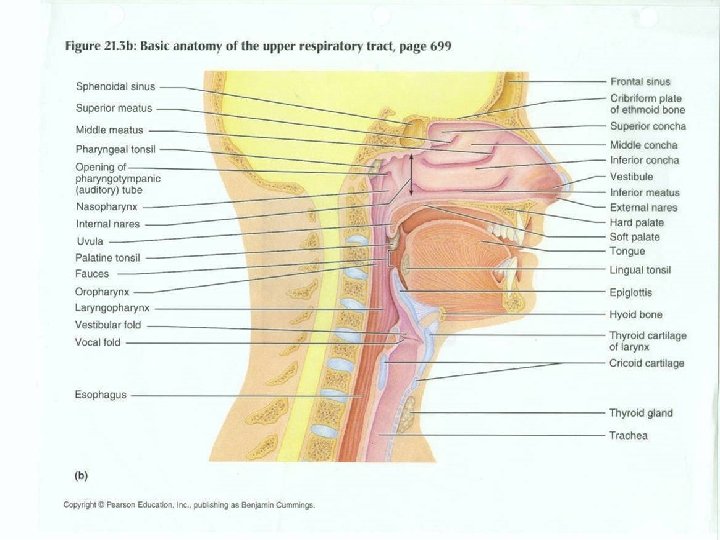
• Funnel shaped tube about 13 cm • Reaches from internal nares to inferior cartilage of larynx • Wall is composed of skeletal muscle • Functions as passageway for food and air • Muscles arranged into two layers ( outer circular and inner longitudinal)
• Nasopharynx 1. superior portion 2. contains opening of Eustachian tube 3. contains pharyngeal tonsils 4. Contains pseudostratified columnar epithelium
Pharynx continued • Oropharynx 1. lies posterior to oral cavity 2. fauces ( openings from the mouth) 3. has both respiratory and digestive functions 4. lined with nonkeratinized stratified squamous epithelial cells 5. has two pairs of tonsils (palatine and Lingual)
• Laryngopharynx 1. begins at level of hyoid bone 2. connects esophagus to larynx
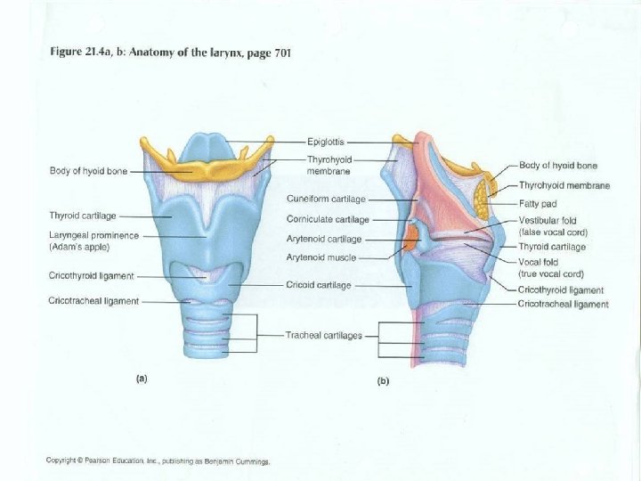
• Short passageway that connects laryngopharynx with trachea • Composed of nine pieces of cartilage 1. thyroid cartilage a. hyaline cartilage b. larger in males 2. Artenoid cartilage a. influences position & tension of vocal cords b. attach vocal cords and intrinsic pharyngeal muscles
• Elastic cartilage covered with epithelium • Stem is attached to anterior rim of thyroid cartilage • During swallowing pharynx and larynx rise a. pharynx widens to receive food b. elevation of larynx causes epiglottis to form lid over glottis • Glottis a. folds of mucous membranes (vocal cords) b. closing directs food into esophagus
• Ventricular folds 1. superior pair 2. false vocal cords • Vocal folds 1. inferior pair 2. push against ventricular folds to hold breath under pressure
• Elastic ligaments 1. tighten by muscles attached to artenoid cartilage 2. greater air pressure the louder the sound 3. pull vocal folds into airway • Pitch caused by tension of vocal folds • Sound originates from vibration of vocal folds
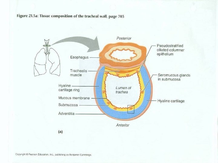
• Anterior to esophagus • Layers of tracheal walls 1. mucosa a. Pseudostratified ciliated columnar epithelium b. provides protection against dust 2. submucosa a. areolar connective tissue b. rings of C shaped cartilage
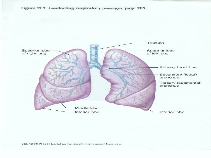
• Division of the trachea • Goes to right and left lung • Corina 1. formed from last tracheal cartilage 2. most sensitive area for producing cough reflex 3. Widening and distortion indicates carcinoma in the lymph nodes
• Changes in bronchi as they branch 1. Ciliated columnar epithelial cells change into nonciliated cuboidal 2. cartilage decreases 3. smooth muscle increases
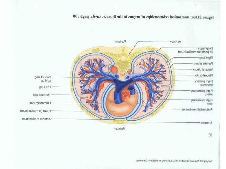
• Pleural membrane 1. enclose and protect each lung 2. parietal plura a. superficial layer b. lines wall of thoracic cavity 3. visceral pleura a. covers the lung b. deep layer
• Pleural cavity 1. space between parietal and visceral pleura 2. secretes fluid to decrease friction
• Extended from diaphragm just slightly superior to clavicle • Lie against ribs • Base (broad inferior portion) • Apex (narrow superior portion) • Mediastinal surface of each lung contains hilus (vessels and bronchi pass into lungs) • Left lung has cardiac notch • Right lung is shorter because of liver pushing on diaphragm
• • Lobes are separated by fissures Right lung has three lobes Left lung has two lobes Branchiopulmonary segment contains small compartment called lobules a. contain lymphatic vessels, arterioles, a venule, and a branch of terminal branchioles b. terminal bronchioles subdivide into respiratory bronchioles
• Respiratory bronchioles divide into alveolar ducts • Respiratory passages from the trachea branch about 25 times
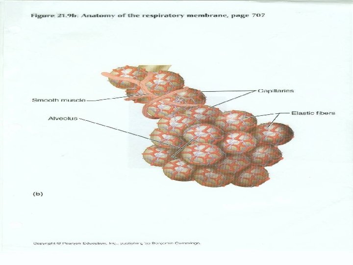
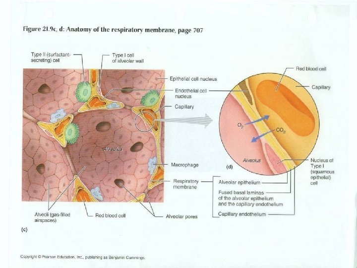
• Cup shaped out pouching, lined with simple squamous epithelium • Alveolar sacs are two or more alveoli that share same opening • Consists of two types of epithelial cells 1. simple squamous a. form most of lining b. gas exchange takes place
2. Septal cells a. cuboidal epithelial cells b. secretes fluid to keep spaces between cells and air moist • Respiratory membrane layers 1. alveolar walls (epithelial cells) 2. epithelial basement membrane 3. capillary basement membrane 4. enothelial cells of capillary
• Alveolar macrophages 1. remove dust and debris 2. wandering phagocytes
• Gas exchange between lungs and atmosphere • Inspiration 1. air flows in when pressure decreases 2. lungs expand 3. controlled by contraction of diaphragm • Expiration 1. also due to pressure gradient 2. elastic recoil of chest