PHARYNX Prepared by Ass Lecturer Sara Mostafa Description

PHARYNX Prepared by/ Ass. Lecturer: Sara Mostafa

Description The pharynx is situated behind the nasal cavities, the mouth, and the larynx Ø It may be divided into nasal, oral, and laryngeal parts Ø Its upper, wider end lying under the skull Ø Its lower, narrow end becoming continuous with the esophagus opposite the sixth cervical vertebra Ø
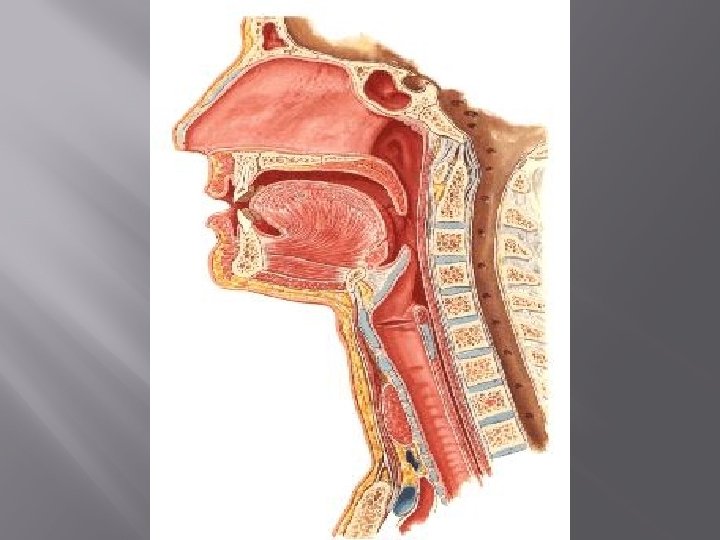

Description The pharynx has a musculomembranous wall, which is deficient anteriorly Ø Here, it is replaced by the posterior openings into the nose (choanae), the opening into the mouth, and the inlet of the larynx Ø By means of the auditory tube, the mucous membrane is also continuous with that of the tympanic cavity Ø

Muscles of the Pharynx Wall of the pharynx consist of the superior, middle, and inferior constrictor muscles Ø Fibers of these muscles run in a somewhat circular direction Ø Stylopharyngeus and salpingopharyngeus muscles Ø Their fibers run in a somewhat longitudinal direction Ø
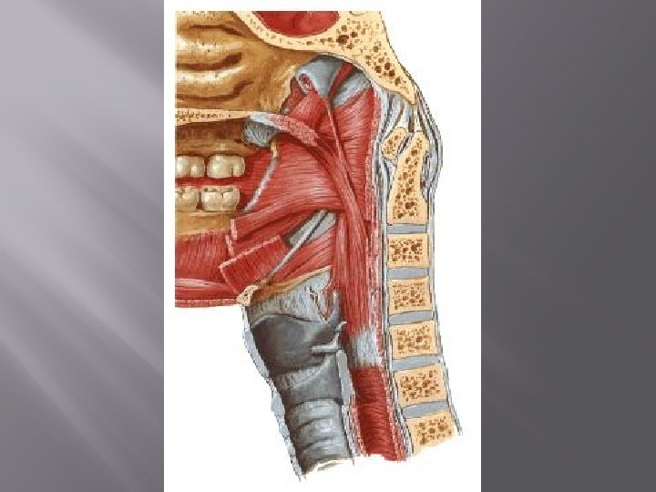
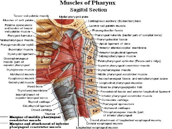

Muscles of the Pharynx The three constrictor muscles extend around the pharyngeal wall to be inserted into a fibrous band or raphe Ø The raphe extends from the pharyngeal tubercle on the basilar part of the occipital bone of the skull down to the esophagus Ø The three constrictor muscles overlap each other so that the middle constrictor lies on the outside of the lower part of the superior constrictor and the inferior constrictor lies outside the lower part of the middle constrictor Ø
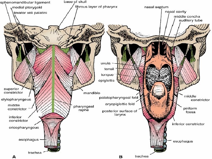

Muscles of the Pharynx The lower part of the inferior constrictor, which arises from the cricoid cartilage, is called the cricopharyngeus muscle Ø The fibers of the cricopharyngeus pass horizontally around the lowest and narrowest part of the pharynx and act as a sphincter Ø

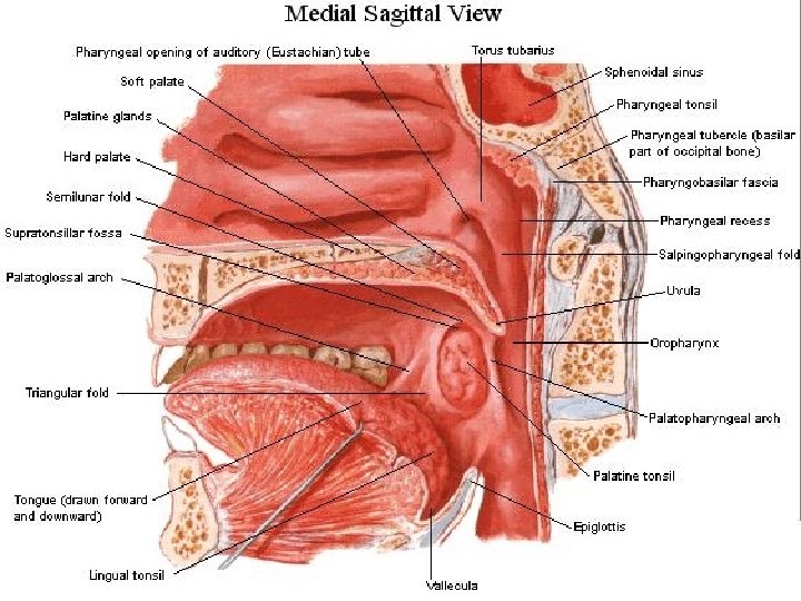
Nasal Pharynx This lies above the soft palate and behind the nasal cavities Ø In the submucosa of the roof is a collection of lymphoid tissue called the pharyngeal tonsil Ø The pharyngeal isthmus is the opening in the floor between the soft palate and the posterior pharyngeal wall Ø On the lateral wall is the opening of the auditory tube, the elevated ridge of which is called the tubal elevation Ø

Nasal Pharynx The pharyngeal recess is a depression in the pharyngeal wall behind the tubal elevation Ø The salpingopharyngeal fold is a vertical fold of mucous membrane covering the salpingopharyngeus muscle Ø

Oral Pharynx This lies behind the oral cavity Ø The floor is formed by the posterior one third of the tongue and the interval between the tongue and epiglottis Ø In the midline is the median glossoepiglottic fold Ø on each side the lateral glossoepiglottic fold Ø The depression on each side of the median glossoepiglottic fold is called the vallecula Ø

Oral Pharynx On the lateral wall on each side are the palatoglossal and the palatopharyngeal arches or folds and the palatine tonsils between them Ø The palatoglossal arch is a fold of mucous membrane covering the palatoglossus muscle Ø The interval between the two palatoglossal arches is called the oropharyngeal isthmus Ø It marks the boundary between the mouth and pharynx Ø

Oral Pharynx The palatopharyngeal arch is a fold of mucous membrane covering the palatopharyngeus muscle Ø The recess between the palatoglossal and palatopharyngeal arches is occupied by the palatine tonsil Ø

Lymphoid Tissue of Pharynx At the junction of the mouth with the oral part of the pharynx, and the nose with the nasal part of the pharynx, are collections of lymphoid tissue Ø They are of considerable clinical importance Ø The palatine tonsils and the nasopharyngeal tonsils are the most important Ø
- Slides: 17