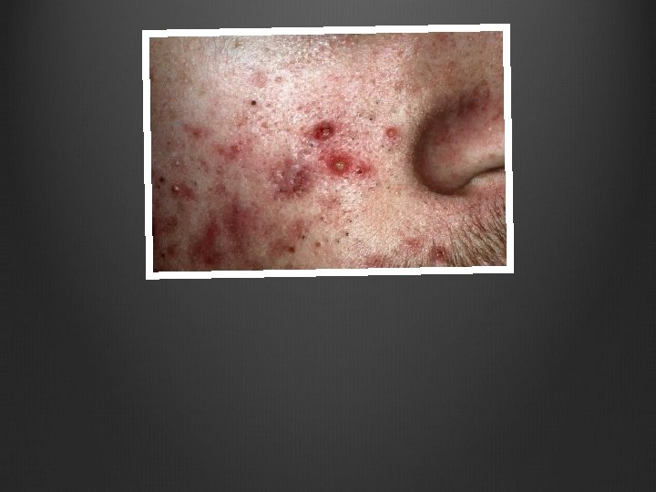
Skin Pathology IV Acne Vulgaris More severe in


 • Same general features as a Dermatofibroma. • DIFFERENCEs: •")


 Horn Cysts")
 Horn Cysts")


 Vacuoles: “Fat Trapping” Not Collagen Trapping like Dermatofibromas")


- Slides: 79
Skin Pathology IV
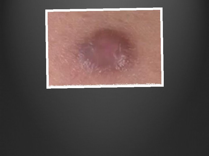
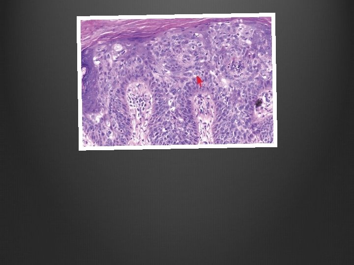
Acne Vulgaris • More severe in Males. • Can be caused by: • Drugs • Occupational Exposures • Cosmetics • Tropical Climates 13 -cis-retinoic acid: a synthetic Vitamin A derivative that has strong anti-sebaceous action and shows remarkable improvement. Causes Birth Defects. • Types: • Non-Inflammatory: • Open Comedones: Small, follicular papules containing a central, black keratin plug (blackheads) • Closed Comedones: Follicular papules without a visible central plug. • Inflammatory: • Erythematous • Propionibacterium Acnes: Administration of Antibiotics to individuals with inflammatory acne.
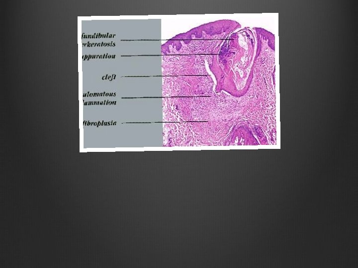
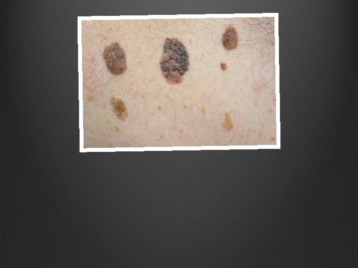
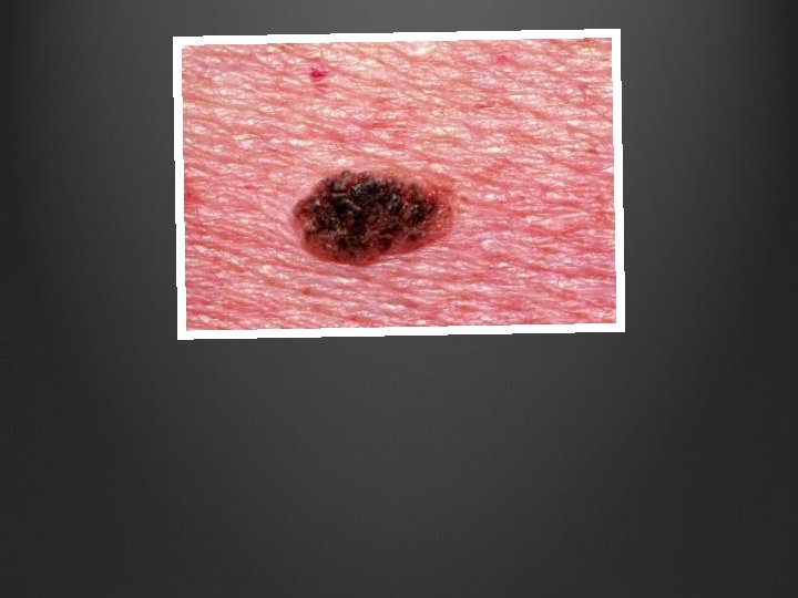
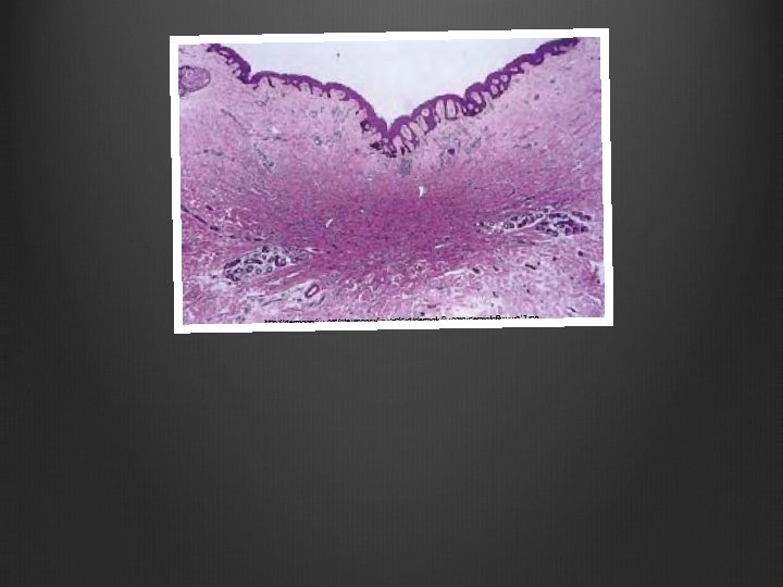
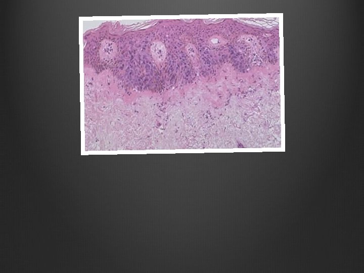
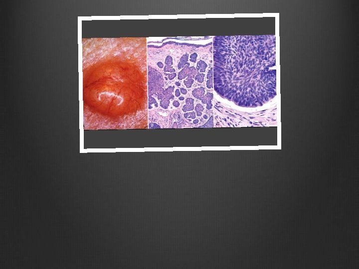
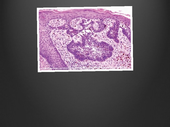
Seborrheic Keratosis • Common Epidermal Tumors • Round, Flat, Coin-Like “tan, waxy plaques” that vary in diameter. • Genetic: FGFR 3, Receptor Tyrosine Kinase • Histology: • Hyperkeratosis • Keratin-Filled Horn Cysts • Invagination Cysts of Keratin • Variable melanin pigmentation.
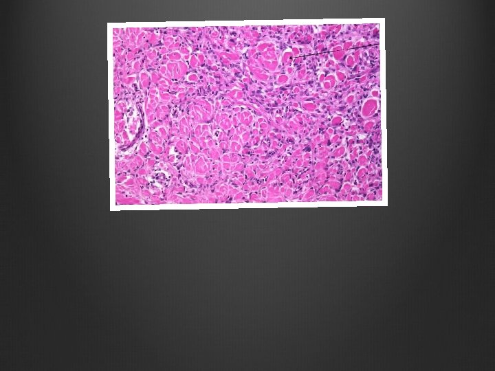
Acanthosis Nigricans • Thickened, Hyperpigmented Skin with a “velvet-like texture” • Appears in flexural areas. • Genetic: FGFR 3, Receptor Tyrosine Kinase • 80% are benign. • 20% are malignant gastrointestinal adenocarcinoma
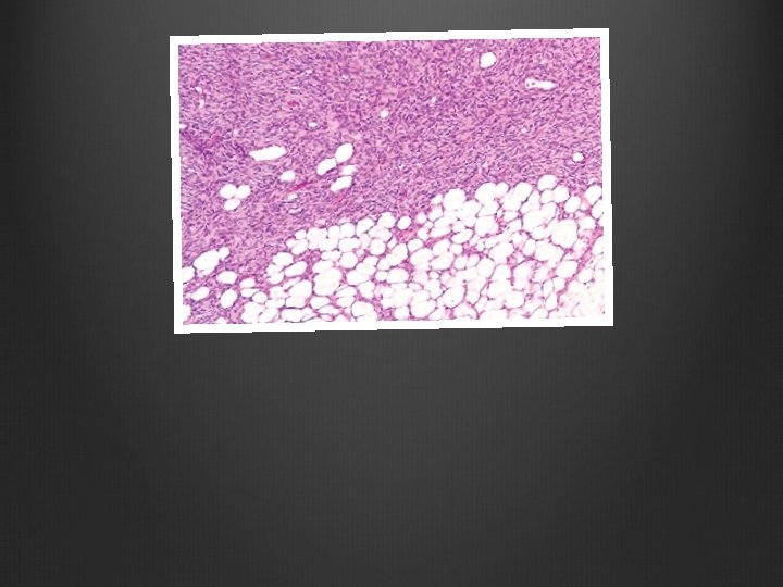
Dermatofibroma • Also called Benign Fibrous Histiocytoma • Usually a history of previous trauma. • Firm, Tan-Brown Papules, can be >1 cm • Dimple-Sign: When sqeezed from edges, center dimples • Histology: • Pseudoepitheliomatous Hyperplasia: overlying the lesion • Epidermis has hyperpigmented Basal Layer • Peripheral Collagen Trapping – at edge of lesion • Usually stays confined to the Dermis • Stains: • Factor XIIIa – Positive • CD 34 - Negative
Dermatofibroma Sacoma Protuberans (DFSP) • Same general features as a Dermatofibroma. • DIFFERENCEs: • DFSP shows vacuoles or “fat trapping” on histology, which is clear. Where Dermatofibroma has “Collagen Trapping”, which is pink. • DFSP is: • Factor XIIIa Negative • CD 34 Positive
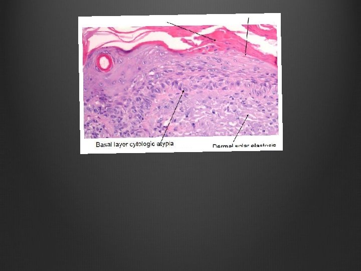
Actinic Keratosis • Caused by UV Light • Considered Pre-Cancerous Squamous Cell Carcinoma • Tan-Brown or Pink-Red Lesions with a rough, yellowish-brown or white scale. • Can also look horned. • Histology: • Hyperkeratosis with Hyperplasia • Dysplasia of the Epidermal Basal Layer (from the bottom, upward) • Not Full Thickness!!! • Full Thickness = Squamous Cell Carcinoma in situ • Elastosis: The superficial epidermis contains thickened, blue-gray elastic fibers caused by UV-damaged Fibroblasts. • Normal skin appendage areas.
Keratoacanthoma • Arises from hair follicles in sun exposed areas in >50 year olds • Description: • Dome-Shaped, Crusty Lesions with Central, Keratin-Filled Crater that mimics a welldifferentiated squamous cell carcinoma. • Histology: • Volcano • Tx: • Can spontaneously regress in 3 -4 months • Should be treated as though it is a Squamous Cell Carcinoma
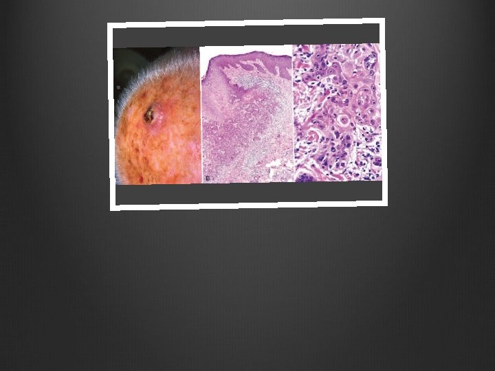
Squamous Cell Carcinoma • Cause: • DNA Damage from UV Light Exposure • Immunosuppression • Squamous Cell Carcinoma in situ: • Not invaded through the Basement Membrane. • Appear as sharply defined, red, scaling plaques. • Invasive Lesions: • Nodular, Hyperkeratotic Scales • Ulceration/Necrosis • Invade the Basement Membrane • Histology: • Variable degrees of differentiation. • Keratin Pearls • May need immunostains to confirm the lineage.
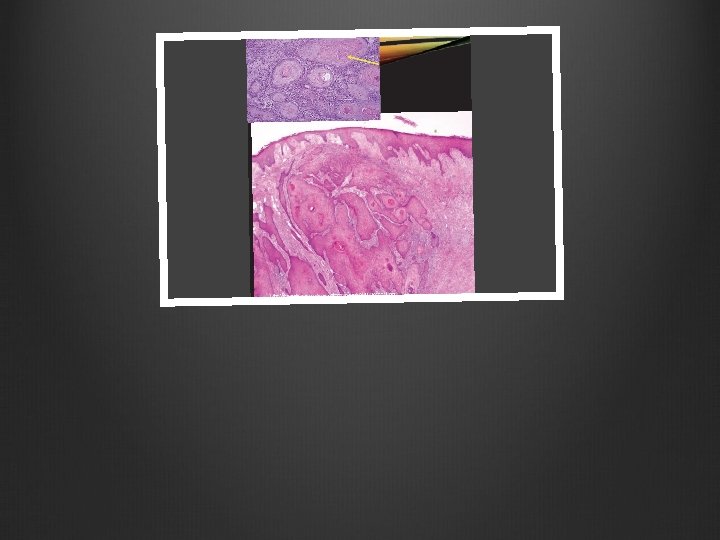
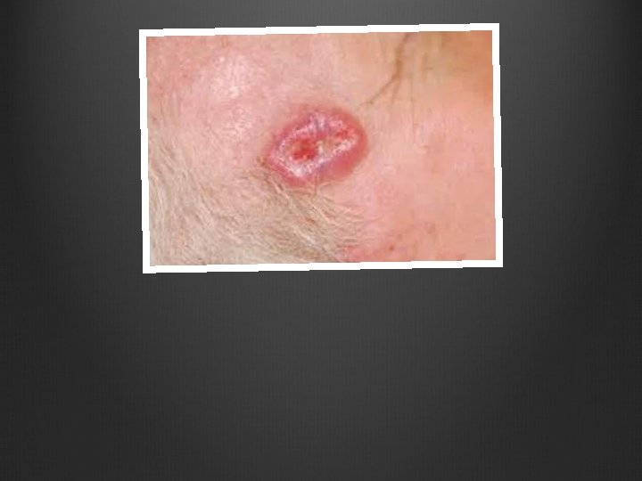
Basal Cell Carcinoma • Cause: • Mutations that activate the Hedgehog Pathway. • Sun-Exposed sites in fair-skinned, elderly patients. • Epidemiology • Locally aggressive cutaneous tumor. • Most common invasive cancer in humans. • Slow-Growing, Rarely Metastasize • Histology: • Variable types • Pearly Papules with Telangiectasis (dilated blood vessels) • Can have local invasion of bone or facial sinuses after many years of neglect = Rodent Ulcers • Blue “basaloid” Cells. • Peripheral Palisades • Clefting: separation of tumor from surrounding stroma • Embedded in mucinous Basal Cell Matrix = Basal Cell Stroma
Nevoid Basal Cell Carcinoma Syndrome • Rare, Genetic Syndrome • Causes Basal Cell Carcinomas at a very young age (12 years old) • Gene: PTCH • Tumor Suppressor Gene • Born with a germline loss of function mutation in one PTCH Allele. • Second Hit is required, by chance, causing early carcinoma formation.
Acne Vulgaris Treatment: 13 -cis-retinoic acid. Side Effects: Serious birth defects
Acne Vulgaris Treatment: 13 -cis-retinoic acid. Side Effects: Serious birth defects
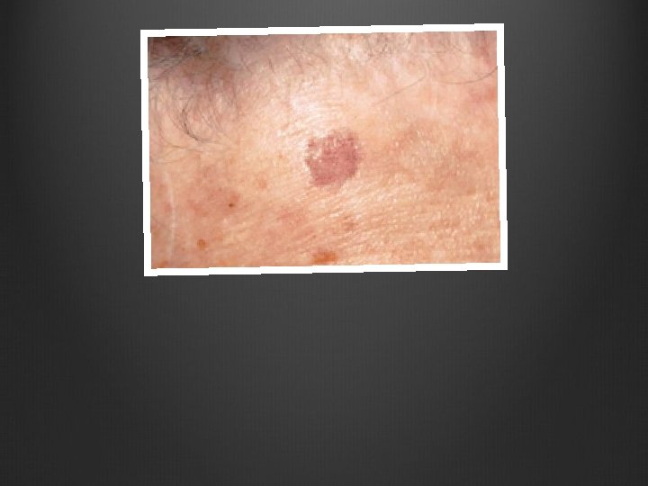
Seborrheic Keratosis Tan-Brown Waxy Plaques that vary in diameter.
Sebhorrheic Keratosis Tan-Brown Waxy Plaques that vary in diameter.
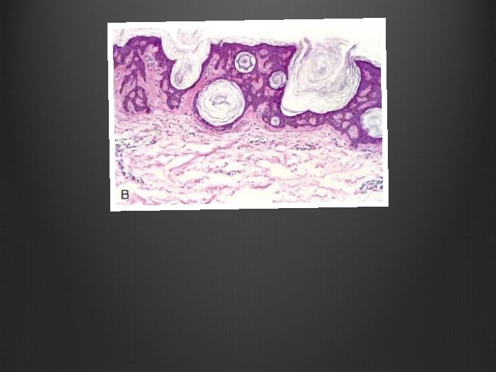
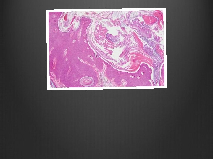
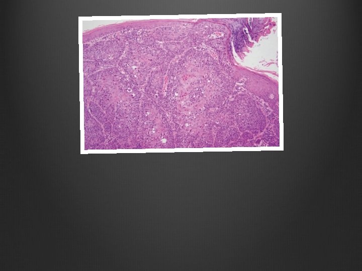
Sebhorrheic Keratosis Invaginations of Keratin (Invagination Cysts) Horn Cysts
Sebhorrheic Keratosis Invaginations of Keratin (Invagination Cysts) Horn Cysts
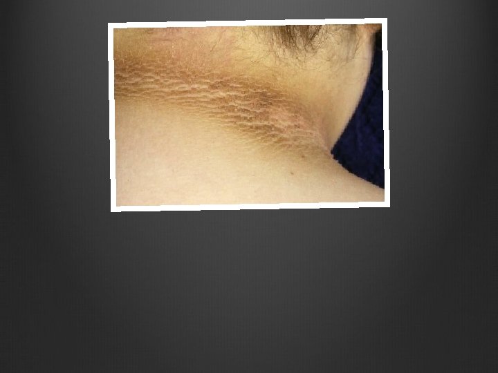
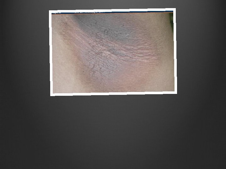
Acanthosis Nigricans Velvet-Like, Thickened, Hyperpigmented Skin Flexural Areas
Acanthosis Nigricans Velvet-Like, Thickened, Hyperpigmented Skin Flexural Areas
Dermatofibroma Dimple Sign: when squeezed from edges, center dimples. Factor XIIIa Positive CD 34 Negative
Dermatofibroma Dimple Sign: when squeezed from edges, center dimples. Factor XIIIa Positive CD 34 Negative
Dermatofibroma Peripheral Collagen Trapping Factor XIIIa Positive CD 34 Negative
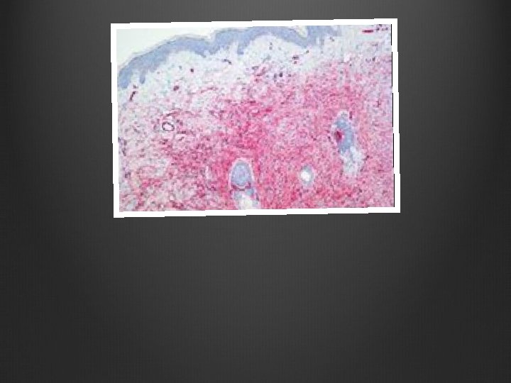
Dermatofibroma Sarcoma Protuberans (DFSP) Vacuoles: “Fat Trapping” Not Collagen Trapping like Dermatofibromas
Dermatofibroma Sarcoma Protuberans CD 34 + Stain Factor XIIIa Negative
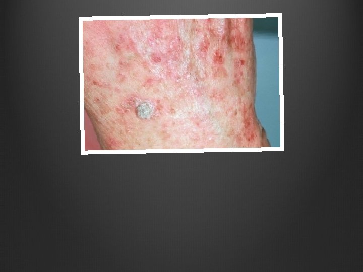
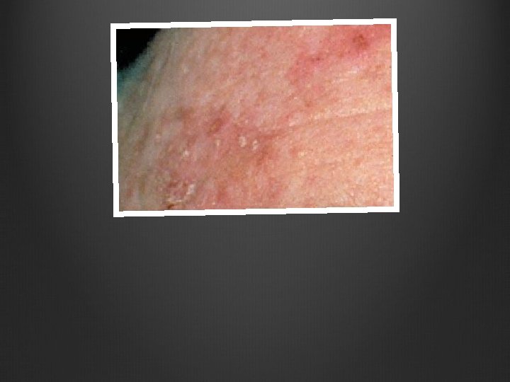
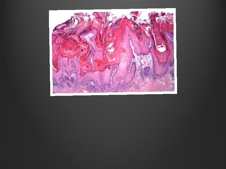
Actinic Keratosis UV Pre-Cancerous Squamous Cell Carcinoma Tan-Brown or Pink-Red lesions with scales/horns.
Actinic Keratosis UV Pre-Cancerous Squamous Cell Carcinoma Tan-Brown or Pink-Red lesions with scales/horns.
Actinic Keratosis UV Pre-Cancerous Squamous Cell Carcinoma Tan-Brown or Pink-Red lesions with scales/horns.
Actinic Keratosis UV Pre-Cancerous Squamous Cell Carcinoma Tan-Brown or Pink-Red lesions with scales/horns.
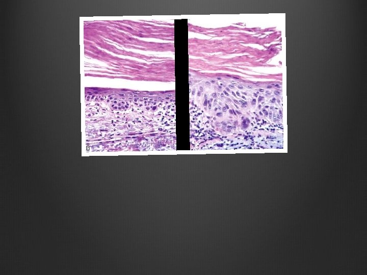
Actinic Keratosis UV Pre-Cancerous Squamous Cell Carcinoma Not Full Thickness Hyperkeratosis, Hyperplasia, Parakeratosis, Dyspasia of Basal Cell Layer Elastosis
Actinic Keratosis UV Pre-Cancerous Squamous Cell Carcinoma Not Full Thickness Hyperkeratosis, Hyperplasia, Parakeratosis, Dyspasia of Basal Cell Layer Elastosis, Normal Skin Appendage Areas
Actinic Keratosis UV Pre-Cancerous Squamous Cell Carcinoma Not Full Thickness Hyperkeratosis, Hyperplasia, Parakeratosis, Dyspasia of Basal Cell Layer Elastosis
Acinic Keratosis UV Pre-Cancerous Squamous Cell Carcinoma Not Full Thickness Hyperkeratosis, Hyperplasia, Parakeratosis, Dyspasia of Basal Cell Layer Elastosis
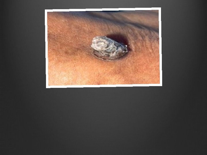
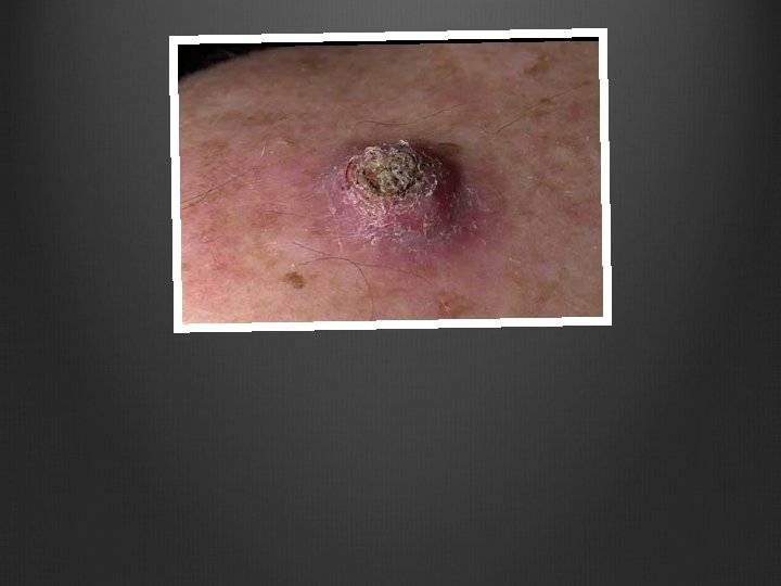
Keratoancthoma Volcano Should be treated as though it is a Squamous Cell Carcinoma
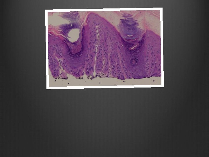
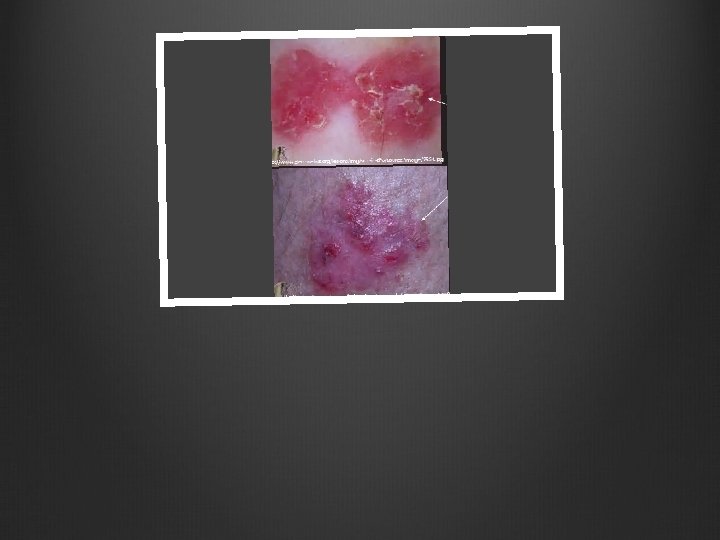
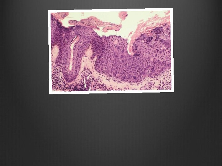
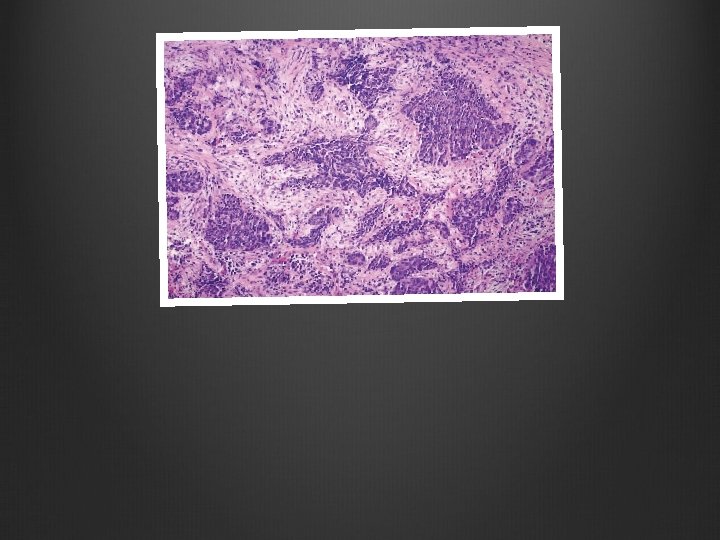
Squamous Cell Carcinoma In Situ Non-Nodular Sharply-Defined, Red, Scaling Plaques
Squamous Cell Carcinoma Invasive Nodular Hyperkeratotic Scales, Ulcerations
Squamous Cell Carcinoma Keratin Pearls Has no invaded the Basement Membrane Full-Thickness Dysplasia – In Situ
Basal Cell Carcinoma Blue “Basaloid” Cells Peripheral Palisades Clefting
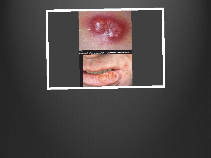
Squamous Cell Carcinoma Keratin Pearls Invaded Basement Membrane Invasive
Squamous Cell Carcinoma Keratin Pearls Invaded Basement Membrane Invasive
Keratoacanthoma Volcano Should be treated as though it is a Squamous Cell Carcinoma
Squamous Cell Carcinoma Keratin Pearls Invaded Basement Membrane Invasive Poorly Differentiated
Basal Cell Carcinoma Blue “Basaloid” Cells Peripheral Palisades Clefting
Basal Cell Carcinoma Pearly Papules containing dilated Sub-Epidermal Blood Vessels. Can have ulcerations.
Squamous Cell Carcinoma Keratin Pearls Invaded Basement Membrane Invasive
Squamous Cell Carcinoma Keratin Pearls Has no invaded the Basement Membrane Full-Thickness Dysplasia – In Situ