
Fluid Electrolyte and Acid Base Balance Chapter 27

Fluid, Electrolyte and Acid. Base Balance Chapter 27
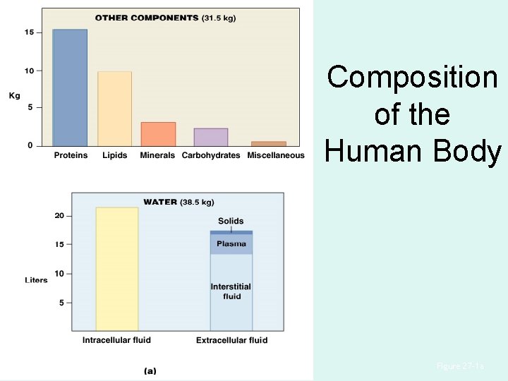
Composition of the Human Body Figure 27– 1 a
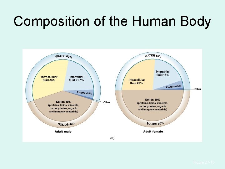
Composition of the Human Body Figure 27– 1 b
Water content varies with age & tissue type Fat has the lowest water content (~20%). Bone is close behind (~22 – 25%). Skeletal muscle is highest at ~65%.
Fluid Compartments • ECF (extra cellular fluid) and ICF (intracellular fluid) are called fluid compartments: – because they behave as distinct entities – are separated by cell membranes and active transport
Water Composition • Is 60% percent of male body weight • Is 50% percent of female body weight • Mostly in intracellular fluid
Water Exchange • Water exchange between ICF and ECF occurs across cell membranes by: – osmosis – diffusion – carrier-mediated transport
Major Subdivisions of ECF • Interstitial fluid of peripheral tissues • Plasma of circulating blood
Minor Subdivisions of ECF • • Lymph, perilymph, and endolymph Cerebrospinal fluid (CSF) Synovial fluid Serous fluids (pleural, pericardial, and peritoneal) • Aqueous humor
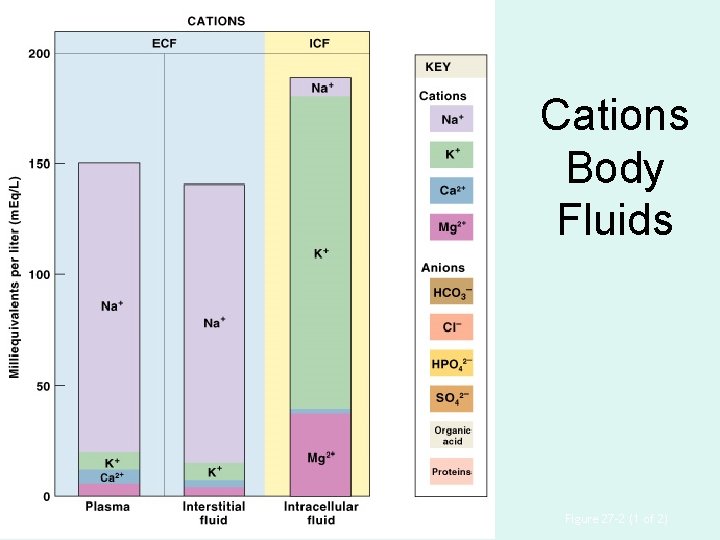
Cations Body Fluids Figure 27– 2 (1 of 2)
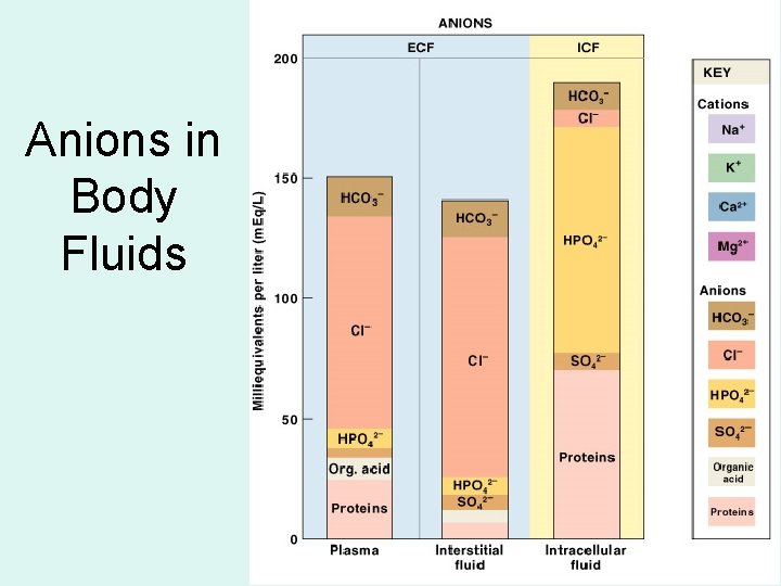
Anions in Body Fluids Figure 27– 2 (2 of 2)
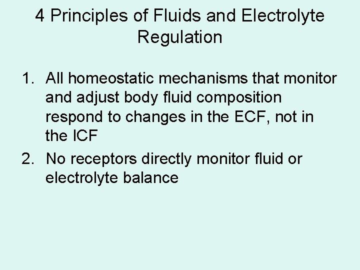
4 Principles of Fluids and Electrolyte Regulation 1. All homeostatic mechanisms that monitor and adjust body fluid composition respond to changes in the ECF, not in the ICF 2. No receptors directly monitor fluid or electrolyte balance
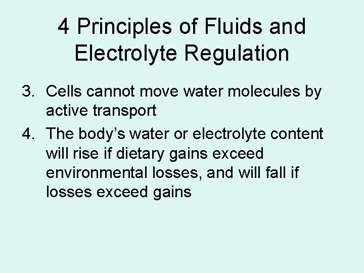
4 Principles of Fluids and Electrolyte Regulation 3. Cells cannot move water molecules by active transport 4. The body’s water or electrolyte content will rise if dietary gains exceed environmental losses, and will fall if losses exceed gains
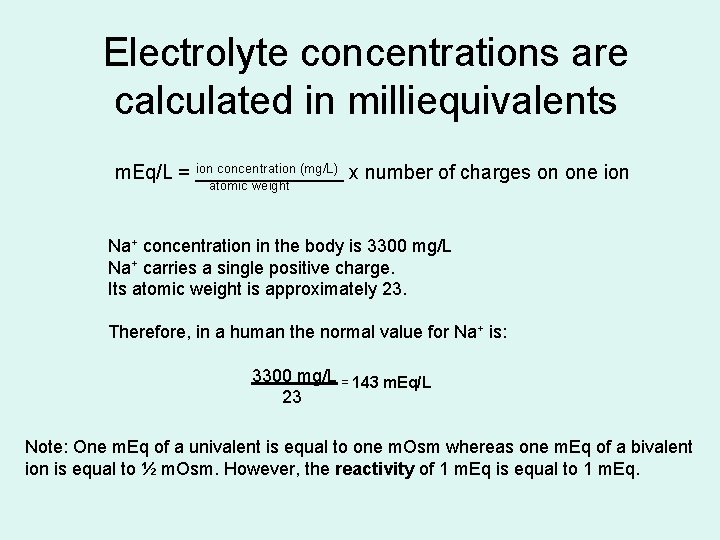
Electrolyte concentrations are calculated in milliequivalents m. Eq/L = ion concentration (mg/L) x number of charges on one ion atomic weight Na+ concentration in the body is 3300 mg/L Na+ carries a single positive charge. Its atomic weight is approximately 23. Therefore, in a human the normal value for Na+ is: 3300 mg/L = 143 m. Eq/L 23 Note: One m. Eq of a univalent is equal to one m. Osm whereas one m. Eq of a bivalent ion is equal to ½ m. Osm. However, the reactivity of 1 m. Eq is equal to 1 m. Eq.
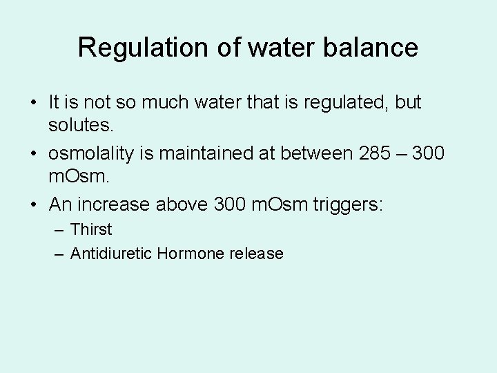
Regulation of water balance • It is not so much water that is regulated, but solutes. • osmolality is maintained at between 285 – 300 m. Osm. • An increase above 300 m. Osm triggers: – Thirst – Antidiuretic Hormone release
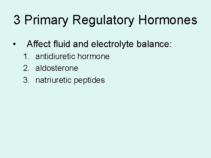
3 Primary Regulatory Hormones • Affect fluid and electrolyte balance: 1. antidiuretic hormone 2. aldosterone 3. natriuretic peptides
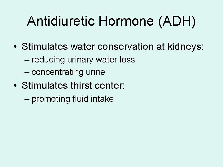
Antidiuretic Hormone (ADH) • Stimulates water conservation at kidneys: – reducing urinary water loss – concentrating urine • Stimulates thirst center: – promoting fluid intake
ADH Production • Osmoreceptors in hypothalamus: – monitor osmotic concentration of ECF • Change in osmotic concentration: – alters osmoreceptor activity • Osmoreceptor neurons secrete ADH
ADH Release (1 of 2) • Axons of neurons in anterior hypothalamus: – release ADH near fenestrated capillaries – in posterior lobe of pituitary gland
ADH Release (2 of 2) • Rate of release varies with osmotic concentration: – higher osmotic concentration increases ADH release
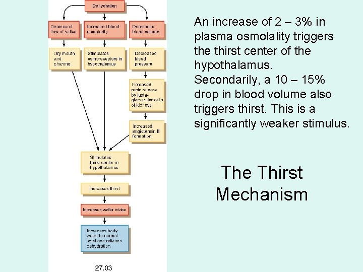
An increase of 2 – 3% in plasma osmolality triggers the thirst center of the hypothalamus. Secondarily, a 10 – 15% drop in blood volume also triggers thirst. This is a significantly weaker stimulus. The Thirst Mechanism
Aldosterone • Is secreted by adrenal cortex in response to: – rising K+ or falling Na+ levels in blood – activation of renin–angiotensin system • Determines rate of Na+ absorption and K+ loss: – along DCT and collecting system
Dehydration Chronic dehydration leads to oliguria. Severe dehydration can result in hypovolemic shock. Causes include: • Hemorrhage • Burns • Vomiting • Diarrhea • Sweating • Diuresis, which can be caused by diabetes insipidus, diabetes mellitus and hypertension (pressure diuresis).
Overhydration (Hypotonic hydration) • Also called water excess • Occurs when excess water shifts into ICF: – distorting cells – changing solute concentrations around enzymes – disrupting normal cell functions
Causes of Overhydration • Ingestion of large volume of fresh water • Injection into bloodstream of hypotonic solution • Endocrine disorders: – excessive ADH production
Causes of Overhydration • Inability to eliminate excess water in urine: – chronic renal failure – heart failure – cirrhosis
Signs of Overhydration + • Abnormally low Na concentrations (hyponatremia) • Effects on CNS function (water intoxication)
Hyponatremia • Hyponatremia results in: – Cerebral edema (brain swelling) – Sluggish neural activity – Convulsions, muscle spasms, deranged behavior. • Treated with I. V. hypertonic mannitol or something similar.
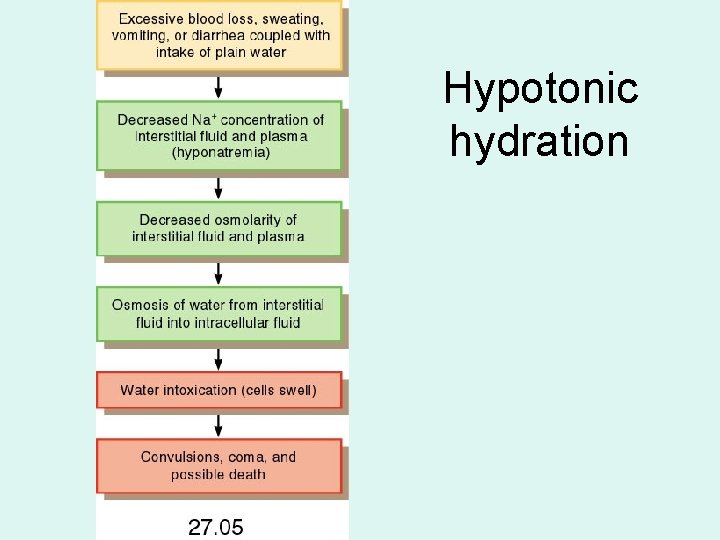
Hypotonic hydration
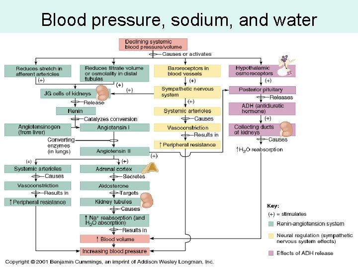
Blood pressure, sodium, and water
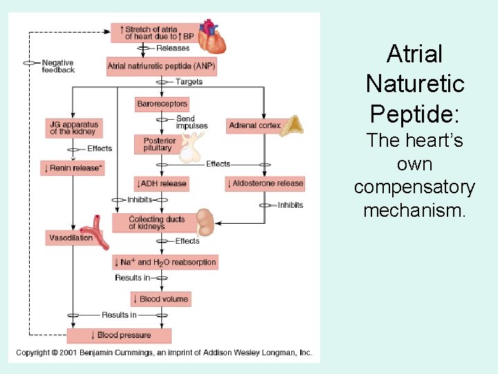
Atrial Naturetic Peptide: The heart’s own compensatory mechanism.
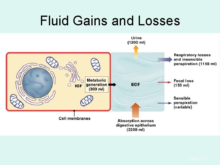
Fluid Gains and Losses Figure 27– 3
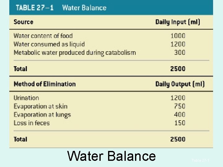
Water Balance Table 27– 1
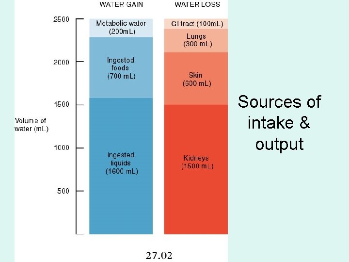
Sources of intake & output
Importance of Electrolyte Balance • Electrolyte concentration directly affects water balance • Concentrations of individual electrolytes affect cell functions
Sodium • Is the dominant cation in ECF • Sodium salts provide 90% of ECF osmotic concentration: – sodium chloride (Na. Cl) – sodium bicarbonate
Normal Sodium Concentrations • In ECF: – about 140 m. Eq/L • In ICF: – is 10 m. Eq/L or less
Potassium • Is the dominant cation in ICF
Normal Potassium Concentrations • In ICF: – about 160 m. Eq/L • In ECF: – is 3. 8– 5. 0 m. Eq/L
2 Rules of Electrolyte Balance 1. Most common problems with electrolyte balance are caused by imbalance between gains and losses of sodium ions 2. Problems with potassium balance are less common, but more dangerous than sodium imbalance
Sodium Balance in ECF 1. Sodium ion uptake across digestive epithelium 2. Sodium ion excretion in urine and perspiration
Sodium Balance in ECF + • Typical Na gain and loss: – is 48– 144 m. Eq (1. 1– 3. 3 g) per day • If gains exceed losses: – total ECF content rises • If losses exceed gains: – ECF content declines
+ Changes in ECF Na Content • Do not produce change in concentration • Corresponding water gain or loss keeps concentration constant
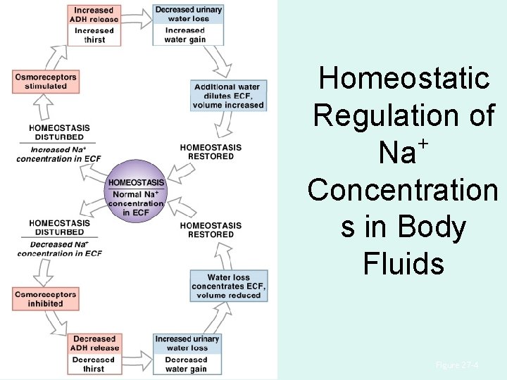
Homeostatic Regulation of + Na Concentration s in Body Fluids Figure 27– 4
+ Na Balance and ECF Volume + • Na regulatory mechanism changes ECF volume: – keeps concentration stable • When Na+ losses exceed gains: – ECF volume decreases (increased water loss) – maintaining osmotic concentration
Large Changes in ECF Volume • Are corrected by homeostatic mechanisms that regulate blood volume and pressure • If ECF volume rises: – blood volume goes up • If ECF volume drops: – blood volume goes down
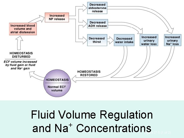
Fluid Volume Regulation + and Na Concentrations Figure 27– 5 (1 of 2)
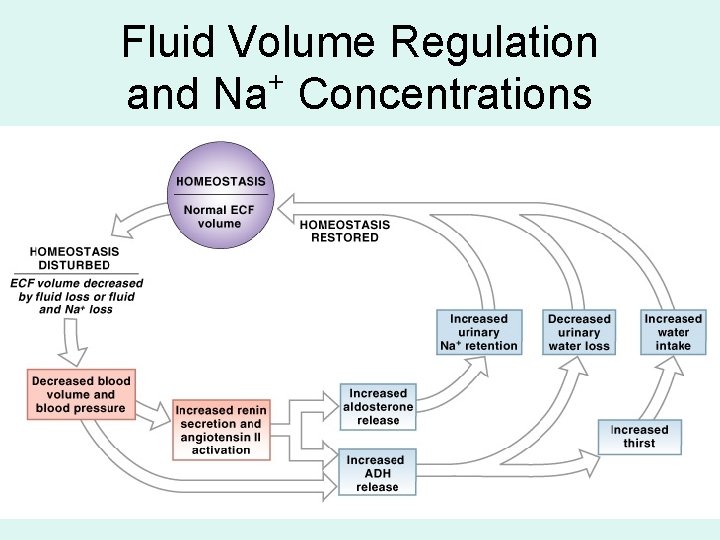
Fluid Volume Regulation + and Na Concentrations
Homeostatic Mechanisms (1 of 2) • A rise in blood volume: – elevates blood pressure • A drop in blood volume: – lowers blood pressure
Homeostatic Mechanisms (2 of 2) • Monitor ECF volume indirectly by monitoring blood pressure: – baroreceptors at carotid sinus, aortic sinus, and right atrium
+ Abnormal Na Concentrations in ECF • Hyponatremia: – body water content rises (overhydration) – ECF Na+ concentration < 130 m. Eq/L • Hypernatremia: – body water content declines (dehydration) – ECF Na+ concentration > 150 m. Eq/L
ECF Volume • If ECF volume is inadequate: – blood volume and blood pressure decline – renin–angiotensin system is activated – water and Na+ losses are reduced – ECF volume increases
Plasma Volume • If plasma volume is too large: – venous return increases: • stimulating natriuretic peptides (ANP and BNP) • reducing thirst • blocking secretion of ADH and aldosterone – salt and water loss at kidneys increases – ECF volume declines
Potassium Balance • 98% of potassium in the human body is in ICF • Cells expend energy to recover potassium ions diffused from cytoplasm into ECF
Processes of Potassium Balance 1. Rate of gain across digestive epithelium 2. Rate of loss into urine
Potassium Loss in Urine • Is regulated by activities of ion pumps: – along distal portions of nephron and collecting system – Na+ from tubular fluid is exchanged for K+ in peritubular fluid
Potassium Loss in Urine • Are limited to amount gained by absorption across digestive epithelium (about 50– 150 m. Eq (1. 9– 5. 8 g)/day
3 Factors in Tubular + Secretion of K 1. Changes in concentration of ECF: – higher ECF concentration increases rate of secretion
3 Factors in Tubular + Secretion of K 2. Changes in p. H: – low ECF p. H lowers peritubular fluid p. H – H+ rather than K+ is exchanged for Na+ in tubular fluid – rate of potassium secretion declines
3 Factors in Tubular + Secretion of K 3. Aldosterone levels: – affect K+ loss in urine – ion pumps reabsorb Na+ from filtrate in exchange for K+ from peritubular fluid • High K+ plasma concentrations stimulate aldosterone
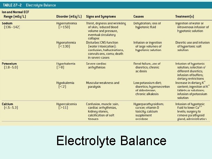
Electrolyte Balance
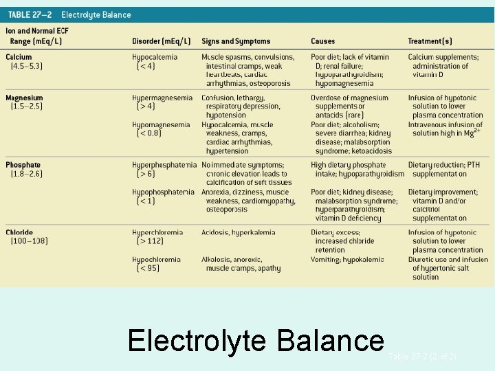
Electrolyte Balance Table 27– 2 (2 of 2)
Calcium • Is most abundant mineral in the body: – 1– 2 kg (2. 2– 4. 4 lb) – 99% deposited in skeleton
Functions of Calcium Ion • • Muscular and neural activities Blood clotting Cofactors for enzymatic reactions Second messengers + 2 (Ca )
Hormones and Calcium Homeostasis • Parathyroid hormone (PTH) and calcitriol: – raise calcium concentrations in ECF • Calcitonin: – opposes PTH and calcitriol
Calcium Absorption • At digestive tract and reabsorption along DCT: – are stimulated by PTH and calcitriol
Calcium Ion Loss • In bile, urine or feces: – is very small (0. 8– 1. 2 g/day) – about 0. 03% of calcium reserve in skeleton
Hypercalcemia • Exists if Ca 2+ concentration in ECF is > 11 m. Eq/L • Is usually caused by hyperparathyroidism: – resulting from oversecretion of PTH • Other causes: – malignant cancers (breast, lung, kidney, bone marrow) – excessive calcium or vitamin D supplementation
Hypocalcemia • Exists if Ca 2+ concentration in ECF is < 4 m. Eq/L • Is much less common than hypercalcemia • Is usually caused by chronic renal failure • May be caused by hypoparathyroidism: – undersecretion of PTH – vitamin D deficiency
Magnesium (1 of 3) • Is an important structural component of bone • The adult body contains about 29 g of magnesium • About 60% is deposited in the skeleton
Magnesium (2 of 3) • Is a cofactor for important enzymatic reactions: – phosphorylation of glucose – use of ATP by contracting muscle fibers
Magnesium (3 of 3) • Is effectively reabsorbed by PCT • Daily dietary requirement to balance urinary loss: – about 24– 32 m. Eq (0. 3– 0. 4 g)
Magnesium Ions + 2 (Mg ) • In body fluids are primarily in ICF: – Mg 2+ concentration in ICF is about 26 m. Eq/L – ECF concentration is much lower
Phosphate Ions (1 of 3) • Are required for bone mineralization • About 740 g PO 43— is bound in mineral salts of the skeleton • Daily urinary and fecal losses: – about 30– 45 m. Eq (0. 8– 1. 2 g)
Phosphate Ions (2 of 3) • In ICF, PO 4 3— is required for: – formation of high-energy compounds – activation of enzymes – synthesis of nucleic acids
Phosphate Ions (3 of 3) • In plasma, PO 4 3 —: – is reabsorbed from tubular fluid along PCT – stimulated by calcitriol • Plasma concentration is 1. 8– 2. 6 m. Eq/L
— Chloride Ions (Cl ) • Are the most abundant anions in ECF • Plasma concentration is 100– 108 m. Eq/L • ICF concentrations are usually low
— Chloride Ions (Cl ) • Are absorbed across digestive tract with Na+ • Are reabsorbed with Na+ by carrier proteins along renal tubules • Daily loss is small: – 48– 146 m. Eq (1. 7– 5. 1 g)
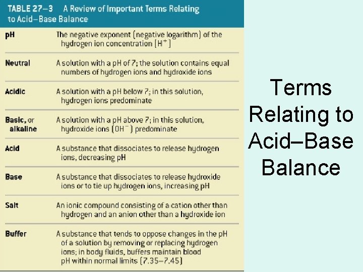
Terms Relating to Acid–Base Balance
Strong or Weak • Strong acids and strong bases: – dissociate completely in solution • Weak acids or weak bases: – do not dissociate completely in solution – some molecules remain intact
Acidosis • Physiological state resulting from abnormally low plasma p. H • Acidemia: – plasma p. H < 7. 35
Alkalosis • Physiological state resulting from abnormally high plasma p. H • Alkalemia: – plasma p. H > 7. 45
Acidosis and Alkalosis • Affect all body systems: – particularly nervous and cardiovascular systems • Both are dangerous: – but acidosis is more common – because normal cellular activities generate acids
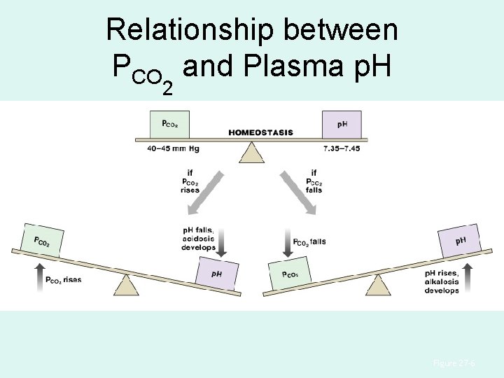
Relationship between PCO and Plasma p. H 2 Figure 27– 6
3 Types of Acids in the Body 1. Volatile acids - Can leave solution and enter the atmosphere (ex: Carbonic acid) 2. Fixed acids - Are acids that do not leave solution. Once produced they remain in body fluids until eliminated by kidneys 3. Organic acids -
Sulfuric Acid and Phosphoric Acid • Are most important fixed acids in the body • Are generated during catabolism of: – amino acids – phospholipids – nucleic acids
Organic Acids • Produced by aerobic metabolism: – are metabolized rapidly – do not accumulate • Produced by anaerobic metabolism (e. g. , lactic acid) – build up rapidly
What’ a “buffer” • Acids are proton donors • Bases are proton acceptors – Strong acids & bases dissociate completely • Acid buffer systems are comprised of compounds that resist p. H changes by accepting protons from solutions containing strong acids. • Base buffer systems accept OH- ions from solutions. (Not discussed in the text).
Buffers • Are dissolved compounds that stabilize p. H: – by providing or removing H+ • Weak acids: – can donate H+ • Weak bases: – can absorb H+
A Buffer System • Consists of a combination of: – a weak acid – and the anion released by its dissociation • The anion functions as a weak base
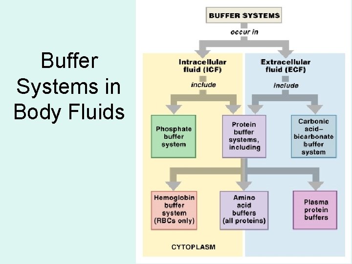
Buffer Systems in Body Fluids Figure 27– 7
3 Major Buffer Systems 1. Protein buffer systems: – help regulate p. H in ECF and ICF – interact extensively with other buffer systems 2. Carbonic acid–bicarbonate buffer system: – most important in ECF 3. Phosphate buffer system: – buffers p. H of ICF and urine
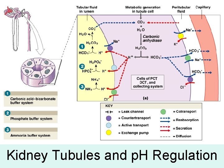
Kidney Tubules and p. H Regulation
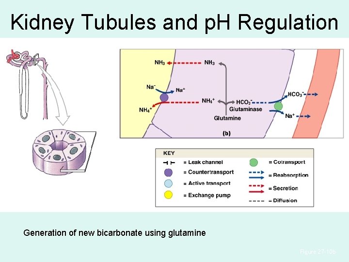
Kidney Tubules and p. H Regulation Generation of new bicarbonate using glutamine Figure 27– 10 b
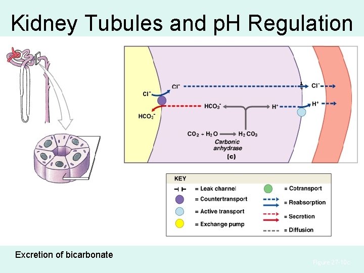
Kidney Tubules and p. H Regulation Excretion of bicarbonate Figure 27– 10 c
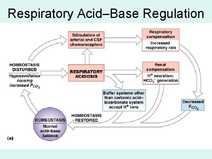
Respiratory Acid–Base Regulation
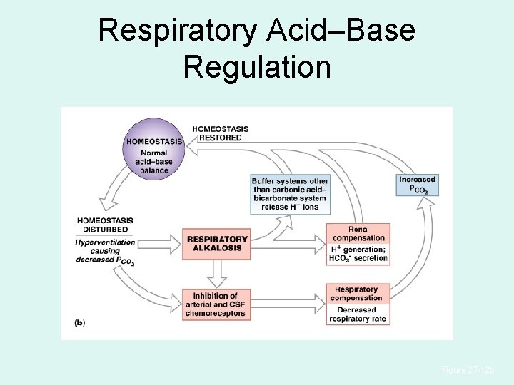
Respiratory Acid–Base Regulation Figure 27– 12 b
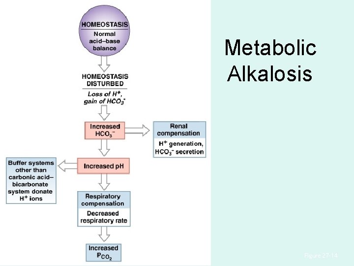
Metabolic Alkalosis Figure 27– 14
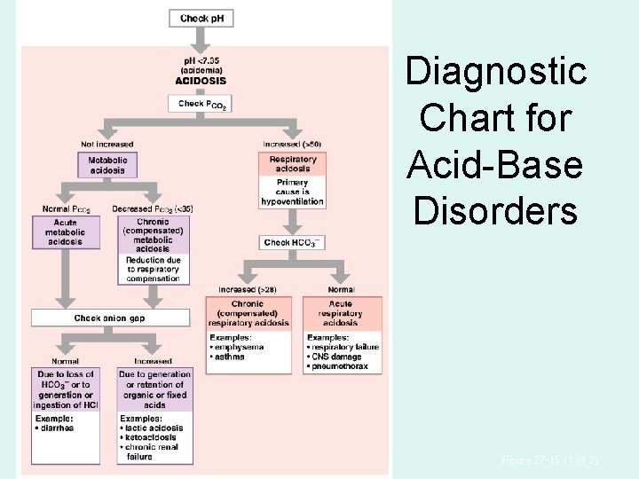
Diagnostic Chart for Acid-Base Disorders Figure 27– 15 (1 of 2)
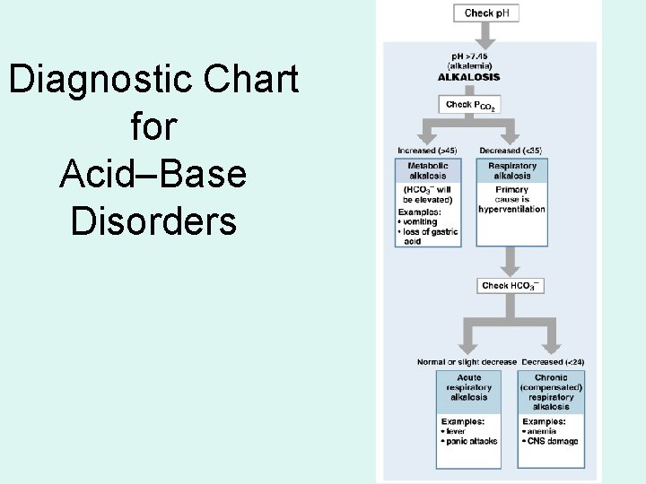
Diagnostic Chart for Acid–Base Disorders
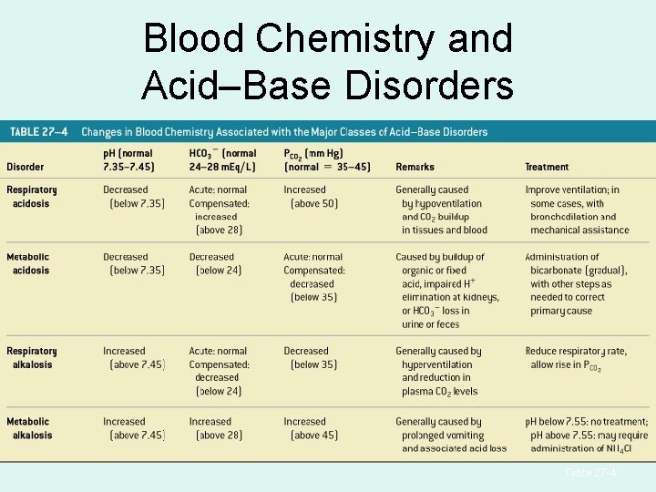
Blood Chemistry and Acid–Base Disorders Table 27– 4
Enjoy
- Slides: 102