Skeletal System Review Sheet Test Date is 1122




- Slides: 56
Skeletal System Review Sheet Test Date is 11/22
Diagrams that will be on the Exam
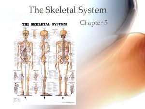
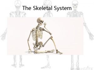
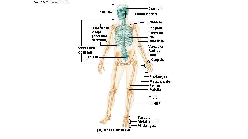
Figure 5. 8 a The human skeleton. Skull Cranium Facial bones Clavicle Thoracic cage (ribs and sternum) Vertebral column Sacrum Scapula Sternum Rib Humerus Vertebra Radius Ulna Carpals Phalanges Metacarpals Femur Patella Tibia Fibula (a) Anterior view Tarsals Metatarsals Phalanges
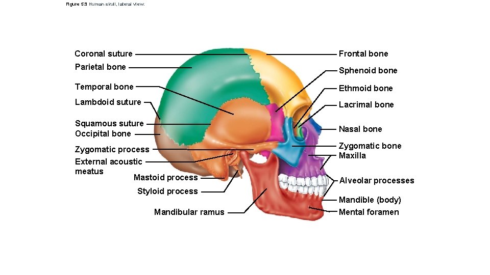
Figure 5. 9 Human skull, lateral view. Coronal suture Frontal bone Parietal bone Sphenoid bone Temporal bone Ethmoid bone Lambdoid suture Lacrimal bone Squamous suture Occipital bone Nasal bone Zygomatic process Zygomatic bone Maxilla External acoustic meatus Mastoid process Styloid process Mandibular ramus Alveolar processes Mandible (body) Mental foramen
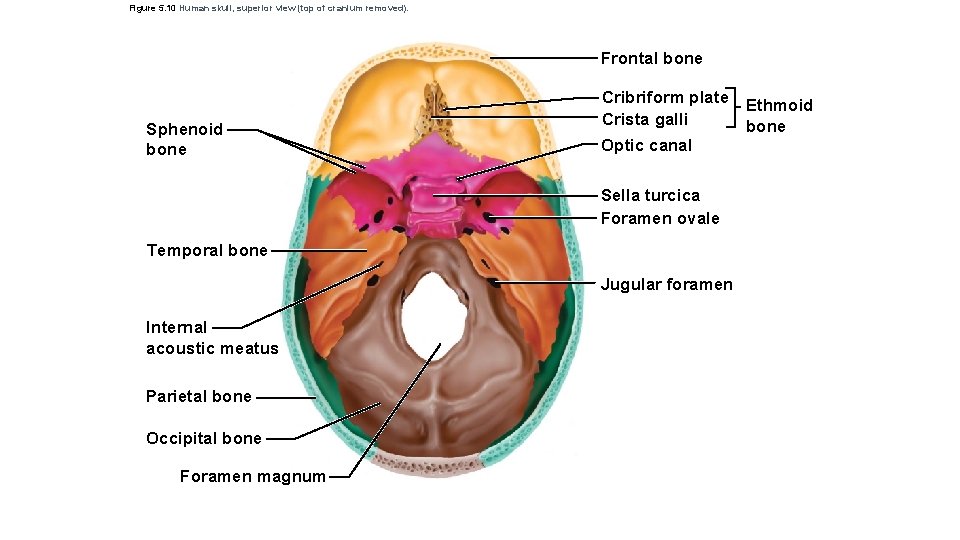
Figure 5. 10 Human skull, superior view (top of cranium removed). Frontal bone Sphenoid bone Cribriform plate Ethmoid Crista galli bone Optic canal Sella turcica Foramen ovale Temporal bone Jugular foramen Internal acoustic meatus Parietal bone Occipital bone Foramen magnum
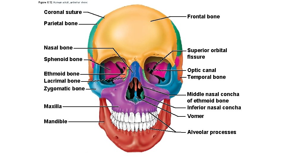
Figure 5. 12 Human skull, anterior view. Coronal suture Frontal bone Parietal bone Nasal bone Sphenoid bone Ethmoid bone Lacrimal bone Zygomatic bone Maxilla Mandible Superior orbital fissure Optic canal Temporal bone Middle nasal concha of ethmoid bone Inferior nasal concha Vomer Alveolar processes
Figure 5. 23 c Bones of the shoulder girdle. Suprascapular notch Coracoid process Superior angle Spine Acromion Glenoid cavity at lateral angle Medial border Lateral border (c) Right scapula, posterior aspect
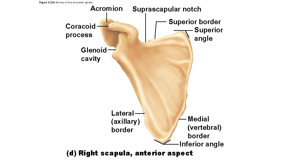
Figure 5. 23 d Bones of the shoulder girdle. Acromion Suprascapular notch Superior border Superior angle Coracoid process Glenoid cavity Lateral (axillary) border Medial (vertebral) border Inferior angle (d) Right scapula, anterior aspect
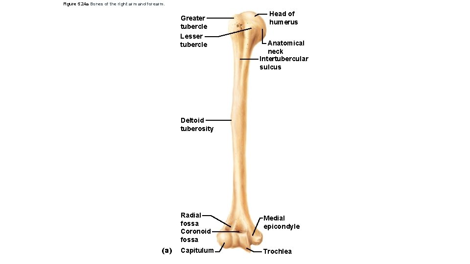
Figure 5. 24 a Bones of the right arm and forearm. Greater tubercle Lesser tubercle Head of humerus Anatomical neck Intertubercular sulcus Deltoid tuberosity (a) Radial fossa Coronoid fossa Medial epicondyle Capitulum Trochlea
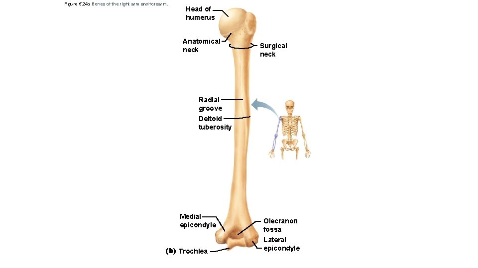
Figure 5. 24 b Bones of the right arm and forearm. Head of humerus Anatomical neck Surgical neck Radial groove Deltoid tuberosity Medial epicondyle (b) Trochlea Olecranon fossa Lateral epicondyle
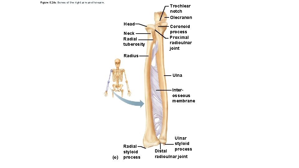
Figure 5. 24 c Bones of the right arm and forearm. Trochlear notch Olecranon Head Neck Radial tuberosity Coronoid process Proximal radioulnar joint Radius Ulna Interosseous membrane (c) Radial styloid process Ulnar styloid process Distal radioulnar joint
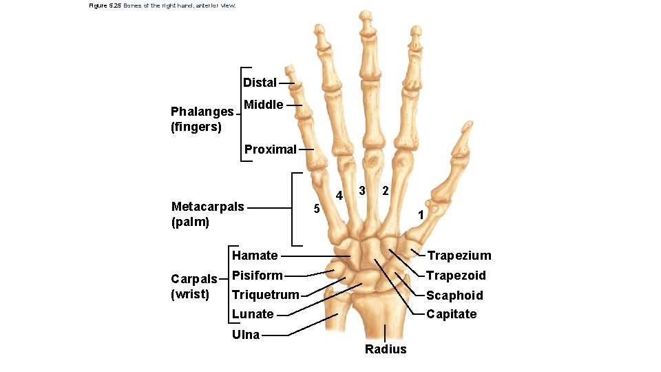
Figure 5. 25 Bones of the right hand, anterior view. Distal Phalanges (fingers) Middle Proximal Metacarpals (palm) Carpals (wrist) 5 4 3 2 1 Hamate Trapezium Pisiform Triquetrum Trapezoid Scaphoid Capitate Lunate Ulna Radius
Figure 5. 26 a The bony pelvis. Iliac crest Sacroiliac joint Ilium Coxal bone (or hip bone) Sacrum Pubis Pelvic brim Coccyx Ischial spine Acetabulum Ischium (a) Pubic symphysis Pubic arch
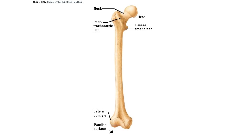
Figure 5. 27 a Bones of the right thigh and leg. Neck Head Intertrochanteric line Lateral condyle Patellar surface (a) Lesser trochanter
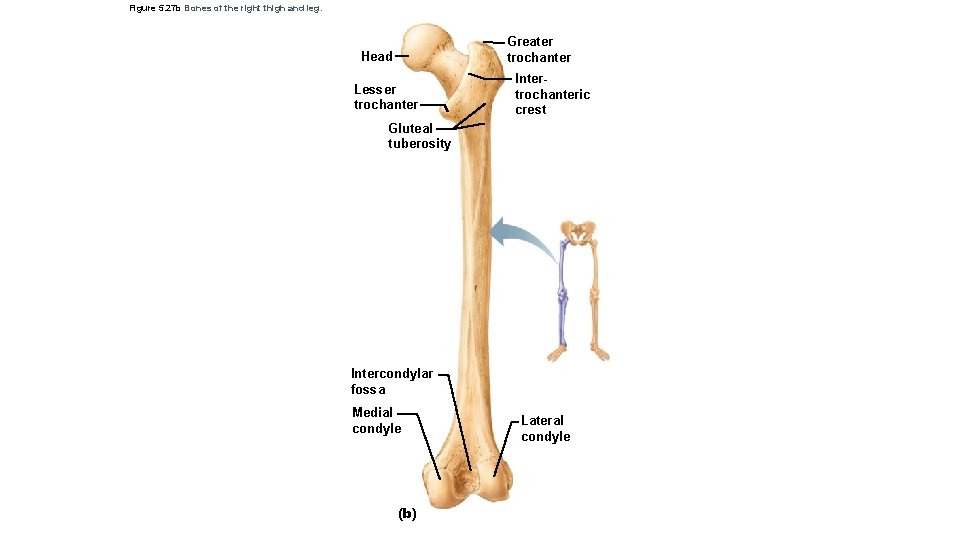
Figure 5. 27 b Bones of the right thigh and leg. Greater trochanter Head Lesser trochanter Intertrochanteric crest Gluteal tuberosity Intercondylar fossa Medial condyle (b) Lateral condyle
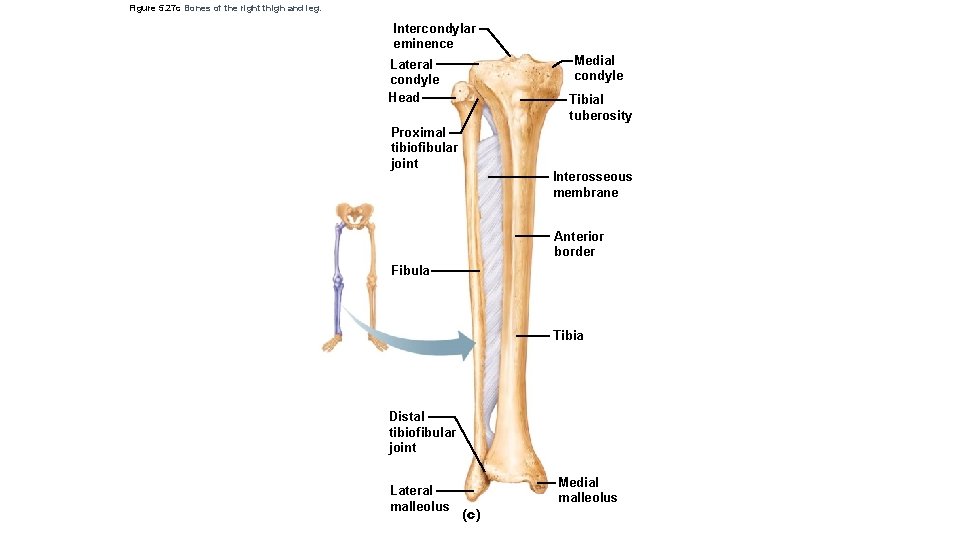
Figure 5. 27 c Bones of the right thigh and leg. Intercondylar eminence Medial condyle Lateral condyle Head Tibial tuberosity Proximal tibiofibular joint Interosseous membrane Anterior border Fibula Tibia Distal tibiofibular joint Lateral malleolus (c) Medial malleolus
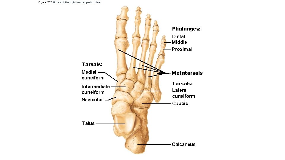
Figure 5. 28 Bones of the right foot, superior view. Phalanges: Distal Middle Proximal Tarsals: Medial cuneiform Intermediate cuneiform Navicular Metatarsals Tarsals: Lateral cuneiform Cuboid Talus Calcaneus
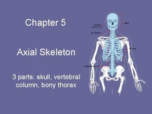
The Axial Skeleton • Forms the longitudinal axis of the body • Divided into three parts 1. ________ 2. _____column 3. _______ © 2015 Pearson Education, Inc.
The Skull • Two sets of bones 1. __________ 2. __________ • Hold eyes in anterior position • Allow facial muscles to express feelings • Bones are joined by _____ • Only the _____ is attached by a freely movable joint © 2015 Pearson Education, Inc.
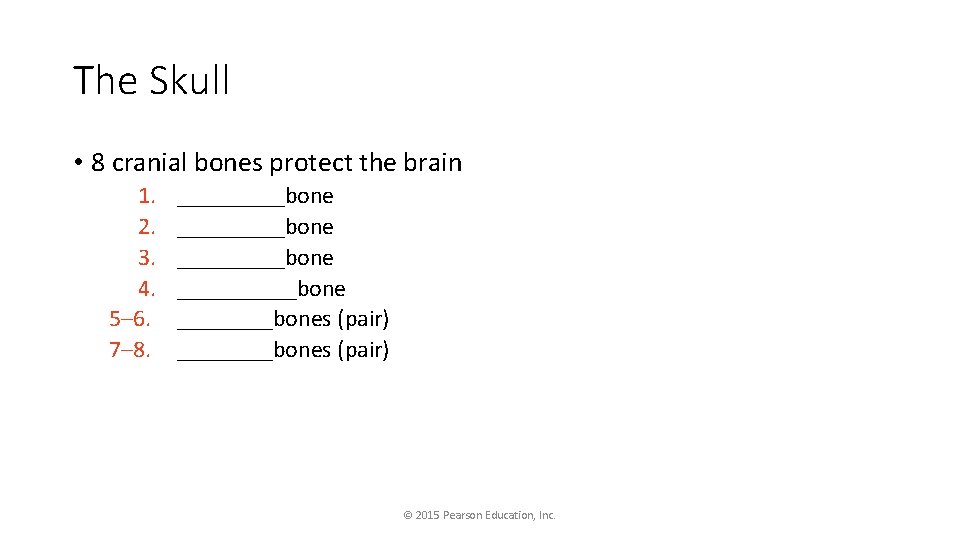
The Skull • 8 cranial bones protect the brain 1. 2. 3. 4. 5– 6. 7– 8. _________bone ________bones (pair) © 2015 Pearson Education, Inc.
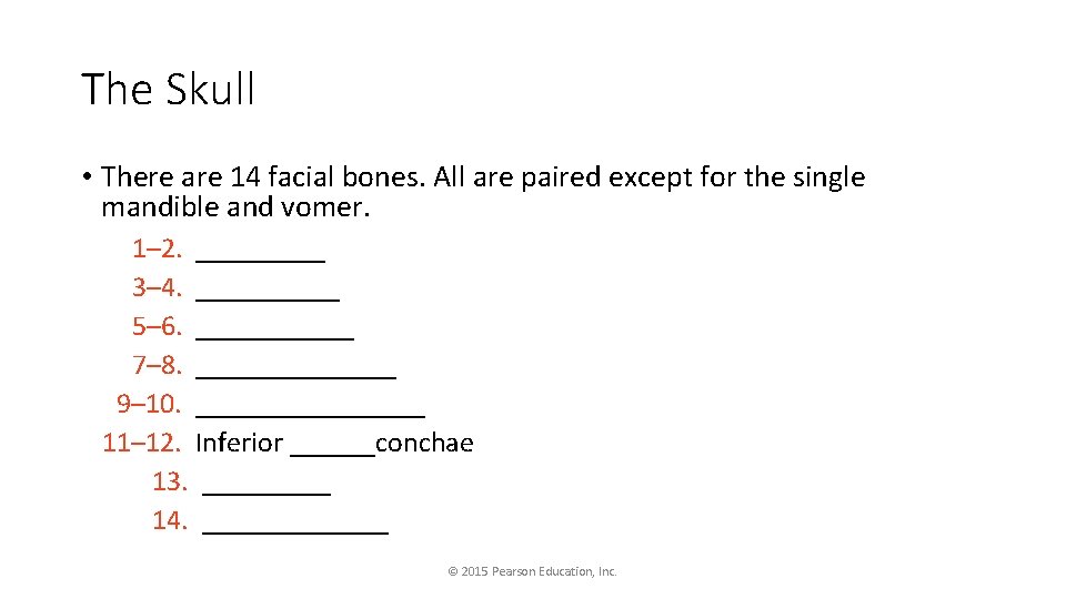
The Skull • There are 14 facial bones. All are paired except for the single mandible and vomer. 1– 2. _____ 3– 4. _____ 5– 6. ______ 7– 8. _______ 9– 10. ________ 11– 12. Inferior ______conchae 13. _____ 14. _______ © 2015 Pearson Education, Inc.
Paranasal Sinuses • Hollow portions of bones surrounding the nasal cavity • Functions of paranasal sinuses • _________ • ______sounds made as we speak © 2015 Pearson Education, Inc.
The Hyoid Bone • Closely related to ______ and ______bones • The only bone that does not ______with another bone • Serves as a movable base for the ______ • Aids in _____and ______ © 2015 Pearson Education, Inc.
The Fetal Skull • The fetal skull is large compared to the infant’s total body length • Fetal skull is 1/4 body length compared to adult skull, which is 1/8 body length • Fontanels are fibrous membranes connecting the cranial bones • Allow skull compression during birth • Allow the brain to grow during later pregnancy and infancy • Convert to bone within 24 months after birth © 2015 Pearson Education, Inc.
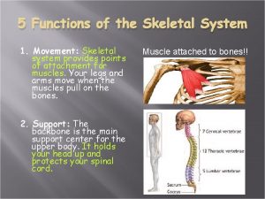
Vertebral Column (Spine) • Vertebral column provides axial support • Extends from skull to the pelvis • 26 single vertebral bones are separated by intervertebral discs • 7 cervical vertebrae are in the neck • 12 thoracic vertebrae are in the chest region • 5 lumbar vertebrae are associated with the lower back © 2015 Pearson Education, Inc.
Vertebral Column (Spine) • 9 vertebrae fuse to form two composite bones • Sacrum formed by the fusion of 5 vertebrae • Coccyx (tailbone) formed by the fusion of 3 to 5 vertebrae © 2015 Pearson Education, Inc.
Vertebral Column (Spine) • Primary curvatures • Spinal curvatures of the thoracic and sacral regions • Present from birth • Form a C-shaped curvature as in newborns • Secondary curvatures • Spinal curvatures of the cervical and lumbar regions • Develop after birth • Form an S-shaped curvature as in adults © 2015 Pearson Education, Inc.
Vertebral Column (Spine) • Parts of a typical vertebra • Body (centrum) • Vertebral arch • Pedicle • Lamina • • Vertebral foramen Transverse processes Spinous process Superior and inferior articular processes © 2015 Pearson Education, Inc.
The Bony Thorax • Forms a cage to protect major organs • Consists of three parts 1. Sternum 2. Ribs • True ribs (pairs 1– 7) • False ribs (pairs 8– 12) • Floating ribs (pairs 11– 12) 3. Thoracic vertebrae © 2015 Pearson Education, Inc.
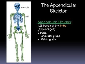
The Appendicular Skeleton • Composed of 126 bones • _____(appendages) • _____girdle • _____ girdle © 2015 Pearson Education, Inc.
The Pectoral (Shoulder) Girdle • Composed of two bones that attach the upper limb to the axial skeletal 1. _______ 2. _____ • Pectoral girdle (2) • Light, poorly reinforced girdle • Allows the upper limb a great deal of freedom © 2015 Pearson Education, Inc.
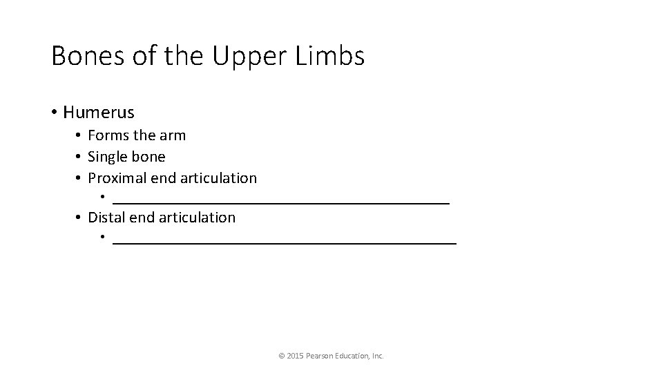
Bones of the Upper Limbs • Humerus • Forms the arm • Single bone • Proximal end articulation • _________________________ • Distal end articulation • _________________________ © 2015 Pearson Education, Inc.
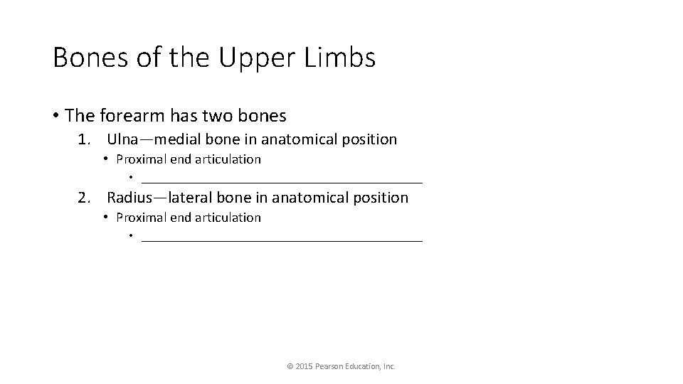
Bones of the Upper Limbs • The forearm has two bones 1. Ulna—medial bone in anatomical position • Proximal end articulation • ______________________ 2. Radius—lateral bone in anatomical position • Proximal end articulation • ______________________ © 2015 Pearson Education, Inc.
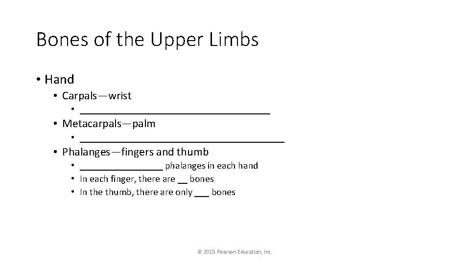
Bones of the Upper Limbs • Hand • Carpals—wrist • ____________________ • Metacarpals—palm • _____________________ • Phalanges—fingers and thumb • _________ phalanges in each hand • In each finger, there are __ bones • In the thumb, there are only ___ bones © 2015 Pearson Education, Inc.
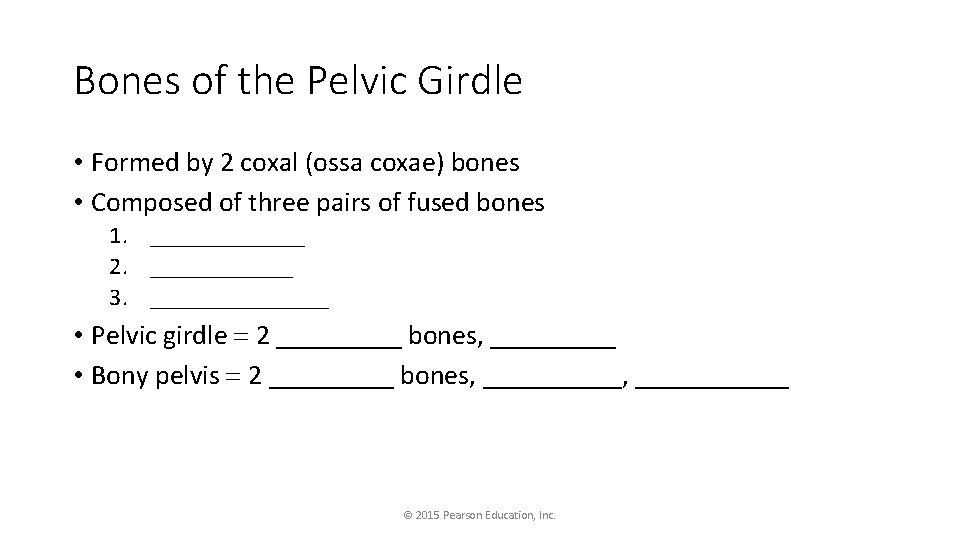
Bones of the Pelvic Girdle • Formed by 2 coxal (ossa coxae) bones • Composed of three pairs of fused bones 1. _______ 2. ______ 3. ________ • Pelvic girdle 2 _____ bones, _____ • Bony pelvis 2 _____ bones, ___________ © 2015 Pearson Education, Inc.
Bones of the Pelvic Girdle • The total weight of the upper body rests on the pelvis • Pelvis protects several organs • ________________ • Part of the ____________ © 2015 Pearson Education, Inc.
Gender Differences of the Pelvis • The ________: • • • Inlet is larger and more circular Pelvis as a whole is shallower, and the bones are lighter and thinner Ilia flare more laterally Sacrum is shorter and less curved Ischial spines are shorter and farther apart; thus, the outlet is larger Pubic arch is more rounded because the angle of the pubic arch is greater © 2015 Pearson Education, Inc.
Bones of the Lower Limbs • The _______—thigh bone • The heaviest, strongest bone in the body • Proximal end articulation • _____________________ • Distal end articulation • _____________________ © 2015 Pearson Education, Inc.
Bones of the Lower Limbs • The lower leg has two bones 1. _____—shinbone; larger and medially oriented • Proximal end articulation • _____________________ 2. _______ - thin and sticklike; lateral to the tibia • __________________ © 2015 Pearson Education, Inc.
Bones of the Lower Limbs • The _____ • Tarsals—___ bones • Two largest tarsals • Calcaneus (heel bone) • Talus • Metatarsals—___ bones form the sole of the foot • Phalanges- _____ bones form the toes © 2015 Pearson Education, Inc.
Arches of the Foot • Bones of the foot are arranged to form three strong arches • Two ______ • One ______ © 2015 Pearson Education, Inc.
Joints • Joints are ______ • Two or more bones meet • Functions of joints • Hold bones together • Allow for mobility • Two ways joints are classified • Functionally • Structurally © 2015 Pearson Education, Inc.
Functional Classification of Joints • Synarthroses • _________ • Amphiarthroses • ___________ • Diarthroses • ___________ © 2015 Pearson Education, Inc.
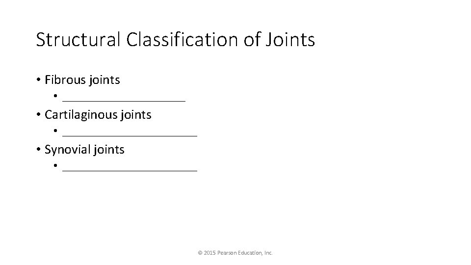
Structural Classification of Joints • Fibrous joints • ___________ • Cartilaginous joints • ____________ • Synovial joints • ____________ © 2015 Pearson Education, Inc.
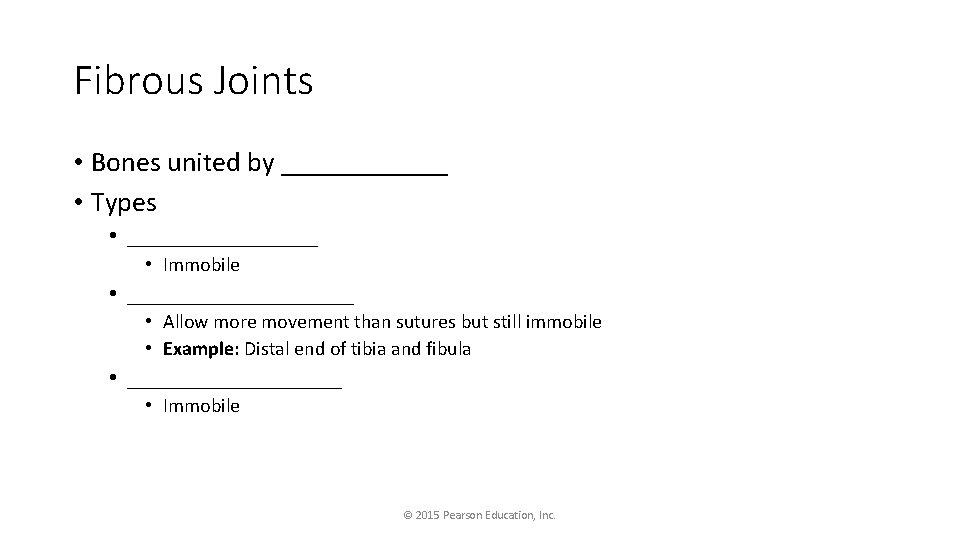
Fibrous Joints • Bones united by ______ • Types • ________ • Immobile • __________ • Allow more movement than sutures but still immobile • Example: Distal end of tibia and fibula • _________ • Immobile © 2015 Pearson Education, Inc.
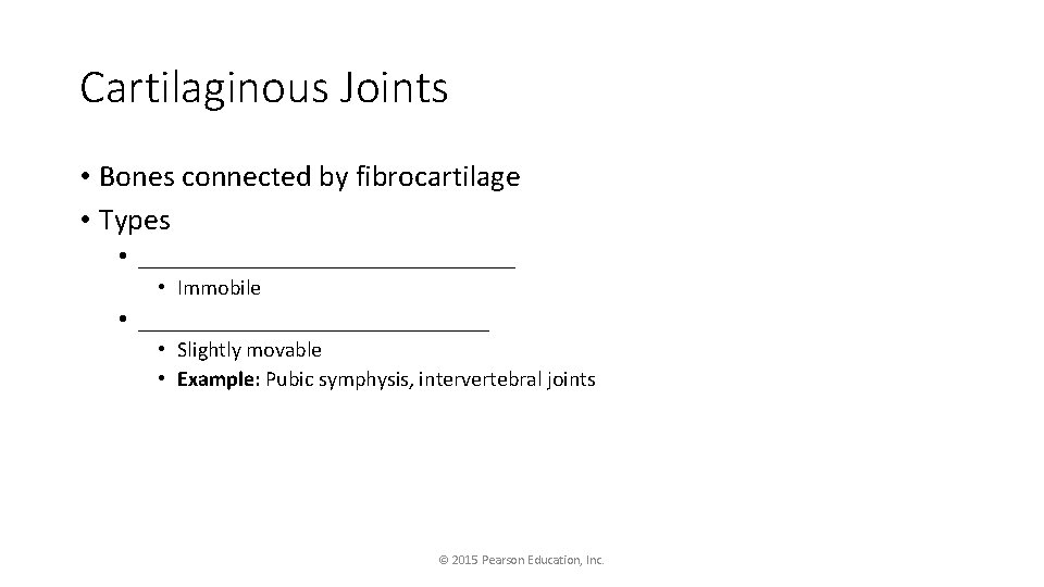
Cartilaginous Joints • Bones connected by fibrocartilage • Types • _______________ • Immobile • ______________ • Slightly movable • Example: Pubic symphysis, intervertebral joints © 2015 Pearson Education, Inc.
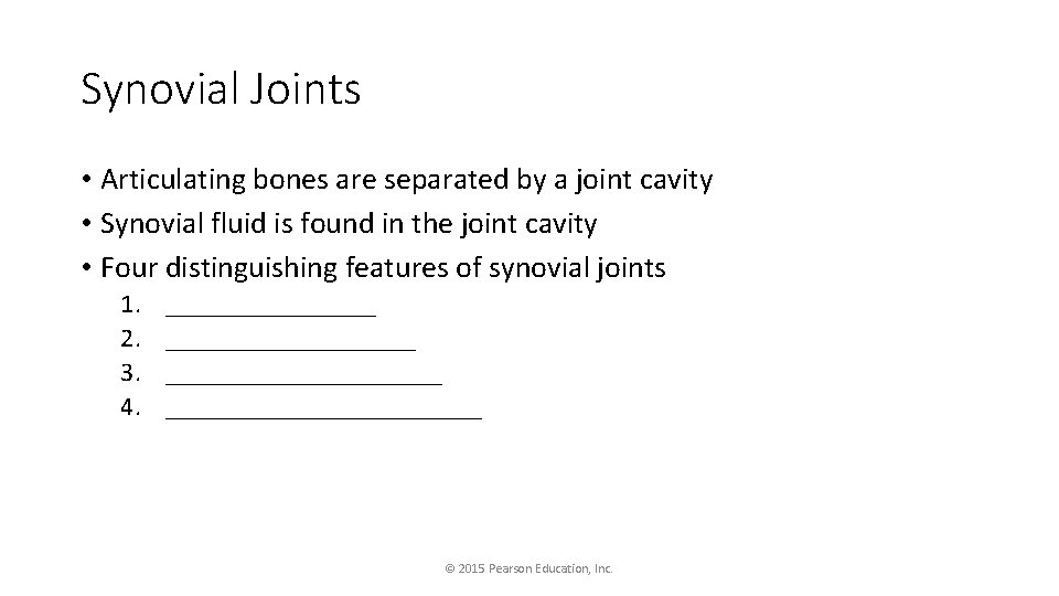
Synovial Joints • Articulating bones are separated by a joint cavity • Synovial fluid is found in the joint cavity • Four distinguishing features of synovial joints 1. 2. 3. 4. ________________________ © 2015 Pearson Education, Inc.
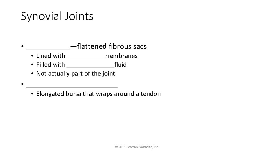
Synovial Joints • ______—flattened fibrous sacs • Lined with ______membranes • Filled with _______fluid • Not actually part of the joint • ____________ • Elongated bursa that wraps around a tendon © 2015 Pearson Education, Inc.
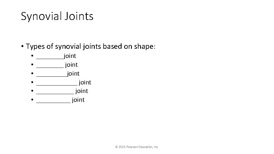
Synovial Joints • Types of synovial joints based on shape: • • • ____joint ____________ joint __________ joint © 2015 Pearson Education, Inc.
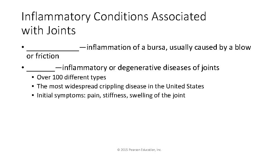
Inflammatory Conditions Associated with Joints • _______—inflammation of a bursa, usually caused by a blow or friction • _______—inflammatory or degenerative diseases of joints • Over 100 different types • The most widespread crippling disease in the United States • Initial symptoms: pain, stiffness, swelling of the joint © 2015 Pearson Education, Inc.
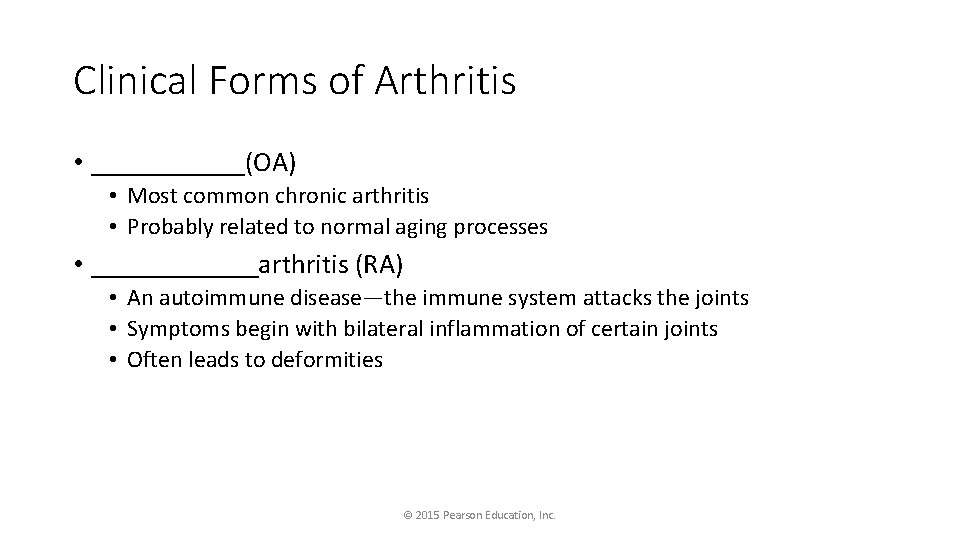
Clinical Forms of Arthritis • ______(OA) • Most common chronic arthritis • Probably related to normal aging processes • ______arthritis (RA) • An autoimmune disease—the immune system attacks the joints • Symptoms begin with bilateral inflammation of certain joints • Often leads to deformities © 2015 Pearson Education, Inc.
Clinical Forms of Arthritis • Gouty arthritis (gout) • Inflammation of joints is caused by a deposition of uric acid crystals from the blood • Can usually be controlled with diet • More common in men © 2015 Pearson Education, Inc.
Skeletal Changes Throughout Life • Fetus • Long bones are formed of _____ cartilage • Flat bones begin as ______membranes • Flat and long bone models are converted to _____ • Birth • ________remain until around age 2 © 2015 Pearson Education, Inc.
Skeletal Changes Throughout Life • Adolescence • Epiphyseal plates become ossified, and long bone growth ends • Size of cranium in relationship to body • _____ old—skull is larger in proportion to the body compared to that of an adult • ________years old—skull is near adult size and proportion • Between ________, the face grows out from the skull © 2015 Pearson Education, Inc.
Skeletal Changes Throughout Life • ___________ • Bone-thinning disease afflicting • 50 percent of women over age 65 • 20 percent of men over age 70 • Disease makes bones fragile, and bones can easily fracture • Vertebral collapse results in kyphosis (also known as “dowager’s hump”) • Estrogen aids in health and normal density of a female skeleton © 2015 Pearson Education, Inc.