Pulmonary Ventilation Dr amna tahir Learning outcomes At


 Fresh air “Old air” Alveolus")


- Slides: 34
Pulmonary Ventilation Dr amna tahir
Learning outcomes At the end of this lecture you are able to get the idea of • Pulmonary ventilation • introduction of different pressure that normally involved changed in normal respiration • Muscles and movements related to respiration • Alveolar ventilation • DEAD SPACE, its types , advantages and disadvantages
Copyright © 2007 Lippincott Williams & Wilkins. Mc. Ardle, Katch, and Katch: Exercise Physiology: Energy, Nutrition, and Human Performance, Sixth Edition
Copyright © 2007 Lippincott Williams & Wilkins. Mc. Ardle, Katch, and Katch: Exercise Physiology: Energy, Nutrition, and Human Performance, Sixth Edition
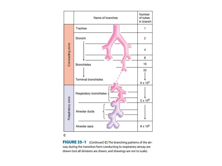
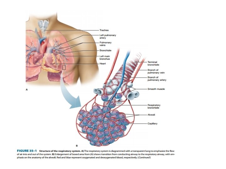
Respiratory Zone Copyright © 2008 Pearson Education, Inc. , publishing as Benjamin Cummings. Figure 16. 3 (3 of 3)
Anatomy of the Respiratory Zone Copyright © 2008 Pearson Education, Inc. , publishing as Benjamin Cummings. Figure 16. 5 a
Copyright © 2007 Lippincott Williams & Wilkins. Mc. Ardle, Katch, and Katch: Exercise Physiology: Energy, Nutrition, and Human Performance, Sixth Edition
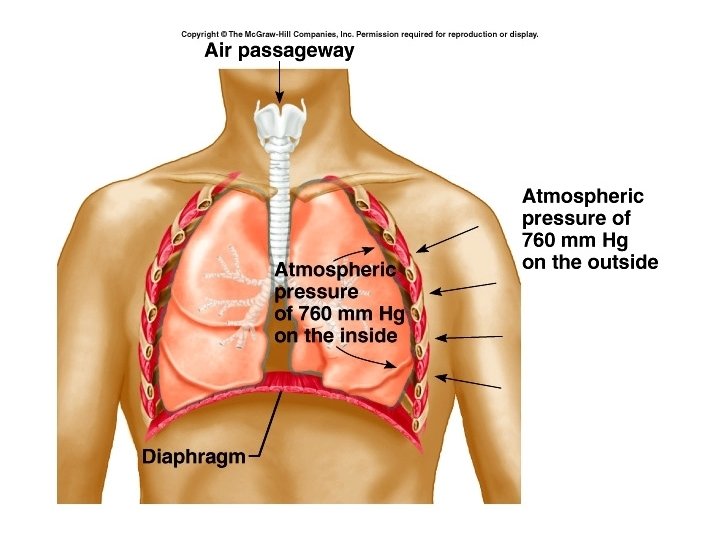
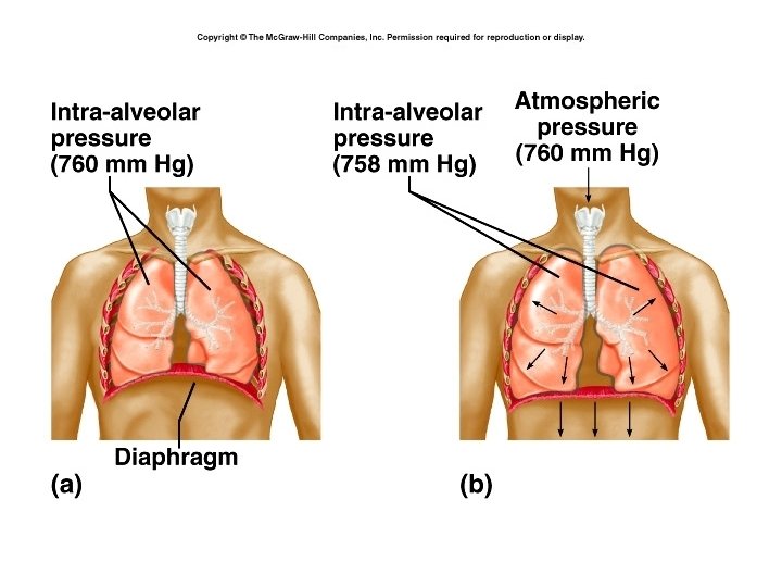
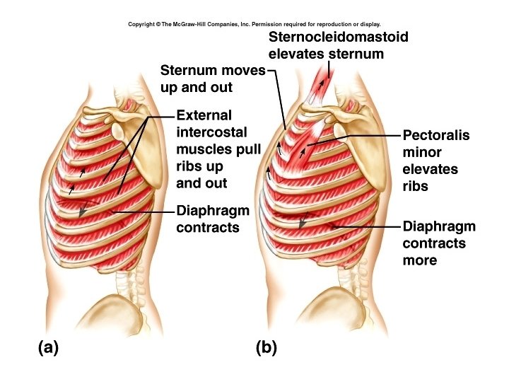
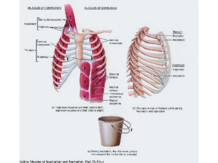
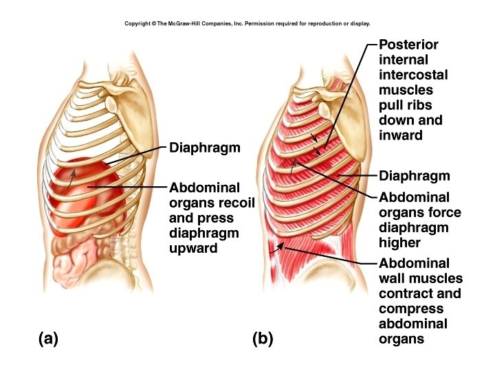
PULMONARY VENTILATION: MECHANISM – Pressure gradients are established by changes in the size of the thoracic cavity that are produced by contraction and relaxation of muscles (Figures 24 -4 and 24 -5) – Boyle’s law: the volume of gas varies inversely with pressure at a constant temperature – Inspiration: contraction of the diaphragm and external intercostals produces inspiration; as they contract, the thoracic cavity becomes larger (Figures 24 -6 and 24 -7) • Expansion of the thorax results in decreased intrapleural pressure, leading to decreased alveolar pressure • Air moves into the lungs when alveolar pressure drops below atmospheric pressure • Compliance: ability of pulmonary tissues to stretch, thus making inspiration possible 12
Chest Wall and Pleural Sac Copyright © 2008 Pearson Education, Inc. , publishing as Benjamin Cummings. Figure 16. 7
Pulmonary Pressures Copyright © 2008 Pearson Education, Inc. , publishing as Benjamin Cummings. Figure 16. 8 a–b
20
21
22
23
24
Volume and Pressure Changes Copyright © 2008 Pearson Education, Inc. , publishing as Benjamin Cummings. Figure 16. 13
Spirometry Copyright © 2008 Pearson Education, Inc. , publishing as Benjamin Cummings. Figure 16. 15
27
Minute Ventilation Total volume of air entering and leaving respiratory system each minute – Minute ventilation = VT x RR – Normal respiration rate = 12 breaths/min – Normal VT = 500 m. L – Normal minute ventilation = • 500 m. L x 12 breaths/min = 6000 m. L/min Copyright © 2008 Pearson Education, Inc. , publishing as Benjamin Cummings.
Dead Space and Ventilation Conducting zone (anatomical dead space) Fresh air “Old air” Alveolus Expiration (c) Copyright © 2008 Pearson Education, Inc. , publishing as Benjamin Cummings. Inspiration (a) CO 2 Exchange with blood (b) Figure 16. 17
Alveolar Ventilation – Volume of air reaching the gas exchange areas per minute – Alveolar ventilation = (VT x RR) – (DSV x RR) – Normal = 4200 m. L/min (500 m. L/br x 12 br/min) – (150 m. L/br X 12 br/min) Copyright © 2008 Pearson Education, Inc. , publishing as Benjamin Cummings.
Respiratory Rate and Ventilation Copyright © 2008 Pearson Education, Inc. , publishing as Benjamin Cummings. Table 16. 1
Definitions of Dead Space Anatomic Dead Space Physiologic Dead Space Low Blood Flow Copyright © 2006 by Elsevier, Inc.
Thank you