Preoperative planning Principle of treatment for Mid face


 Internal Fixation 1. Suspension Wires")


 2. Zygomaticomaxillary (Lefort –II) 3.")


 0. 3 – 0. 6 mm Used for")


- Slides: 39
Pre-operative planning Principle of treatment for Mid face #: CONSERVATIVE OPERATIVE/SURGICAL: REDUCTION and FIXATION
Indications for Closed Reduction: Non displaced fracture, Grossly comminuted fractures, Fractures exposed by significant loss of overlying soft tissues, Edentulous maxillary fractures, In children with developing dentition. Indications for open reduction: Displaced fractures, Multiple fractures of the facial bones, Fractures of the edentulous maxilla with severe displacement, Delay of treatment and interposition of soft tissues between non-contacting displaced fracture segments, Specific systemic conditions contraindicating IMF.
fractures 1. 2. 3. 4. 5. 6. 7. 8. 9. 10.
Classification of methods of Maxillary Fracture Fixation A ) Internal Fixation 1. Suspension Wires 2. Direct Osteosynthesis B) External Fixation 1. Craniomandibular 2. Craniomaxillary
Suspension Wires – non-rigid osteosynthesis i. Frontal-central or laterally placed ii. Circumzygomatic iii. Zygomatic iv. Circumpalatal/palatal screw v. Infraorbital vi. Piriform Aperture vii. Peralveolar
Suspension Wires- Circumzygomatic wiring by Obwegeser.
Suspension Wires. Circumzygomatic wiring by Obwegeser
Suspension Wires- Orbital rim wiring
Suspension Wires- Piriform aperture wiring
Summary of Suspension wiring according to fracture site Type of Suspension Wire Type of Le Fort Fracture 1. Frontal a. Central Le Fort III & II b. Lateral Le Fort III & II 2. Circumzygomatic Le Fort I & II 3. Zygomatic Le Fort I 4. Infraorbital Le Fort I 5. Piriform Aperture Le Fort I
Disadvantages of Suspension Wiring Incomplete fixation of fractured fragments Insufficient visualization of fractures by closed reduction Compression against the cranial base No 3 -dimensional stability Patients dislike intra-oral splints as it hinders oral hygiene maintainence.
Internal Fixation Direct Osteosynthesis - 1. Interosseous Wires. 2. Plates and Screws.
Direct osteosynthesis Intraosseous Wires 1. Maxillary (Lefort –I ) 2. Zygomaticomaxillary (Lefort –II) 3. Frontonasal (Le. Fort –II &III) 4. Zygomaticofrontal (Lefort III) 5. Zygomatic bone (comminuted)
Non rigid type of osteosynthesis No 3 dimensional stability, it provides only monoplane traction. IMF is always needed Interfragmentary pressure can not be controlled. Under functional stress, wire loses rigidity, direction control and surface contact. Delayed healing because of micromovement at fracture site.
Direct osteosynthesis 2. Plates & Screws for midface fractures - Stainless steel mini-plating system Titanium mini-plating system Vitallium, Cobalt chromium, molybdenum alloy plates Bioresorbable plating system.
Miniplates and screws These are monocortical, semi-rigid fixation device which provide 3 D stability. Designs: X, H, L, T, Y Thickness: 0. 6 -1 mm Type of metals: Stainless steel, Titanium, Vitallium Advantages: Easily adaptable, Monocortical, Functional
Micro plates Harle & duker(1975; Luhr(1979) 0. 3 – 0. 6 mm Used for : FN region , Frontal bone, Frontal process of maxilla Sites of application: Linear/T/Y plate at FN region, Long curve plate for frontal process of maxilla or frontal bone Mesh fixation Used for retention and alignment of small fragments or bone grafts.
Bone plate osteosynthesis Advantages – 1. Simple & less intraoperative time 2. Intraoral approach is sufficient 3. Postoperative IMF is not needed or period of IMF is reduced. 4. Three dimensional stability and early return of function.
DEFINITIVE TREATMENT LEFORT I FRACTURE LEFORT III FRACTURE
DEFINITIVE TREATMENT LEFORT I FRACTURE SURGICAL APPROACH- MAXILLARY VESTIBULAR 1. 3. 4. 2.
LEFORT II FRACTURE SURGICAL APPROACH- A – Subciliary incision B – Sub tarsal incision C - Infraorbital incision D - Extension of Subciliary incision
Maxillary vestibular approach can also be taken for Le. Fort II fracture
CORONAL APPROACH GLABELLA APPROACH
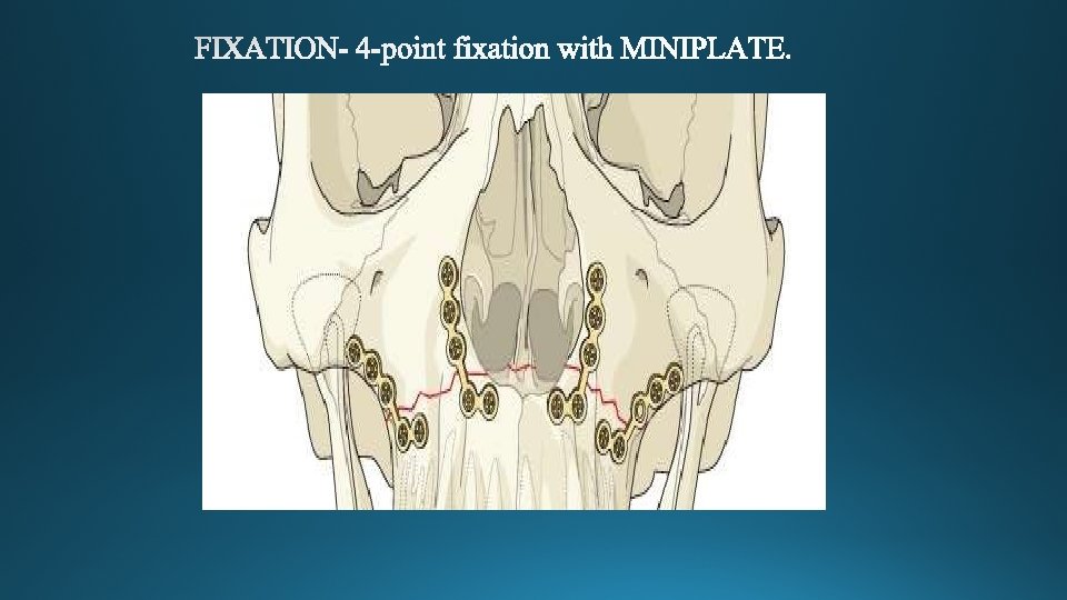
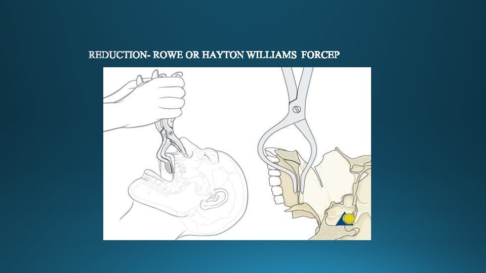
FIXATION-
STAGE III. DEFINITIVE TREATMENT LEFORT III FRACTURE- SURGICAL APPROACH- Existing Laceration
A. Lateral eyebrow approach B. Upper-eyelid approach GLABELLA APPROACH
Coronal approach PREAURICULAR APPROACH
Zygoma hook
FIXATION- 3 -point fixation
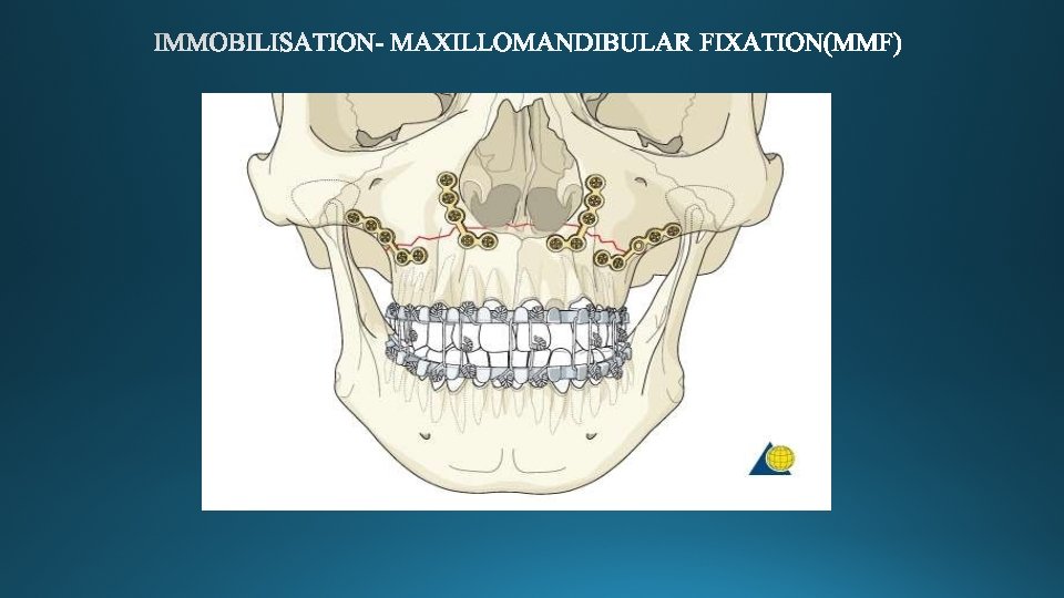
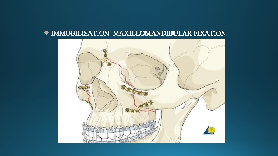
IMMOBILISATION- MAXILLOMANDIBULAR FIXATION if required
Principles of Maxillary Reconstruction Miniplates can bridge gaps of up to approximately 0. 5 cms Gaps >0. 5 cms – bone grafts Bone grafts bridging the gap should be wedged underneath the plate & held in place with screws fixed from plate directly into the graft.
Immediate Bone Grafting Buttress reinforcement retained by plates or screws can assist in restoring maxillary height & preventing Contour deficiencies. Rib graft Iliac crest Calvaria Mandibular bone graft Alloplastic bone graft
Le fort fractures are common in the trauma patient. They require accurate radiologic diagnosis and surgical management to prevent severe functional debilities and cosmetic deformity. A thorough understanding of the anatomy, craniofacial buttresses and treatment options will give the maxillofacial surgeon the optimal tools for achieving a successful result.