POSTPARTUM HAEMORRHAGE Dr Reza Nasr MD MRCOG DFFP




- Slides: 55
POSTPARTUM HAEMORRHAGE Dr. Reza Nasr MD MRCOG DFFP Consultant in Obstetrics and Gynaecology University of London
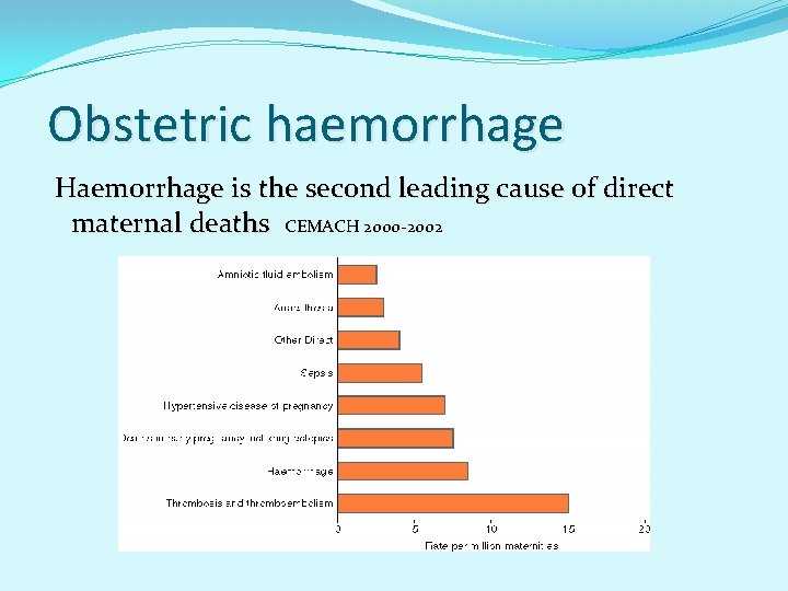
Obstetric haemorrhage Haemorrhage is the second leading cause of direct maternal deaths CEMACH 2000 -2002
WHY DELAY? DENIAL: amount of blood loss underestimated/ ignored until patient very unstable DENIAL: expecting the bleeding “to stop soon” DELAY: hard to get the obstetrician back to the bedside for evaluation DELAY: repetitive use of the same procedure or medication (eg D&C, methergine) rather than moving up the protocol
Postpartum hemorrhage (PPH) is commonly defined as blood loss exceeding 500 m. L following vaginal birth and 1, 000 m. L following cesarean Definitions vary, however, and diagnosis of PPH is subjective and often based on inaccurate estimates of blood loss.
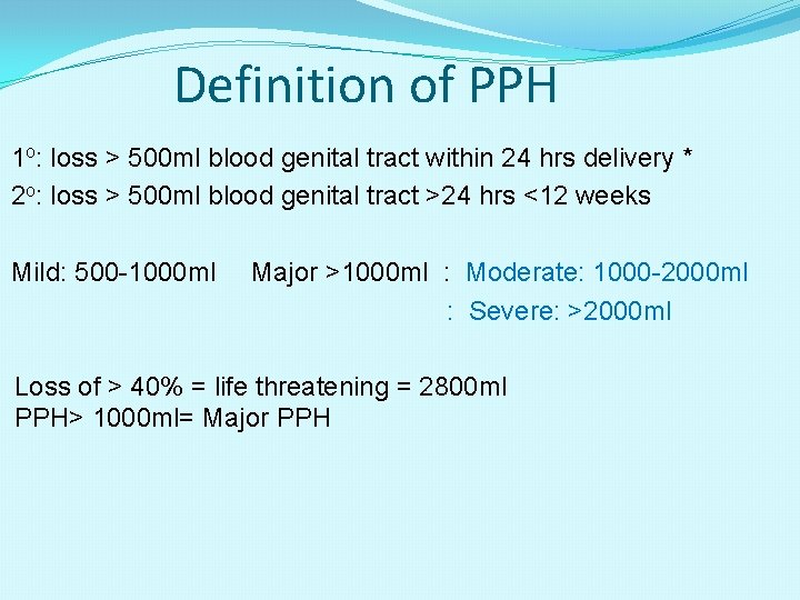
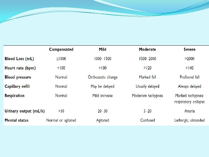
Definition of PPH 1 o: loss > 500 ml blood genital tract within 24 hrs delivery * 2 o: loss > 500 ml blood genital tract >24 hrs <12 weeks Mild: 500 -1000 ml Major >1000 ml : Moderate: 1000 -2000 ml : Severe: >2000 ml Loss of > 40% = life threatening = 2800 ml PPH> 1000 ml= Major PPH
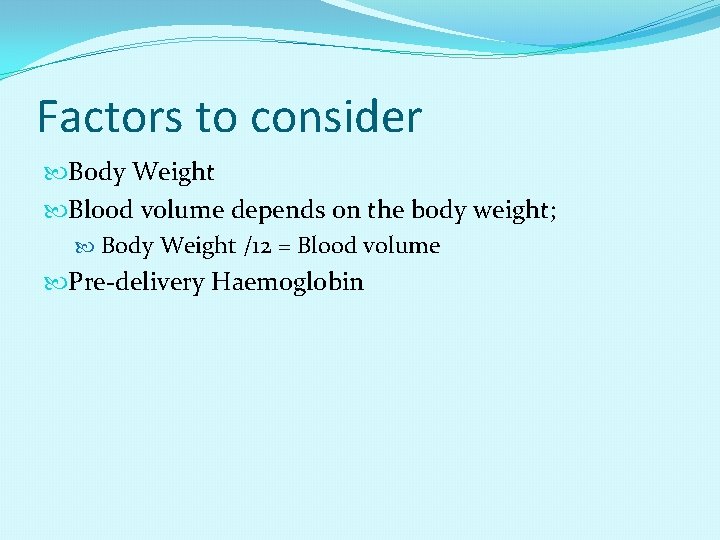
Factors to consider Body Weight Blood volume depends on the body weight; Body Weight /12 = Blood volume Pre-delivery Haemoglobin
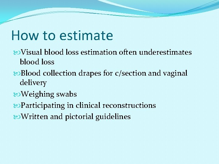
How to estimate Visual blood loss estimation often underestimates blood loss Blood collection drapes for c/section and vaginal delivery Weighing swabs Participating in clinical reconstructions Written and pictorial guidelines
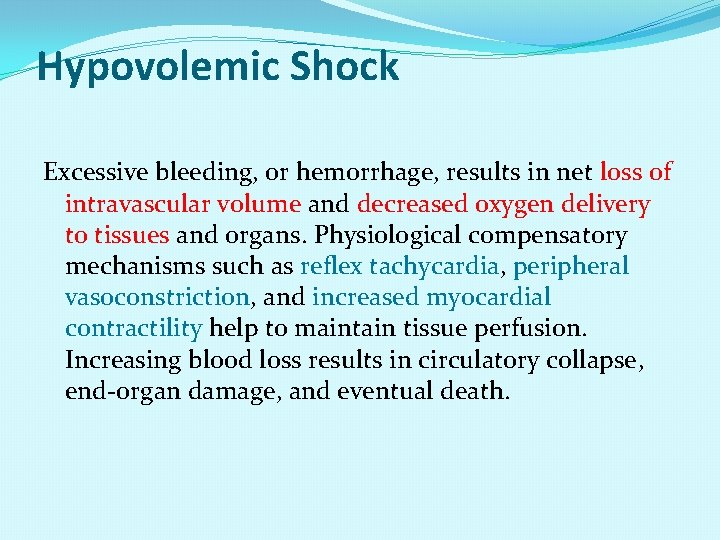
Hypovolemic Shock Excessive bleeding, or hemorrhage, results in net loss of intravascular volume and decreased oxygen delivery to tissues and organs. Physiological compensatory mechanisms such as reflex tachycardia, peripheral vasoconstriction, and increased myocardial contractility help to maintain tissue perfusion. Increasing blood loss results in circulatory collapse, end-organ damage, and eventual death.
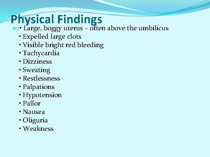
Physical Findings • Large, boggy uterus – often above the umbilicus • Expelled large clots • Visible bright red bleeding • Tachycardia • Dizziness • Sweating • Restlessness • Palpations • Hypotension • Pallor • Nausea • Oliguria • Weakness
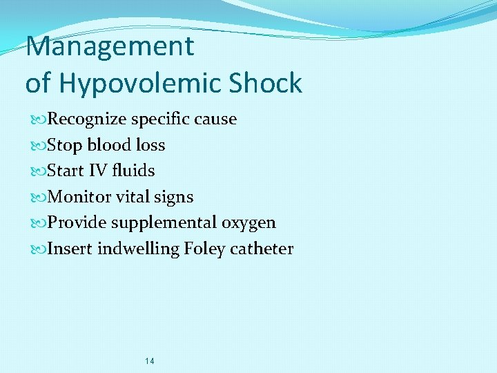
Management of Hypovolemic Shock Recognize specific cause Stop blood loss Start IV fluids Monitor vital signs Provide supplemental oxygen Insert indwelling Foley catheter 14
symptoms of hemorrhage or shock from blood loss may be hidden by the normal plasma volume increases that occur during pregnancy.
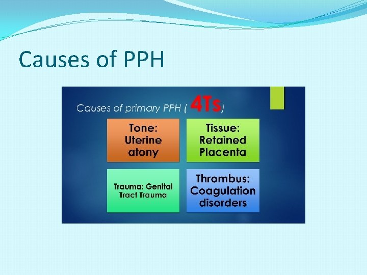
Causes of PPH
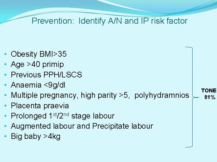
Prevention: Identify A/N and IP risk factor • • • Obesity BMI>35 Age >40 primip Previous PPH/LSCS Anaemia <9 g/dl Multiple pregnancy, high parity >5, polyhydramnios Placenta praevia Prolonged 1 st/2 nd stage labour Augmented labour and Precipitate labour Big baby >4 kg TONE 81%
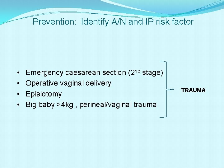
Prevention: Identify A/N and IP risk factor • • Emergency caesarean section (2 nd stage) Operative vaginal delivery Episiotomy Big baby >4 kg , perineal/vaginal trauma TRAUMA
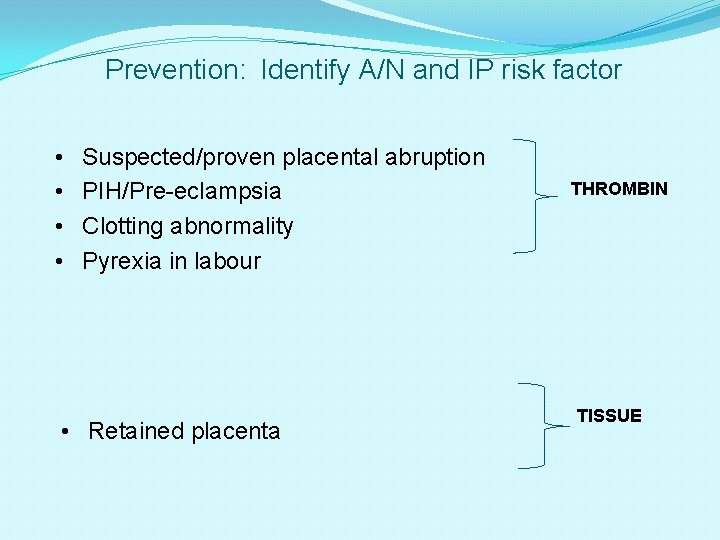
Prevention: Identify A/N and IP risk factor • • Suspected/proven placental abruption PIH/Pre-eclampsia Clotting abnormality Pyrexia in labour • Retained placenta THROMBIN TISSUE
Majority of cases (2/3 rds) : NO risk factors Offer prophylactic oxytocics to all for 3 rd stage
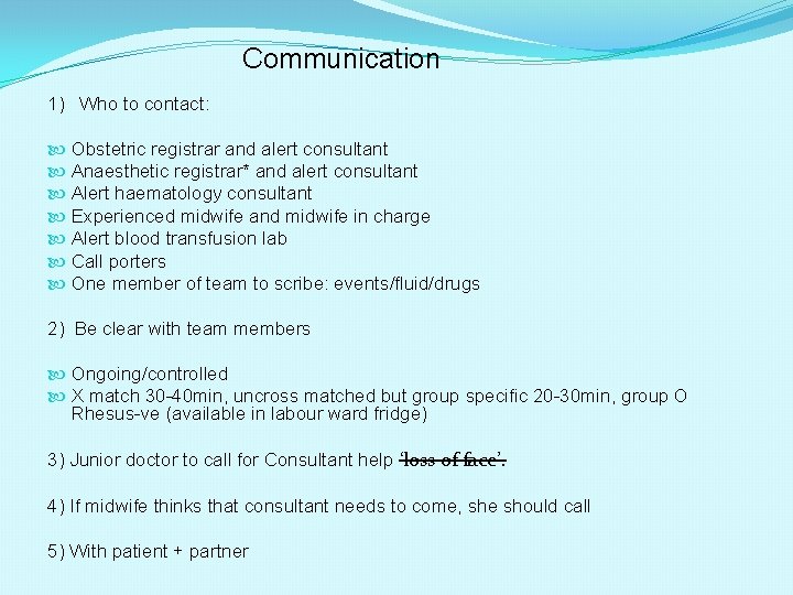
Communication 1) Who to contact: Obstetric registrar and alert consultant Anaesthetic registrar* and alert consultant Alert haematology consultant Experienced midwife and midwife in charge Alert blood transfusion lab Call porters One member of team to scribe: events/fluid/drugs 2) Be clear with team members Ongoing/controlled X match 30 -40 min, uncross matched but group specific 20 -30 min, group O Rhesus-ve (available in labour ward fridge) 3) Junior doctor to call for Consultant help ‘loss of face’. 4) If midwife thinks that consultant needs to come, she should call 5) With patient + partner
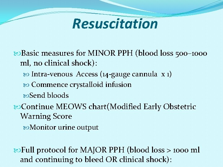
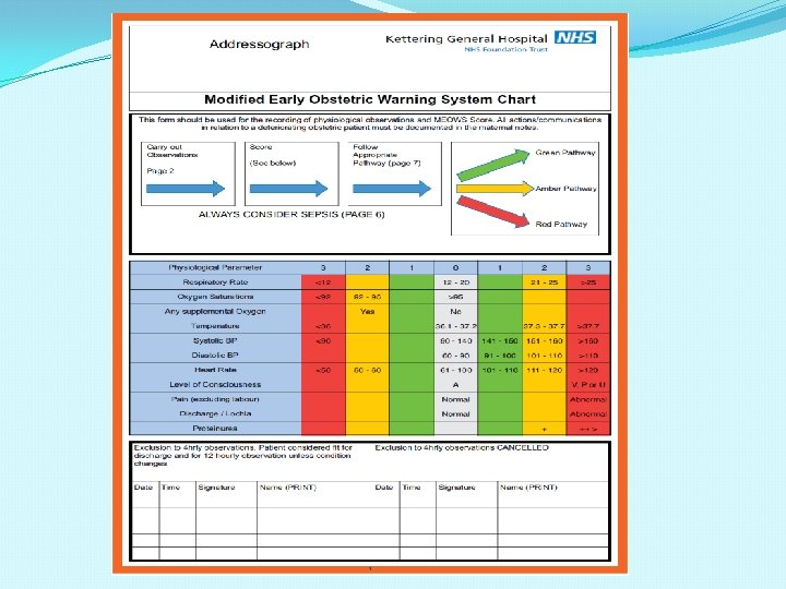
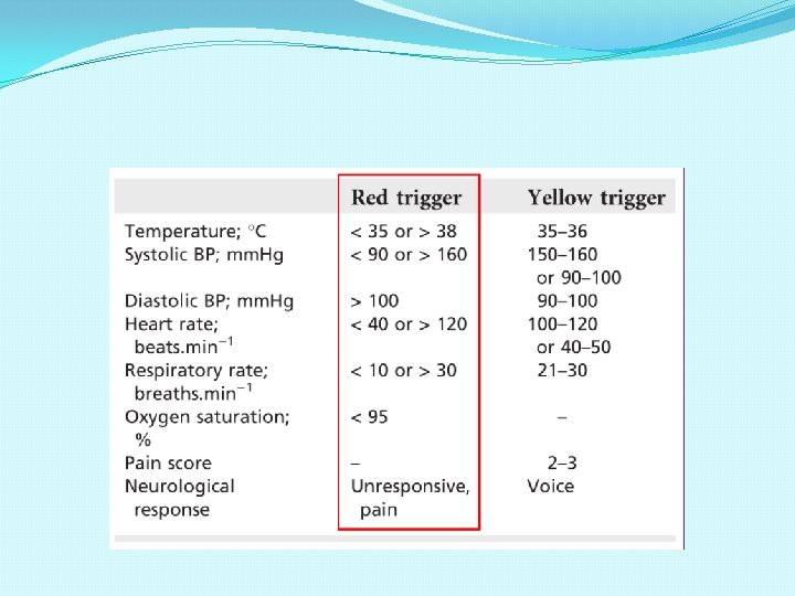
Resuscitation Basic measures for MINOR PPH (blood loss 500– 1000 ml, no clinical shock): Intra-venous Access (14 -gauge cannula x 1) Commence crystalloid infusion Send bloods Continue MEOWS chart(Modified Early Obstetric Warning Score Monitor urine output Full protocol for MAJOR PPH (blood loss > 1000 ml and continuing to bleed OR clinical shock):
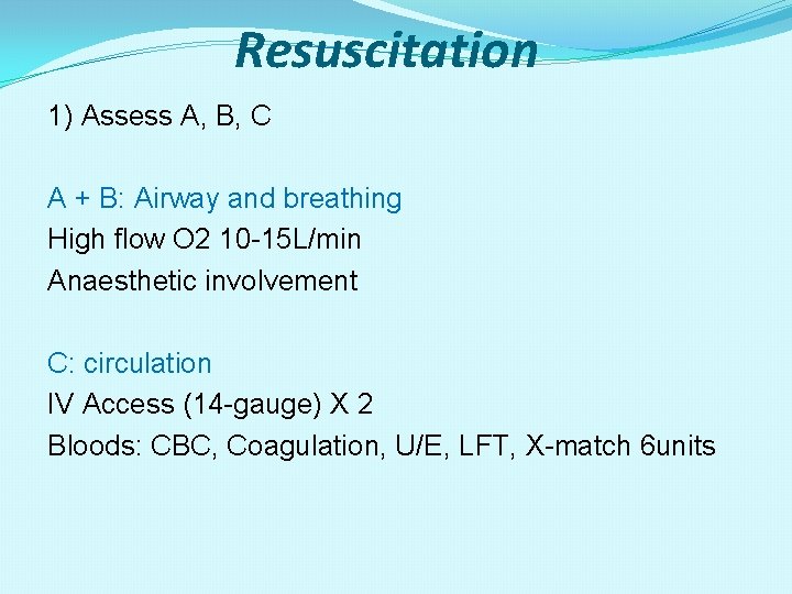
Resuscitation 1) Assess A, B, C A + B: Airway and breathing High flow O 2 10 -15 L/min Anaesthetic involvement C: circulation IV Access (14 -gauge) X 2 Bloods: CBC, Coagulation, U/E, LFT, X-match 6 units
Resuscitation 2) Fluids: Hartmann’s (2 L) Colloid /(1 -2 L) up to 3. 5 L Blood If cross-matched blood is still unavailable, give uncrossmatched group -specific blood OR give ‘O Rh. D negative’ blood FFP (4 every 4 -6 units blood or if Pt/APTT >1. 5 x normal) PLT (if <50 000) Cryoprecipitate (if fibrinogen <1 g/L)
Resuscitation 3) Target: • Hb>8 g/dl • PLT>75 000 • INR, PT and activated PTT<1. 5 x normal • Fibrinogen >1 g/L
Resuscitation Position flat. Keep the woman warm using appropriate available measures. Transfuse blood as soon as possible. Until blood is available, infuse up to 3. 5 litres of warmed crystalloid Hartmann’s solution (2 litres) and/or colloid (1. 5 litres) as rapidly as required. The best equipment available should be used to achieve rapid warmed infusion of fluids. Special blood filters should NOT be used, as they slow infusions.
women with known risk factors for PPH should not be delivered in a hospital without a blood bank on site
What blood components? When the blood loss reaches about 4. 5 litres (80% of blood volume) and large volumes of replacement fluids have been given, there will be clotting factor defects and blood components should be given. up to 1 litre of fresh frozen plasma (FFP) and 10 units of cryoprecipitate (two packs) may be given empirically in the face of relentless bleeding, while awaiting the results of coagulation studies FFP 1 unit : 200 -300 mls CRYOPRECIPITATE : almost exclusively a treatment for low fibrinogen levels e. g. in DIC Cryo and FFP needs some time to thaw, ask early
Recombinant activated factor VII will not work if there is no fibrinogen and effectiveness may also be suboptimal with severe thrombocytopenia (less than 20000). Therefore, fibrinogen should be above 1 g/l and platelets greater than 20000 before factor VII a is given. factor VII needs fibrinogen and platelet to work
Tranexamic Acid Is there a use for antifibrinolytic drugs? Although evidence is conflicting, there is a consensus view that fibrinolytic inhibitors (such as tranexamic acid) seldom, if ever, have a place in the management of obstetric haemorrhage.
Investigation and monitoring in Minor PPH Basic measures for MINOR PPH (blood loss 500– 1000 ml, no clinical shock and bleeding ceasing): Consider venepuncture (20 ml) for: group and screen full blood count coagulation screen including fibrinogen pulse and blood pressure recording every 15 minutes.
Full Protocol for MAJOR PPH (blood loss greater than 1000 ml and continuing to bleed OR clinical shock): Consider venepuncture (20 ml) for: crossmatch (4 units minimum) full blood count coagulation screen including fibrinogen renal and liver function for baseline Monitor temperature every 15 minutes. Continuous pulse, blood pressure recording and respiratory rate (using oximeter, electrocardiogram and automated blood pressure recording). Foley catheter to monitor urine output. Two peripheral cannulae, 14 - or 16 -gauge. Consider arterial line monitoring (once appropriately experienced staff available for insertion). Consider transfer to intensive therapy unit once the bleeding is controlled or monitoring at high dependency unit on delivery suite, if appropriate. Recording of parameters on a flow chart such as the modified obstetric early warning system (MEOWS)charts. Documentation of fluid balance, blood products and procedures.
CVP / Arterial Line Central venous and direct arterial pressure monitoring should be used when the cardiovascular system is compromised by haemorrhage or disease
Anaesthesia The presence of cardiovascular instability is a relative contraindication to regional anaesthesia. Blockage of the sympathetic system can potentially lead to worsening hypotension due to haemorrhage.
Arresting the bleeding Causes for PPH may be considered to relate to one or more of ‘the four Ts’: tone (abnormalities of uterine contraction) tissue (retained products of conception) trauma (of the genital tract) thrombin (abnormalities of coagulation).
Source of Bleeding Most common cause of primary PPH is uterine atony retained products (placenta, membranes, clots) vaginal/cervical lacerations or haematoma ruptured uterus broad ligament haematoma extragenital bleeding (for example, subcapsular liver rupture) uterine inversion.
ATONY Bimanual compression Empty bladder Syntocinon 5 units IV + /- repeat Ergometrine 500 mcg IM Syntocinon infusion 40 units/500 mls N/saline 10 u/hr Haemobate (Carboprost, PGF 2α) 250 mcg IM every 15 min max 8 doses Haemobate 500 mcg intramyometrium ( D/C by company) Misoprostol 1000 mcg PR
ATONY If pharmacological measures fail to control the haemorrhage, initiate surgical haemostasis sooner rather than later. Intrauterine balloon tamponade is an appropriate first line ‘surgical’ intervention for most women where uterine atony is the only or main cause of haemorrhage.
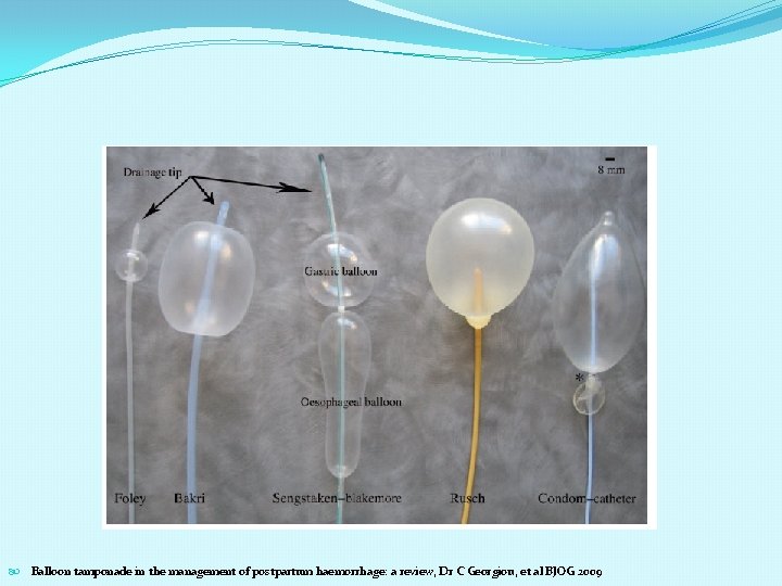
Balloon Tamponade Foley catheter, Bakri balloon, Sengstaken–Blakemore oesophageal catheter and a condom catheter Some of the reports of balloon tamponade describe the intervention as the ‘tamponade test’. A ‘positive test’ (control of PPH following inflation of the balloon) indicates that laparotomy is not required, whereas a ‘negative test’ (continued PPH following inflation of the balloon) is an indication to proceed to laparotomy.
Balloon Tamponde It is not only a treatment Also it is a diagnosis test and taking action for laparotomy
Balloon tamponade in the management of postpartum haemorrhage: a review, Dr C Georgiou, et al BJOG 2009
When to deflate the baloon In most cases, 4– 6 hours of tamponade should be adequate to achieve haemostasis and, ideally, it should be removed during daytime hours, in the presence of appropriate senior staff, should further intervention be necessary. Before its complete removal, the balloon could be deflated but left in place to ensure that bleeding does not reoccur. Routine practice: just less than 24 hours to deflate during the daytime
Haemostatic suturing In the absence of comparative data to demonstrate that any one variant is superior to another, obstetricians are encouraged to familiarise themselves with one technique under the supervision of an experienced colleague. It is recommended that a laminated diagram of the brace technique be kept in theatre.
Internal iliac artery ligation Nevertheless, the available observational data suggest that balloon tamponade and haemostatic suturing may be more effective than internal iliac artery ligation and they are unquestionably easier to perform.
Selective arterial occlusion or embolisation by interventional radiology Interventional radiology may be considered in cases of placenta praevia with accreta if intra-arterial balloons can be placed in the radiology department before the woman goes to theatre for caesarean section
ATONY Resort to hysterectomy SOONER RATHER THAN LATER (especially in cases of placenta accreta or uterine rupture).
Consider use of factor r. VII only after discussion with Consultant haematologist NOVOSEVEN REQUIRES FIBRINOGEN TO WORK, SO GIVE CRYOPRECIPITATE PRIOR TO NOVOSEVEN and PLATELET (if low)
Treatment post PPH • Pulse, BP, RR rate (every 15 min) • Oxygen Saturation • Intra-arterial/CVP line to be considered • Fluid balance chart: Hourly measurements • Blood gases and acid base status as needed • Repeat haematology, clotting screen, biochemistry 6 -12 hourly
Secondary PPH Often associated with endometritis When antibiotics are clinically indicated, a combination of ampicillin (clindamycin if penicillin allergic) and metronidazole is appropriate. In cases of endomyometritis (tender uterus) or overt sepsis, then the addition of gentamicin is recommended. A senior obstetrician should be involved in decisions and performance of any evacuation of retained products of conception as these women are carrying a high risk for uterine perforation.
Investigations of secondary PPH should include high and low vaginal swabs, blood cultures if pyrexial, full blood count, C-reactive protein. A pelvic ultrasound may help to exclude the presence of retained products of conception, although the appearance of the immediate postpartum uterus may be unreliable
Secondary PPH It is generally accepted that secondary PPH is often associated with infection and conventional treatment involves antibiotics and uterotonics. In continuing haemorrhage, insertion of balloon catheter may be effective.
Thank You