
Postpartum Complications Postpartum Physical Assessment B breast U
Postpartum Complications
Postpartum Physical Assessment B - breast U - uterus B - bowels B - bladder L - lochia E - episiotomy
Routine care for the postpartum woman: Health promotion and disease prevention (1) • Give Vitamin A 200, 000 IU. • Provide preventive treatment for hookworm to prevent anemia in endemic areas. • Provide iron/folic acid supplementation for at least 30 days postpartum to prevent and treat anemia.
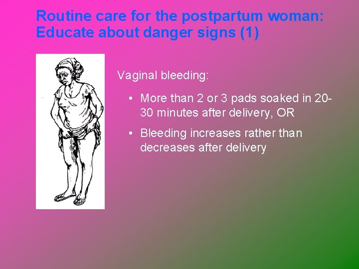
Routine care for the postpartum woman: Educate about danger signs (1) Vaginal bleeding: • More than 2 or 3 pads soaked in 2030 minutes after delivery, OR • Bleeding increases rather than decreases after delivery
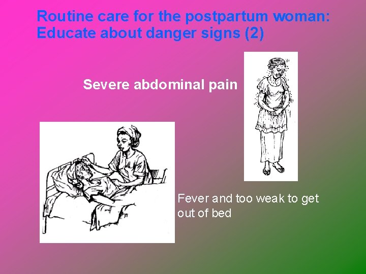
Routine care for the postpartum woman: Educate about danger signs (2) Severe abdominal pain Fever and too weak to get out of bed
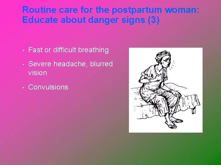
Routine care for the postpartum woman: Educate about danger signs (3) • Fast or difficult breathing • Severe headache, blurred vision • Convulsions
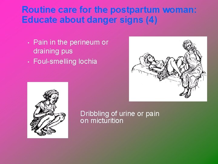
Routine care for the postpartum woman: Educate about danger signs (4) • • Pain in the perineum or draining pus Foul-smelling lochia Dribbling of urine or pain on micturition
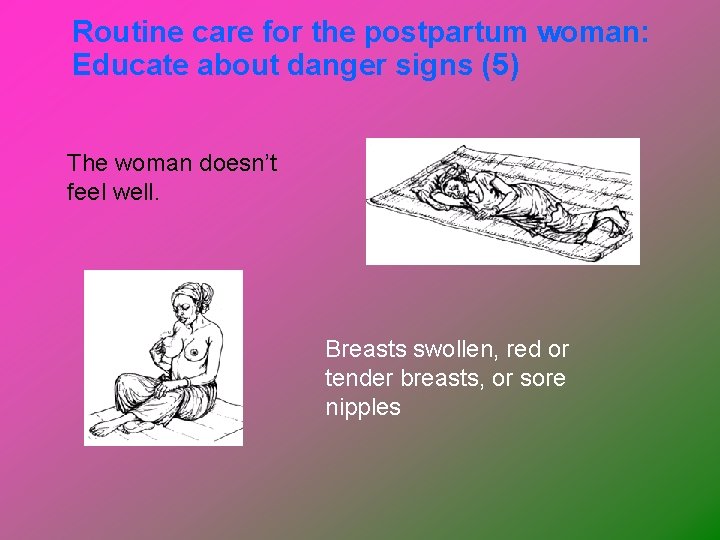
Routine care for the postpartum woman: Educate about danger signs (5) The woman doesn’t feel well. Breasts swollen, red or tender breasts, or sore nipples
Postpartum Hemorrhage (PPH) Definition and incidence Ø PPH traditionally defined as loss of more than: • 500 ml of blood after vaginal birth • 1000 ml after cesarean birth Ø Cause of maternal morbidity and mortality Ø Life-threatening with little warning Ø Often unrecognized until profound symptoms
Etiology of PPH The causes of postpartum hemorrhage can be thought of as the four Ts: Øtone, Øtissue, Øtrauma, Øthrombin
Postpartum Hemorrhage Etiology and risk factors (1) Ø Uterine atony • Marked hypotonia of uterus • Leading cause of PPH, complicating approximately 1 in 20 births • Brisk venous bleeding with impaired coagulation until the uterine muscle contracts 11 of 34
Postpartum Hemorrhage Etiology and risk factors (1) Uterine atony ØMultiple gestation, Øhigh parity, Øprolonged labor Øchorioamnionitis, Øaugmented labor, Ø tocolytic agents
Management of uterine atony Explore the uterine cavity. Inspect vagina and cervix for lacerations. If the cavity is empty, Massage and give methylergonovine 0. 2 mg, the dose can be repeated every 2 to 4 hours. Rectal 800 mcg. Misoprostol is beneficial.
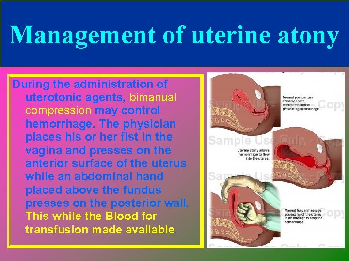
Management of uterine atony During the administration of uterotonic agents, bimanual compression may control hemorrhage. The physician places his or her fist in the vagina and presses on the anterior surface of the uterus while an abdominal hand placed above the fundus presses on the posterior wall. This while the Blood for transfusion made available.
Complications of Puerperium Uterine Atony (Cont’d) • Treatment Uterine compression Oxytocics – Early suckling causes endogenous release of oxytocin – Oxytocin IV/IM 10 units – Methylergonovine – Methyl prostoglandin F
Postpartum Hemorrhage Etiology and risk factors (2) Ш Lacerations of genital tract • Should be suspected if bleeding continues with a firm, contracted fundus • Includes perineal and cervical lacerations as well as pelvic hematomas 16 of 34
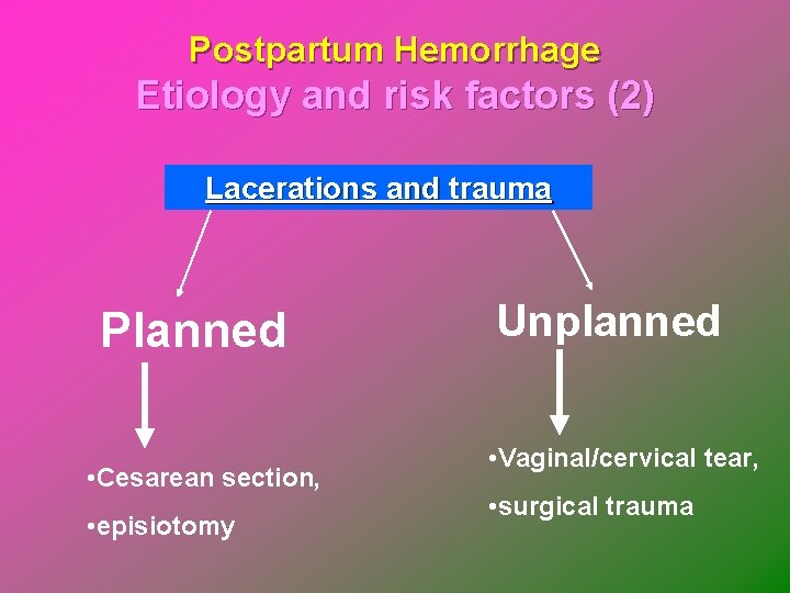
Postpartum Hemorrhage Etiology and risk factors (2) Lacerations and trauma Planned • Cesarean section, • episiotomy Unplanned • Vaginal/cervical tear, • surgical trauma
Postpartum Hemorrhage Genital tract lacerations Management Genital trauma always must be eliminated first if the uterus is firm.
Postpartum Hemorrhage Etiology and risk factors (2) UTERINE RUPTURE Rupture of the uterus is described as complete or incomplete and should be differentiated from dehiscence of a cesarean section scar.
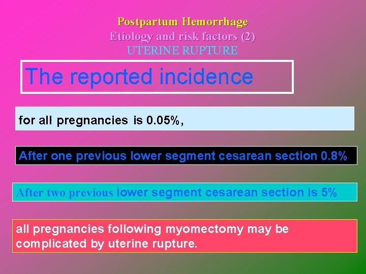
Postpartum Hemorrhage Etiology and risk factors (2) UTERINE RUPTURE The reported incidence for all pregnancies is 0. 05%, After one previous lower segment cesarean section 0. 8% After two previous lower segment cesarean section is 5% all pregnancies following myomectomy may be complicated by uterine rupture.
Postpartum Hemorrhage Etiology and risk factors (2) UTERINE RUPTURE Complete rupture describes a full-thickness defect of the uterine wall and serosa resulting in direct communication between the uterine cavity and the peritoneal cavity.
Postpartum Hemorrhage Etiology and risk factors (2) UTERINE RUPTURE Incomplete rupture describes a defect of the uterine wall that is contained by the visceral peritoneum or broad ligament. In patients with prior cesarean section,
Postpartum Hemorrhage Etiology and risk factors (2) UTERINE RUPTURE dehiscence describes partial separation of the scar with minimal bleeding, with the peritoneum and fetal membranes remaining intact.
Management of Rupture Uterus The identification or suspicion of uterine rupture must be followed by an immediate and simultaneous response from the obstetric team. Surgery should not be delayed owing to hypovolemic shock because it may not be easily reversible until the hemorrhage is controlled.
Management of Rupture Uterus Ø Ø Ø Upon entering the abdomen, aortic compression can be applied to decrease bleeding. Oxytocin should be administered to effect uterine contraction to assist in vessel constriction and to decrease bleeding. Hemostasis can then be achieved by ligation of the hypogastric artery, uterine artery, or ovarian arteries.
Management of Rupture Uterus Ø Ø Ø At this point, a decision must be made to perform hysterectomy or to repair the rupture site. In most cases, hysterectomy should be performed. In selected cases, repair of the rupture can be attempted. When rupture occurs in the body of the uterus, bladder rupture must be ruled out by clearly mobilizing and inspecting the bladder to ensure that it is intact. This avoids injury on repair of the defect as well.
Management of Rupture Uterus Ø Ø A lower segment lateral rupture can cause transection of the uterine vessels. The vessels can retract toward the pelvic side wall, and the site of bleeding must be isolated before placing clamps to avoid injury to the ureter and iliac vessels. Typically, longitudinal tears, especially those in a lateral position, should be treated by hysterectomy, whereas low transverse tears may be repaired.
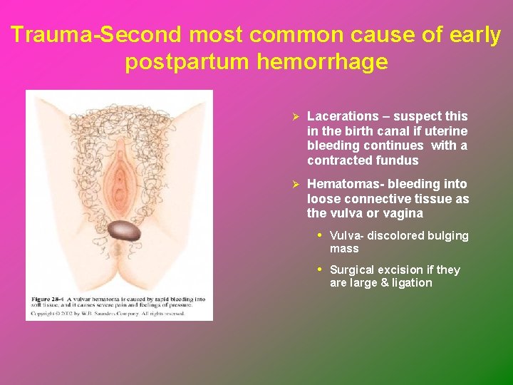
Trauma-Second most common cause of early postpartum hemorrhage Ø Lacerations – suspect this in the birth canal if uterine bleeding continues with a contracted fundus Ø Hematomas- bleeding into loose connective tissue as the vulva or vagina • Vulva- discolored bulging mass • Surgical excision if they are large & ligation
Postpartum Hemorrhage Etiology and risk factors (3) Ø Retained placenta • Nonadherent retained placenta – managed by manual separation and removal by the primary care provider • Adherent retained placenta – may be caused by implantation into defective endometrium 29 of 34
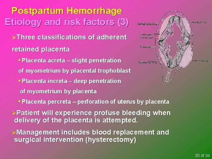
Postpartum Hemorrhage Etiology and risk factors (3) ØThree classifications of adherent retained placenta • Placenta acreta – slight penetration of myometrium by placental trophoblast • Placenta increta – deep penetration of myometrium by placenta • Placenta percreta – perforation of uterus by placenta ØPatient will experience profuse bleeding when delivery of the placenta is attempted. ØManagement includes blood replacement and surgical intervention (hysterectomy) 30 of 34
Postpartum Hemorrhage Etiology and risk factors (4) Inversion of uterus (turning inside out) Ø May be life-threatening Ø A complete inversion protrudes out of the vagina Ø Primary signs – hemorrhage, shock, pain Ø Prevention is the best measure – don’t pull on the umbilical cord unless there is definite separation of the placenta 31 of 34
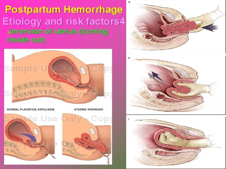
Postpartum Hemorrhage Etiology and risk factors 4 Inversion of uterus (turning inside out)
Postpartum Hemorrhage Etiology and risk factors (5) Subinvolution of uterus – delayed involution of the uterus Ø Usually see late post partum bleeding Ø Causes include retained placental fragments and infection 33 of 34
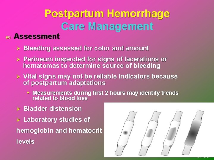
Postpartum Hemorrhage Care Management Assessment Ø Bleeding assessed for color and amount Ø Perineum inspected for signs of lacerations or hematomas to determine source of bleeding Ø Vital signs may not be reliable indicators because of postpartum adaptations • Measurements during first 2 hours may identify trends related to blood loss Ø Bladder distension Ø Laboratory studies of hemoglobin and hematocrit levels 34 of 34
Postpartum Hemorrhage Care Management Plan of care and implementation Ø Initial treatment – fundal massage, expression of clots, relief of bladder distension, IV fluids Ø Medical management • Hypotonic uterus – examine for retained placental fragments, medications, surgical interventions • Bleeding with a contracted uterus – identify and treat underlying cause • Uterine inversion – emergency replacement of the uterus into the pelvic cavity • Subinvolution – medications, surgical intervention 35 of 34
Postpartum Hemorrhage Care Management Plan of care and implementation Ø Nursing interventions • Vital signs, uterine assessment, medication administration, notification of primary care provider • Providing explanations about interventions and need to act quickly • Once stable, ongoing post partum assessments and care • Instructions in increasing dietary iron, protein intake, and iron supplementation • May need assistance with infant care and household activities until strength regained 36 of 34
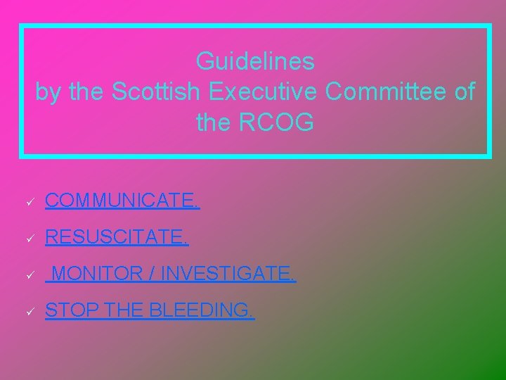
Guidelines by the Scottish Executive Committee of the RCOG ü COMMUNICATE. ü RESUSCITATE. ü ü MONITOR / INVESTIGATE. STOP THE BLEEDING.
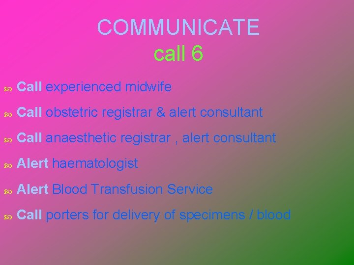
COMMUNICATE call 6 Call experienced midwife Call obstetric registrar & alert consultant Call anaesthetic registrar , alert consultant Alert haematologist Alert Blood Transfusion Service Call porters for delivery of specimens / blood
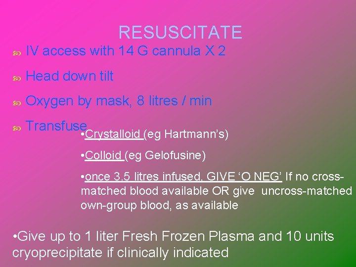
RESUSCITATE IV access with 14 G cannula X 2 Head down tilt Oxygen by mask, 8 litres / min Transfuse • Crystalloid (eg Hartmann’s) • Colloid (eg Gelofusine) • once 3. 5 litres infused, GIVE ‘O NEG’ If no crossmatched blood available OR give uncross-matched own-group blood, as available • Give up to 1 liter Fresh Frozen Plasma and 10 units cryoprecipitate if clinically indicated
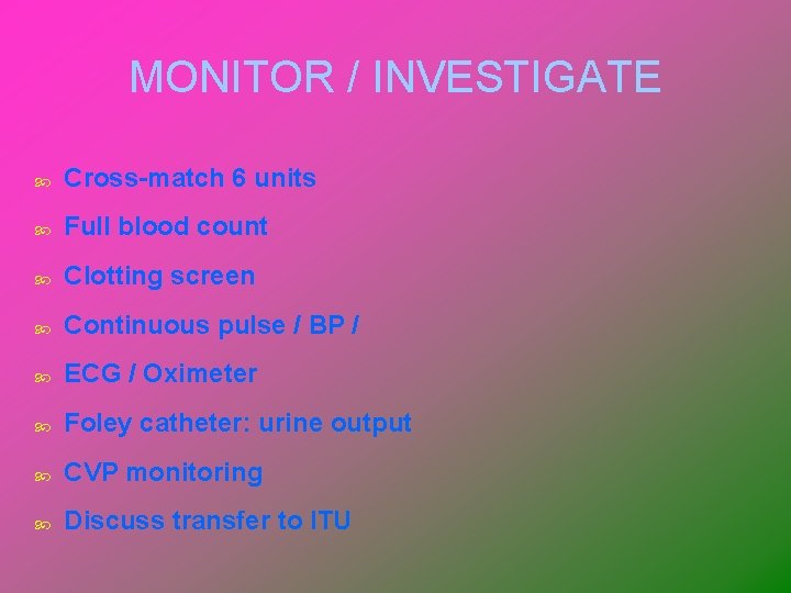
MONITOR / INVESTIGATE Cross-match 6 units Full blood count Clotting screen Continuous pulse / BP / ECG / Oximeter Foley catheter: urine output CVP monitoring Discuss transfer to ITU
STOP THE BLEEDING Exclude causes of bleeding other than uterine atony Ensure bladder empty Uterine compression IV syntocinon 10 units IV ergometrine 500 mg Syntocinon infusion (30 units in 500 ml) IM Carboprost (500 mg) Surgery earlier rather than late Hysterctomy early rather than late (GRADE B)
If conservative measures fail to control haemorrhage, initiate surgical haemostasis SOONER RATHER THAN LATER I. At laparotomy, direct intramyometrial injection of Carboprost (Haemabate) 0. 5 mg II. III. IV. Bilateral ligation of uterine arteries Bilateral ligation of internal iliac (hypogastric arteries) Hysterectomy (GRADE C)
Resort to hysterectomy SOONER RATHER THAN LATER (especially in cases of placenta accreta or uterine rupture) (GRADE C)
Whole blood frequently is used for rapid correction of volume loss because of its ready availability, but component therapy is ideal. A general practice has been to transfuse 1 unit of fresh-frozen plasma for every 3 to 4 units of red cells given to patients who are bleeding profusely
Hemorrhagic (Hypovolemic) Shock Emergency situation in which blood is diverted to the brain and heart Ø May not see signs until post partum patient loses 30% to 40% of blood volume Medical management – restore circulating blood volume and treat underlying cause Nursing interventions – monitor tissue perfusion, see emergency box Fluid or blood replacement therapy 45 of 34
Prophylactic oxytocics should be offered routinely in the management of the third stage of labour as they reduce the risk of PPH by about 60%. (GRADE A)
Coagulopathies Idiopathic thrombocytopenic purpura (ITP) – decreased platelet life span, need to control platelet stability von Willebrand disease—type of hemophilia Disseminated intravascular coagulation (DIC) Pathologic clotting Ø Correction of underlying cause • Removal of fetus • Treatment for infection • Preeclampsia or eclampsia • Removal of placental abruption Ø 47 of 34
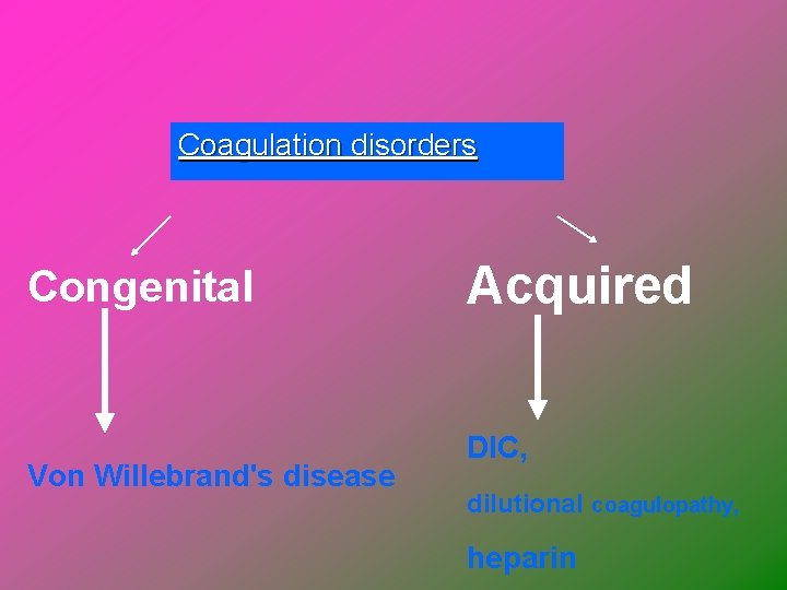
Coagulation disorders Congenital Von Willebrand's disease Acquired DIC, dilutional coagulopathy, heparin
Thromboembolic Disease Results from blood clot caused by inflammation or partial obstruction of vessel May be superficial or deep venous thrombosis or a pulmonary embolus Incidence and etiology Ø Venous stasis Ø Hypercoagulation Clinical manifestations – redness and swelling in the affected extremity, pain, positive Homan’s sign 49 of 34
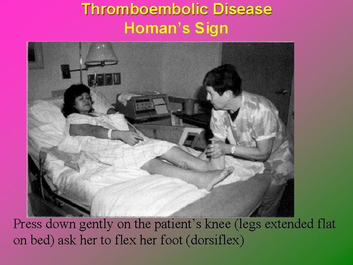
Thromboembolic Disease Homan’s Sign Press down gently on the patient’s knee (legs extended flat on bed) ask her to flex her foot (dorsiflex)
Thromboembolic Disease Medical management Ø Superficial – analgesia, rest/elevation Ø Deep – anticoagulant therapy, bedrest/elevation, Ø Pulmonary embolus – IV heparin therapy Nursing interventions Ø Ø assessment of the affected area, signs of bleeding, personal care, medication administration Teach not to massage affected area!! 51 of 34
Postpartum Infections § Endometritis – malodorous lochia, fever (100. 6), chills, abdominal pain, uterine tenderness, tachycardia and subinvolution § The infection may spread to cause peritonitis and septic pelvic thrombophlebitis § Treat with IV antibiotics § Emotional support 52
Postpartum Infections Puerperal sepsis: any infection of genital canal within 28 days after abortion or birth Most common infecting agents are numerous streptococcal and anaerobic organisms Endometritis Wound infections Urinary tract infections Mastitis 53 of 34
Complications of Puerperium Fever Ø UTI/Pyelonephritis Ø DVT/Thrombophlebitis Ø “Milk fever” (Lasts < 24 hours) Ø Drug reaction Ø Perineal infection(Day five) Ø Pulmonary Atelectasis (48 hours) Ø Mastitis (2 -3 weeks post partum)
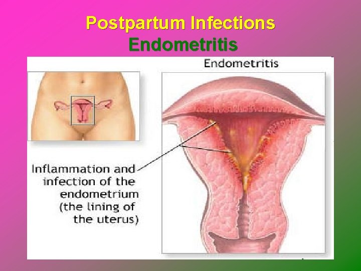
Postpartum Infections Endometritis
Postpartum Endometritis Infection of the decidua (pregnancy endometrium) Incidence Ø <3% after vaginal delivery Ø 10 -50% after cesarean delivery • 5 -15% after scheduled elective cesareans Risk Factors Ø Prolonged labor, prolonged ROM, multiple vaginal exams, internal monitors, maternal DM, meconium, manual removal of placenta, low socioeconomic status
PP Endometritis Polymicrobial, ascending infection Ø Mixture of aerobes and anaerobes from genital tract Ø BV and colonization with GBS increase likelihood of infection Clinical manifestations (occur within 5 days pp) Ø Fever – most common sign Ø Uterine tenderness Ø Foul lochia Ø Leukocytosis Ø Bacteremia – in 10 -20%, usually a single organism
PP Endometritis Workup Ø CBC Ø Blood cultures Ø Urine culture Ø DNA probe for GC/chlamydia Ø Imaging studies if no response to adequate abx in 48 -72 h • CT scan abd/pelvis • US abd/pelvis
PP Endometritis Treatment Ø Broad spectrum IV abx • Clindamycin 900 mg IV q 8 h and • Gentamicin 1. 5 mg/kg IV q 8 h Ø Treat until afebrile for 24 -48 h and clinically improved; oral therapy not necessary Ø Add ampicillin 2 g IV q 4 h to regimen when not improving to cover resistant enterococci Prevention Ø Abx prophylaxis for women undergoing C-section • Cefazolin 1 -2 g IV as single dose
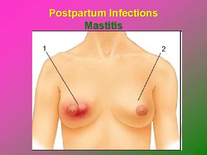
Postpartum Infections Mastitis - A breast infection occurring 1 -2 weeks after childbirth § Engorgement and blocked mild duct increases risk § Fever, localized breast pain, redness, warmth and inflammation § Breastfeeding should continue § Antibiotics § Nurse's role is to support, educate and refer 60
Mastitis Infection of the lactating breast- 2 nd or 3 rd week after birth Caused by S. aureus, often on hands of mother or caregivers Can enter through a crack in the nipple Engorgement & stasis of milk frequently precede mastitis
Mastitis Continued SIGNS & SYMPTOMS: Ø Feels like the flu with fatigue & aching muscles Ø Fever of 101. 1 F Ø Localized area of redness & inflammation THERAPEUTIC MANAGEMENT Ø ATB & decompression of breast by breastfeeding or pumping Ø Bedrest during acute phase Ø Fluids & analgesics for discomfort
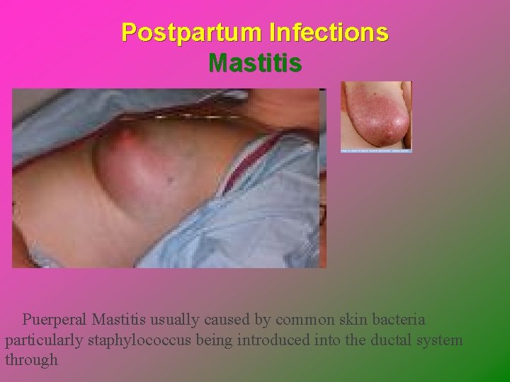
Postpartum Infections Mastitis Puerperal Mastitis usually caused by common skin bacteria particularly staphylococcus being introduced into the ductal system through
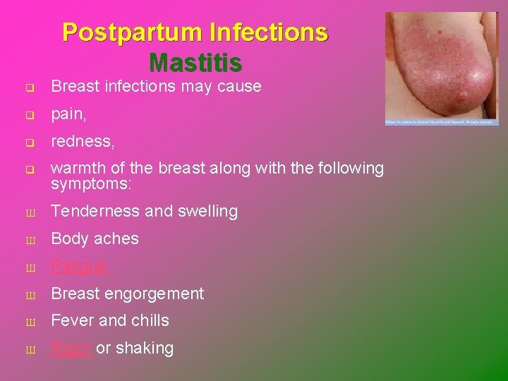
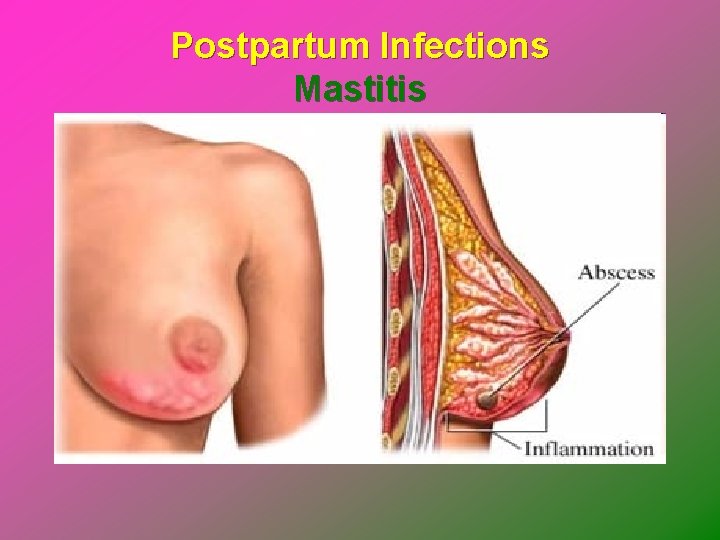
Postpartum Infections Mastitis q Breast infections may cause q pain, q redness, q warmth of the breast along with the following symptoms: Ш Tenderness and swelling Ш Body aches Ш Fatigue Ш Breast engorgement Ш Fever and chills Ш Rigor or shaking
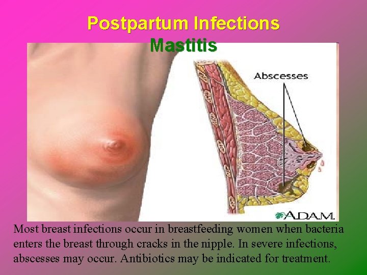
Postpartum Infections Mastitis Most breast infections occur in breastfeeding women when bacteria enters the breast through cracks in the nipple. In severe infections, abscesses may occur. Antibiotics may be indicated for treatment.
Postpartum Infections Mastitis
Postpartum Infections Mastitis
Postpartum Infections Care Management Prevention is the best intervention Ø Hand washing Ø Good maternal perineal hygiene Antibiotic administration Wound management Breast care 68 of 34
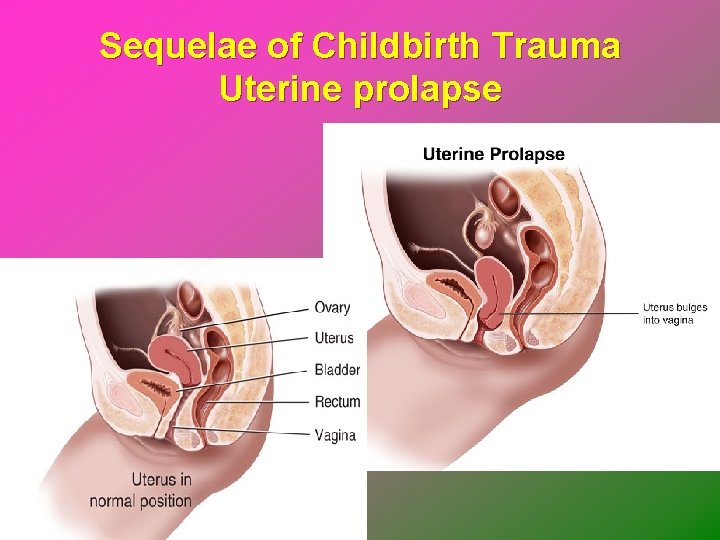
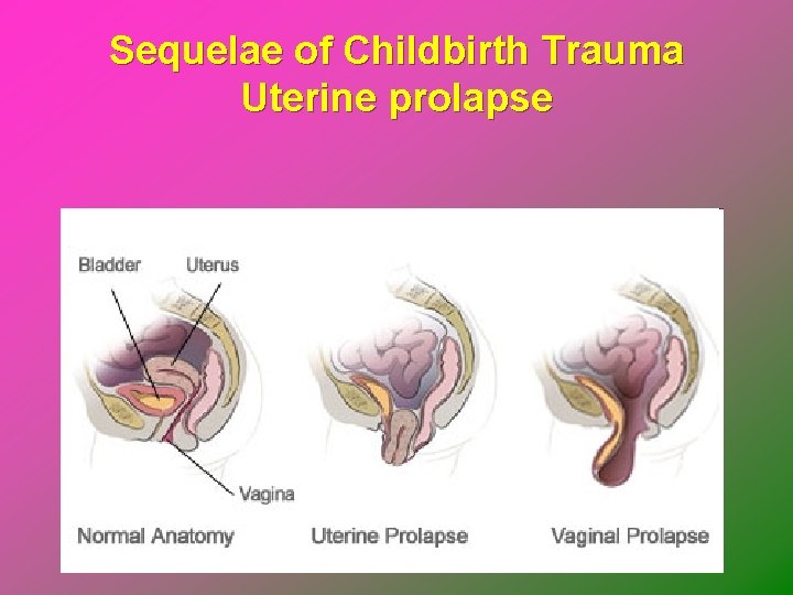
Sequelae of Childbirth Trauma Disorders of uterus and vagina related to pelvic relaxation and urinary incontinence, are often result of childbearing Uterine displacement and prolapse Ø Posterior displacement, or retroversion Ø Retroflexion and anteflexion Ø Prolapse a more serious displacement • Cervix and body of uterus protrude through vagina and vagina is inverted 69 of 34
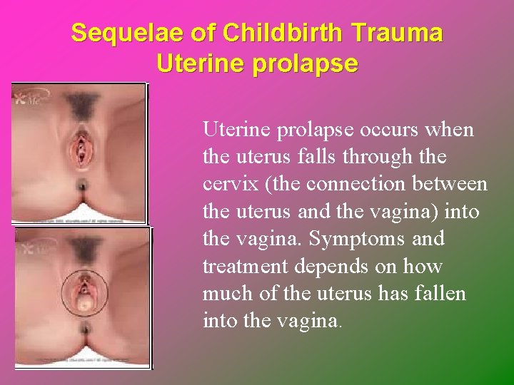
Sequelae of Childbirth Trauma Uterine prolapse occurs when the uterus falls through the cervix (the connection between the uterus and the vagina) into the vagina. Symptoms and treatment depends on how much of the uterus has fallen into the vagina.
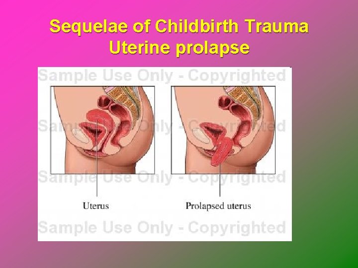
Sequelae of Childbirth Trauma Uterine prolapse
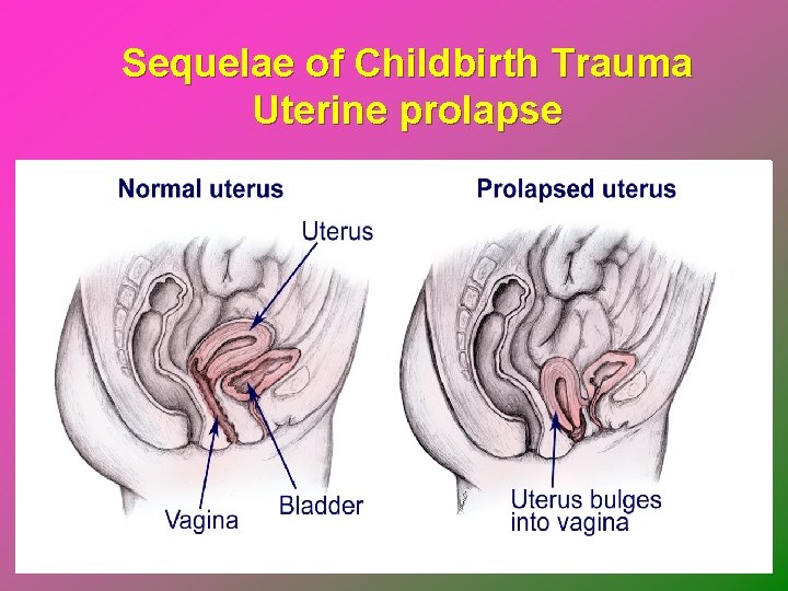
Sequelae of Childbirth Trauma Uterine prolapse
Sequelae of Childbirth Trauma Uterine prolapse
Sequelae of Childbirth Trauma Uterine prolapse
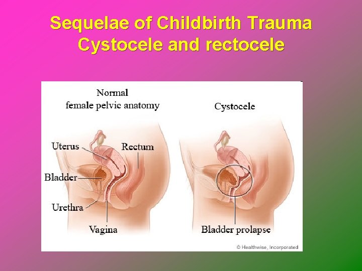
Sequelae of Childbirth Trauma Cystocele and rectocele Ø Cystocele: protrusion of bladder downward into vagina when support structures in vesicovaginal septum are injured Ø Rectocele is herniation of anterior rectal wall through relaxed or ruptured vaginal fascia and rectovaginal septum Urinary incontinence 75 of 34
Sequelae of Childbirth Trauma Cystocele and rectocele
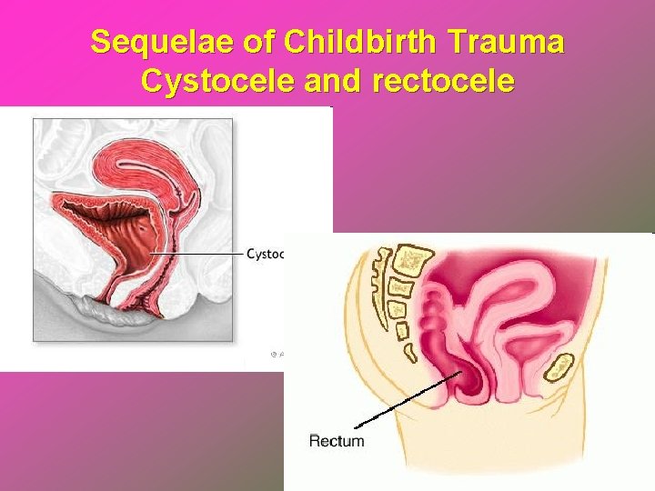
Sequelae of Childbirth Trauma Cystocele and rectocele
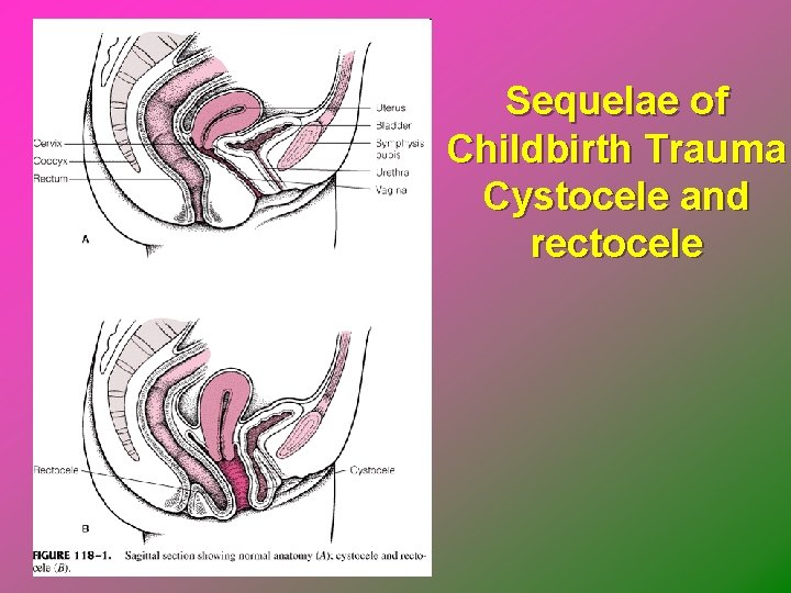
Sequelae of Childbirth Trauma Cystocele and rectocele
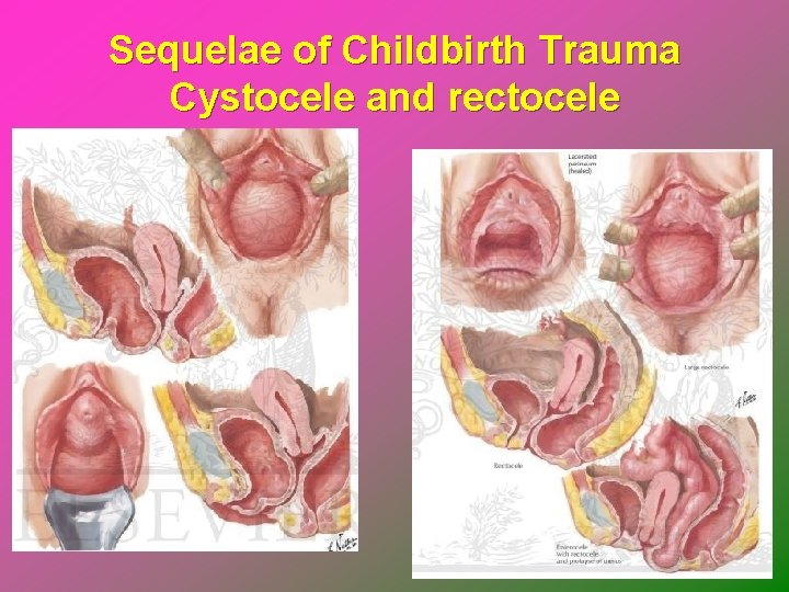
Sequelae of Childbirth Trauma Cystocele and rectocele
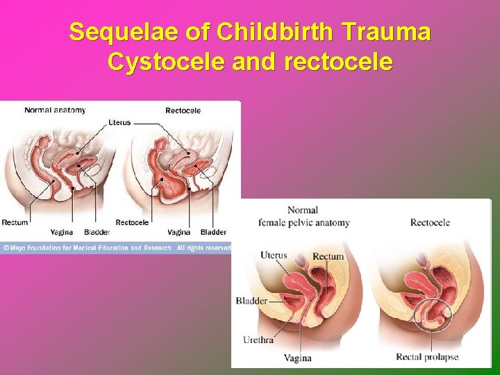
Sequelae of Childbirth Trauma Cystocele and rectocele
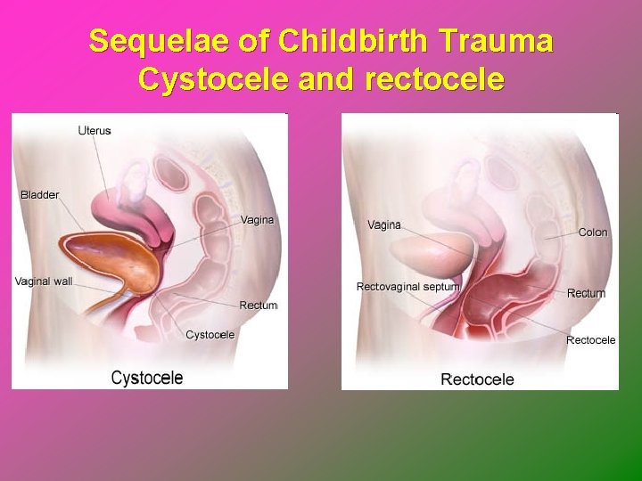
Sequelae of Childbirth Trauma Cystocele and rectocele
Sequelae of Childbirth Trauma Genital fistulas Ø May result from congenital anomaly, gynecologic surgery, obstetric trauma, cancer, radiation therapy, gynecologic trauma, or infection • Vesicovaginal: between bladder and genital tract • Urethrovaginal: between urethra and vagina • Rectovaginal: between rectum or sigmoid colon and vagina 82 of 34
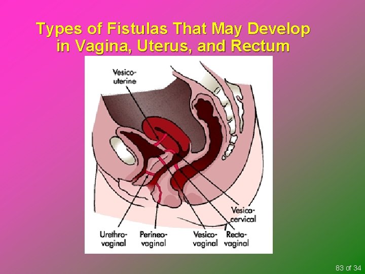
Types of Fistulas That May Develop in Vagina, Uterus, and Rectum 83 of 34
Postpartum Psychologic Complications Mental health disorders in postpartum period have implications for mother, newborn, and entire family Ø Interfere with attachment to newborn and family integration Ø May threaten safety and well-being of mother, newborn, and other children 84 of 34
Postpartum Psychologic Complications Postpartum depression without psychotic features Ø PPD: an intense and pervasive sadness with severe and labile mood swings Ø Treatment options • Antidepressants, anxiolytic agents, and electroconvulsive therapy • Psychotherapy focuses fears and concerns of new responsibilities and roles, and monitoring for suicidal or homicidal thoughts 85 of 34
Postpartum Psychologic Complications Postpartum depression with psychotic features Ø Postpartum psychosis: syndrome characterized by depression, delusions, and thoughts of harming either infant or herself Ø Psychiatric emergency, and may require psychiatric hospitalization Ø Antipsychotics and mood stabilizers such as lithium are treatments of choice 86 of 34
Loss and Grief Losses of what was hoped for, dreamed about, and/or planned Any perception of loss of control during the birthing experience Birth of a child with handicap Maternal death Fetal or neonatal death 87 of 34
Loss and Grief Conceptual model of parental grief Ø Acute distress Ø Intense grief Ø Reorganization Anticipatory grief 88 of 34
Loss and Grief Plan of care and implementation Ø Communicating and care techniques • Actualize the loss • Provide time to grieve • Interpret normal feelings • Allow for individual differences • Cultural and spiritual needs of parents • Physical comfort 89 of 34
Loss and Grief Plan of care and implementation Ø Options for parents • Seeing and holding • Bathing and dressing • Privacy • Visitations: other family members or friends • Religious rituals/funeral arrangements • Special memories • Pictures 90 of 34
Maternal Death Rare for woman to die in childbirth Families are at risk for developing complicated bereavement and altered parenting of surviving baby and other children in family Referral to social services can help combat potential problems before they develop 91 of 34
Key Points Postpartum hemorrhage is most common and serious type of excessive obstetric blood loss Hemorrhagic (hypovolemic) shock is an emergency situation; the perfusion of body organs may become severely compromised and death may ensue Potential hazards of therapeutic interventions may further compromise the woman with hemorrhagic disorders 92 of 34
Key Points Postpartum infection is a major cause of maternal morbidity and mortality Postpartum urinary tract infections are common because of trauma experienced during labor Breast infection affects about 1% of women soon after childbirth 93 of 34
Key Points Structural disorders of uterus and vagina related to pelvic relaxation are often the delayed but direct result of childbearing Understanding of grief responses and bereavement process is fundamental to the nursing process Therapeutic communication and counseling techniques can help families in identifying their feelings and in feeling comfortable in expressing their grief 94 of 34
Key Points Follow-up after discharge is an essential component in providing care to families who have experienced a loss Nurses need to be aware of their own feelings of grief and loss to provide a nonjudgmental environment of care and support for bereaved families 95 of 34
- Slides: 95