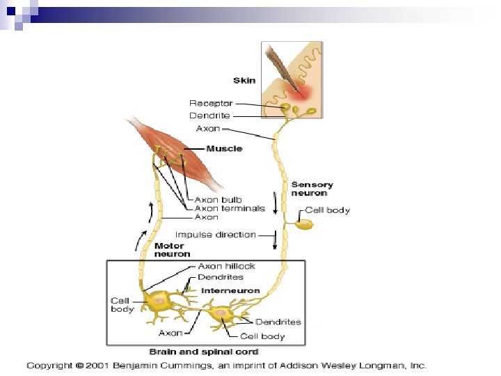
PHYSIOLOGY Nervous System Types of Neurons n Afferent


 1. Acetyl Choline (ACh) – – 2. Made")


- Slides: 91
PHYSIOLOGY Nervous System
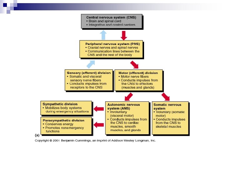
Types of Neurons n Afferent ¨ Sensory n Efferent ¨ Motor n Interneurons also known as association neurons ¨ Between neuron
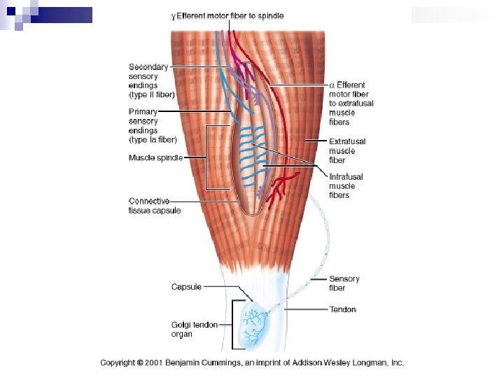
Classes of Sensory Receptors also known as Neurons Mechano-receptors: mechanical forces- stretching alters membrane permeability (1) hair cells* (deflection = depolarization = AP's) ie. lateral line of fish (mechanoreceptor= neuromasts detect water movement, etc) (2) stretch receptors of muscles (3) equilibrium receptor of inner ear (4) receptors of skin (touch, pain, cold, heat). Chemo-receptors: chemicals sense solutes in solvents, taste, smell Osmo-receptors: of hypothalamus which monitors blood osmotic pressure Photo-receptors: light - eye, eyespots, infrared receptors of snakes, etc. Thermo-receptors: radiant (heat) energy Phono-receptors: sound waves Electro-receptors: detect electric currents. . . electric eels, etc. . Nociceptors: pain receptors. . . naked dendrites of skin (epidermis)
Neuroglial Cells of the CNS
Astrocytes In the CNS only n Most abundant Neuroglial Cell n Formation of Synapses n Plays a role in making exchanges between capillaries and neurons n Helps to form the Blood Brain Barrier n ¨ The BBB protects the brain from intruders
Astrocytes
Microglial Cells n Macrophage ¨ Scavenges n apoptotic cells May go bad causing Alzheimer’s Disease ¨ Excessive secretion of Interleukin-1 ¨ Helps to maintain homeostasis in the brain
Ependymal Cells of the CNS
Ependymal Cells Lines ventricles in the brain and the central cavity of the spinal cord n Cells have cilia n ¨ Used to circulate the cerebrospinal fluid
Oligodendrocyte Cells of the CNS
Oligodendrocyte n Oligodendrocytes ¨ Production of myelin in the CNS ¨ Can cover as many as 60 neurons with myelin
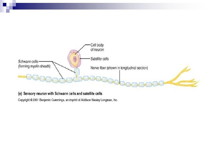
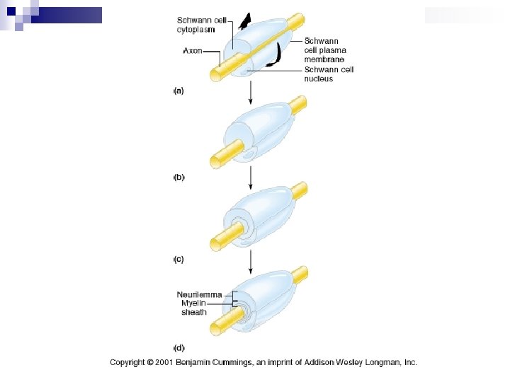
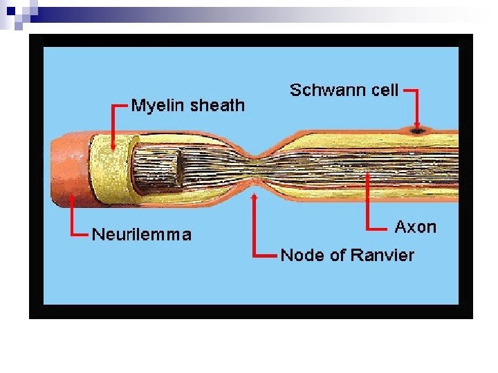
Schwann/Satellite Cells n Schwann Cells ¨ Production of myelin in the PNS ¨ Not able to cover one neuron, must use multiple Schwann Cells ¨ Formation of the Nodes of Ranvier ¨ Produces Neuronal Growth Factor n Satellite Cells ¨ Function unknown
Myelin Sheath n Myelin ¨ Insulates the axon for rapid conduction of action potentials n Nodes of Ranvier ¨ Gray v. White matter in the brain ¨ Multiple Sclerosis is an autoimmune disease
n http: //www. youtube. com/watch? v=Naecv 3 h 868 c
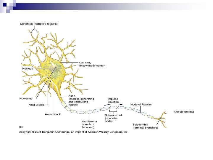
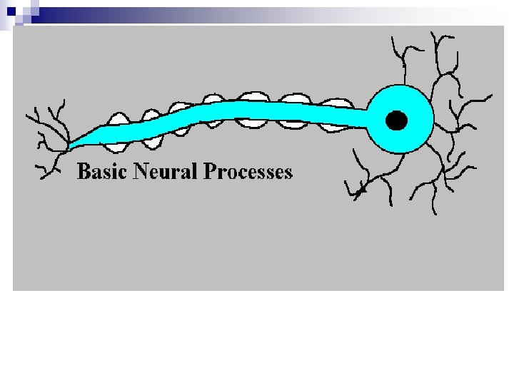
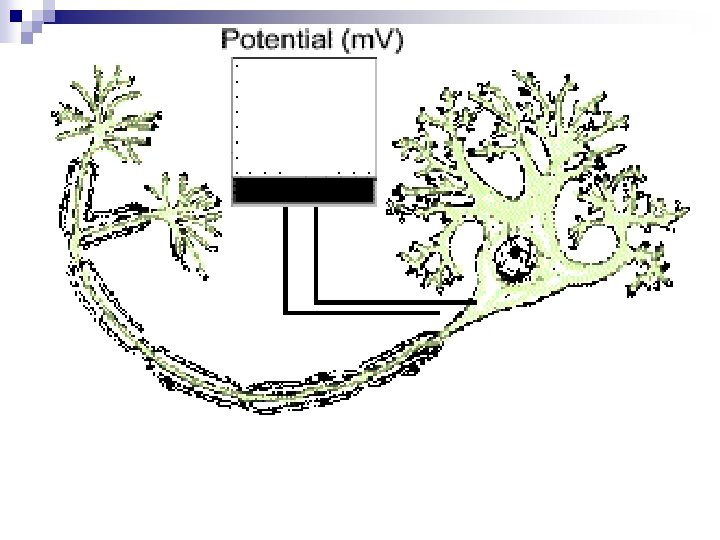
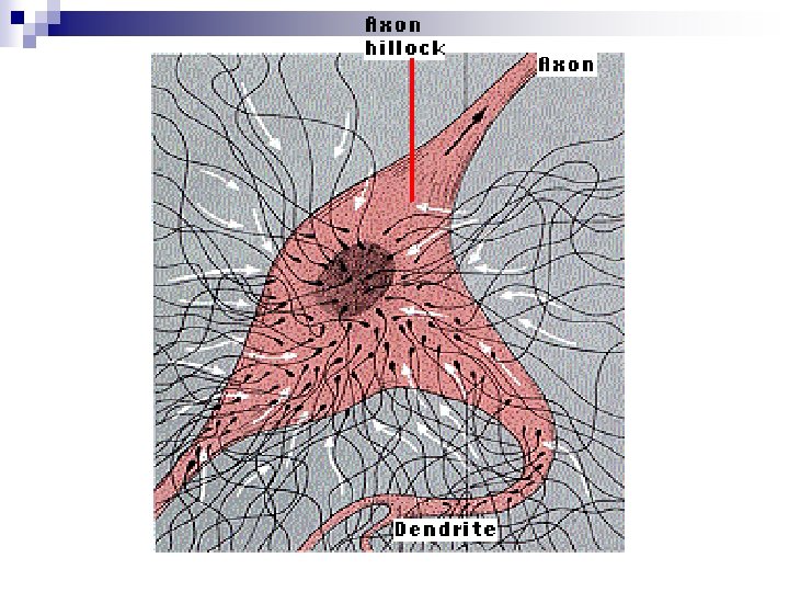
Neuron n Receptive Zone ¨ Where n the Graded Response occurs Cell Body Same information as a regular cell but no centrioles § Amitotic ¨ Contains ligand regulated gates ¨ n Dendrites Projections to help form synapses ¨ Contains ligand regulated gates ¨
Neuron n Conducting Zone ¨ Axon Hillock Begins action potentials n Accumulation of K+ ions n Contains voltage regulated gates for Na+/K+ n ¨ Axon Propagation of action potentials n Contains voltage regulated gates for Na+/K+ n Anterograde vs. Retrograde and Polio n
Secretory Zone ¨ Terminal Boutons Contains voltage regulated gates for Ca+2 n Contains vesicles filled with Neurotransmitter n
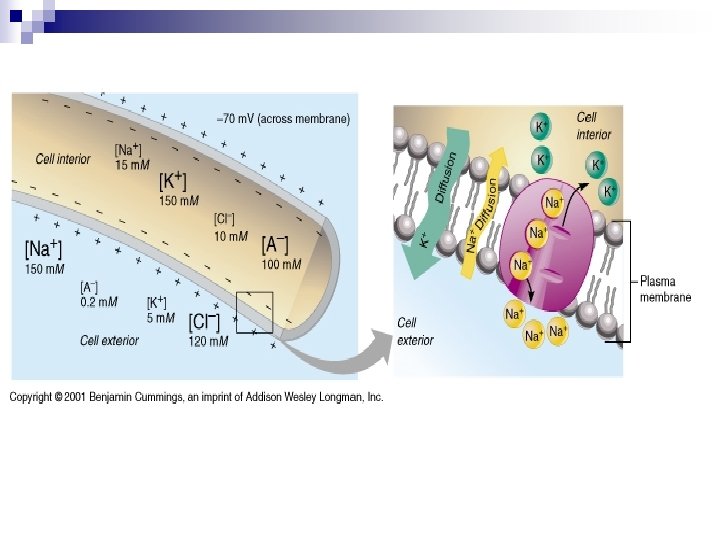
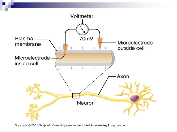
Resting Membrane Potential n n -70 m. V Membrane is said to be polarized ¨ Voltage generated by ionic movement through the membrane n Creates a current ¨ ¨ n n Current = Voltage/ Resistance Current generates a Kinetic Energy More Na+ on the outside of the cell More K+ on the inside of the cell ¨ Diffusion down their electrochemical gradient
Resting Membrane Potential n Maintained by the Na+/K+ATPase pumps ¨ Will not allow the neuron to reach equilibrium across the membrane ¨ Actively transports 3 Na+ out of the cell and 2 K+ into the cell
Graded Response n n Short lived Localized changes in membrane potential ¨ Can depolarize or hyperpolarize n Dependent on IPSP or EPSP n the membrane The magnitude of the graded potential varies directly with the stimulus strength ¨ The stronger stimulus causes greater voltage change and the current flows farther n The current dies out within a few millimeters of its origin ¨ Graded response only signals over a very short distance
Graded Response n Ligand sensitive Na+ gates will open with a stimulus ¨ Na+ diffuses into the cell down its electrochemical gradient n Depolarization of the membrane ¨ K+ is repelled down the membrane towards the axon hillock n K+ can diffuse out of the cell because the plasma membrane is very “leaky”
Action Potentials Begins at the axon hillock n Voltage regulated Na+ and K+ gates n ¨ Along with Na+/K+ATPase pumps along the entire membrane n All or nothing response
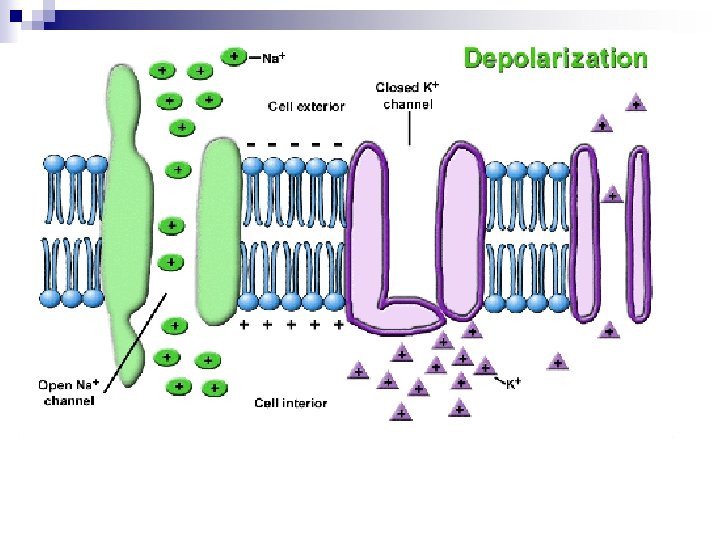
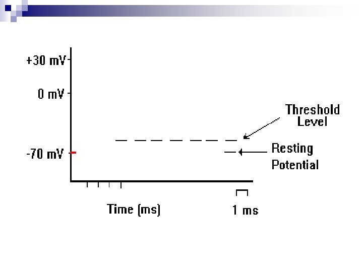
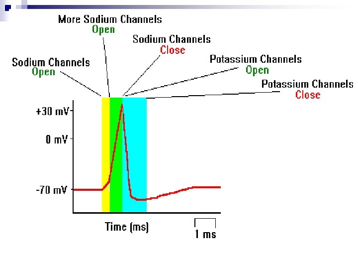
Action Potentials n Depolarization ¨ -50 m. V due to the accumulation of K+ at the axon hillock triggers an action potential ¨ At -50 m. V Na+ voltage regulated gates open n n Na+ diffuses into the cell down its electrochemical gradient Na+ repels K+ down the membrane ¨ Positive Feedback “on” n The more positive the voltage, due to Na+ diffusing into the cell, the more Na+ gates open. This creates a more positive voltage and more Na+ gates open ¨ Positive Feedback n +30 m. V “off”
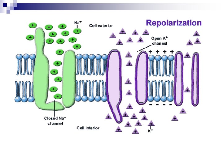
Action Potential n Repolarization ¨ At +30 m. V All Na+ gates close quickly n All K+ gates open n ¨ n K+ diffuses out of the cell down its electrochemical gradient K+ gates close slowly at -70 m. V ¨ K+ continues to diffuse out of the cell until it reaches -90 m. V § All K+ gates are closed
Action Potential n Hyperpolarization ¨ At n -90 m. V the Na+/K+ATPase pump turns on Pumps 3 Na+ out and 2 K+ into the cell ¨ Re-establishes resting membrane potential
Propagation of an Action Potential n As the influx of Na+ repels the K+ down the membrane there is an accumulation of K+ ¨ The K+ accumulation with change the membrane voltage to -50 m. V ¨ The occurs when the previous action potential reaches +30 m. V n Repolarization is chasing Depolarization down the membrane
Refractory Period n Absolute refractory ¨ From the opening of the Na+ channels until the Na+ channels begin to reset to their original resting state ¨ Cannot re-stimulate the neuron during this time n Relative refractory ¨ The n n interval following the absolute refractory period Na+ channels have returned to their resting state K+ channels are still open and repolarizing the membrane ¨ Can re-stimulate the neuron during this time with a great stimulus
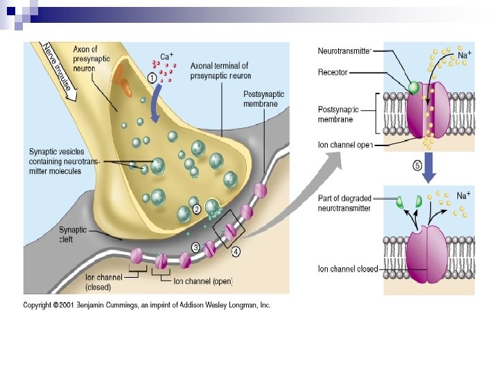
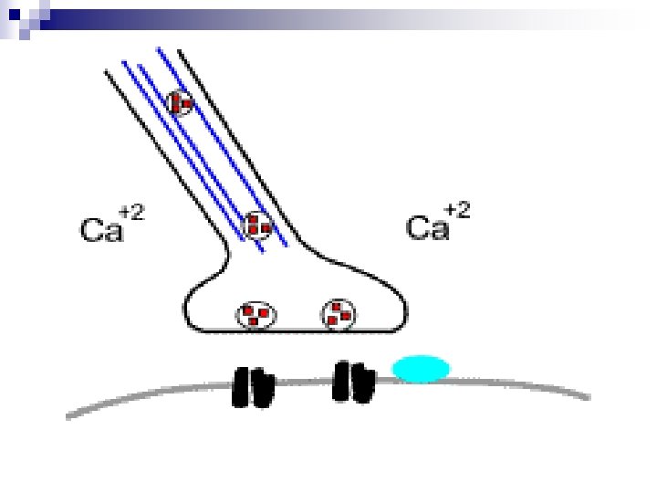
Synapse Presynaptic neuron n Postsynaptic neuron n Synaptic Cleft n ¨ About n 10 angstroms between neurons Synaptic Vesicles ¨ Filled with neurotransmitter
Synapse Voltage regulated Calcium channels n Membrane reaches -50 m. V due the accumulation of K+ n Calcium channels open n ¨ Calcium gradient n diffuses in down its electrochemical 2 Calcium ions bind to the vesicle ¨ The vesicle fuses with the membrane for exocytosis of the NT
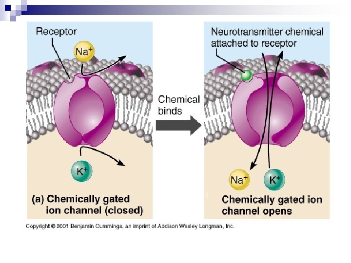
Synapse n The Neurotransmitter crosses the synaptic cleft ¨ NT binds to the receptors on the postsynaptic neuron n Neurotransmitter are removed from the synaptic cleft by: ¨ Reuptake ¨ Phagocytosis ¨ Enzymatic Degradation
Events at the Synapse AP reaches axon terminal Voltage-gated Ca 2+ channels open Ca 2+ entry Ca 2+ = Signal for Neurotransmitter Release Exocytosis of neurotransmitter containing vesicles
1. Axon Diameter
Fig. 8 -18 2. Signal Transduction in Myelinated Axon: Animation Demyelination diseases (E. g. ? )
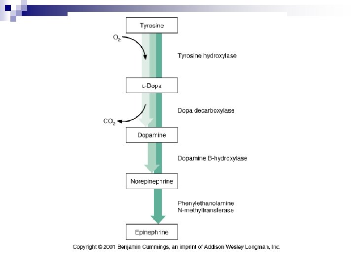
3 Classes of Neurotransmitters (of 7) 1. Acetyl Choline (ACh) – – 2. Made from Acetyl Co. A and choline Synthesized in axon terminal Quickly degraded by ACh-esterase Cholinergic neurons and receptors – Nicotinic (agonistic) and muscarinic (antagonist) Amines – Serotonin (tryptophane) and Histamine (histidine) ¨ – – – • Dopamine and Norepinephrine (tyrosine) Widely used in brain, role in emotional behavior (NE used in ANS) Adrenergic neurons and receptors - and Gases – 1. SSRI = antidepressants NO (nitric oxide) and CO Others: AA, (e. g. , GABA), lipids, peptides, purines
Neurotransmitters n Cholinergic Receptors ¨ Nicotinic ¨ Muscarinic n Catecholamine ¨ Alpha ¨ Beta
Nicotinic Receptors Stimulated by ACh and nicotine, not stimulated by muscarine. n Found at all ganglionic synapses. n Also found at neuromuscular junctions. n A ligand sensitive gate n
Muscarinic Receptors n n n Stimulated by ACh and muscarine, not stimulated by nicotine. Found at target organs when ACh is released by post-ganglionic neurons (all of parasympathetic, and some sympathetic). Stimulated selectively by Muscarine, Bethanechol. Blocked by Atropine. Stimulation causes: ¨ ¨ ¨ ¨ ¨ Increased sweating. Decreased heart rate. Decreased blood pressure due to decreased cardiac output. Bronchoconstriction and increased bronchosecretion. Contraction of the pupils, and contraction of ciliary body for near vision. Tearing and salivation. Increased motility and secretions of the GI system. Urination and defecation. Engorgement of genitalia.
Catecholamine Receptors n n NE and epinephrine, each act on α- and β-adrenergic receptors Two subclasses of α-adrenergic receptors Activation of α 1 -receptors usually results in a slow depolarization linked to the inhibition of K+ channels ¨ activation of α 2 -receptors produces a slow hyperpolarization due to the activation of a different type of K+ channel. ¨ n n There are three subtypes of β-adrenergic receptor Agonists and antagonists of adrenergic receptors ¨ β-blocker propanolol (Inderol®). n However, most of their actions are on smooth muscle receptors, particularly the cardiovascular and respiratory systems
α 1 adrenergic receptors Mainly involved with contraction of smooth muscle n G protein, c. AMP action n
α 2 adrenergic receptors n Three types of receptors ¨ α 2 A, n α 2Β, and α 2 C These receptors have a critical role in regulating neurotransmitter release from sympathetic nerves and from adrenergic neurons in the central nervous system
β 1 adrenergic receptors n Specific actions of the β 1 receptor include: ¨ Increases n cardiac output by raising heart rate and increasing the volume expelled with each beat (increased ejection fraction). ¨ Renin release from juxtaglomerular cells. ¨ Lipolysis in adipose tissue.
β 2 adrenergic receptors n Specific actions of the β 2 receptor include: ¨ Smooth muscle relaxation, e. g. in bronchi. ¨ Relax non-pregnant uterus. ¨ Relax detrusor urinae muscle of bladder wall ¨ Dilate arteries to skeletal muscle ¨ Glycogenolysis and gluconeogenesis ¨ Contract sphincters of GI tract ¨ Thickened secretions from salivary glands. ¨ Inhibit histamine-release from mast cells ¨ Increase renin secretion from kidney
β 3 adrenergic receptors n Specific actions of the β 3 receptor include: ¨ Enhancement ¨ CNS effects of lipolysis in adipose tissue.
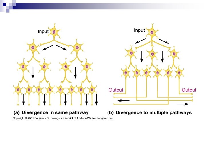
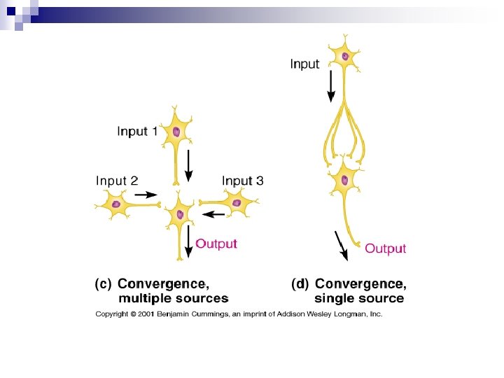
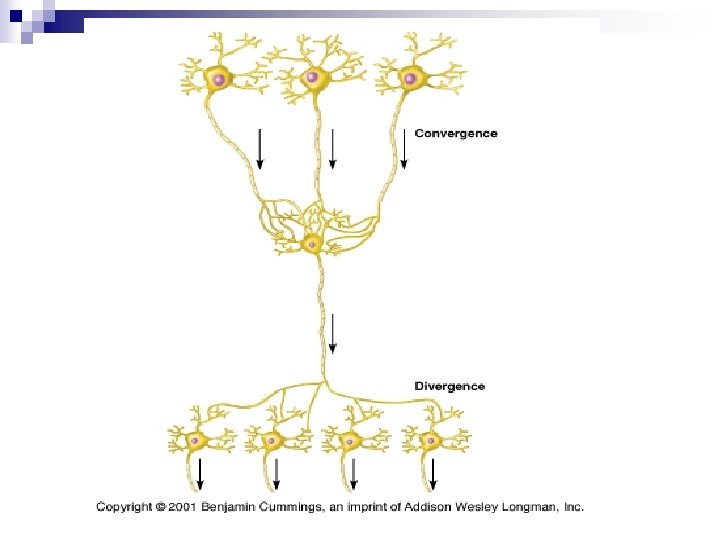
Neurological Communication There’s no one-to-one communication between neurons n May be as many as 500 neurons communicating with a single neuron n ¨ Convergence ¨ Divergence
Postsynaptic Responses Can lead to either EPSP or IPSP Any one synapse can only be either excitatory or inhibitory Fast synaptic potentials Opening of chemically gated ion channel Rapid & of short duration Slow synaptic potentials Involve G-proteins and 2 nd messengers Can open or close channels or change protein composition of neuron
Integration of Neural Information Transfer Multiple graded potentials are integrated at axon hillock to evaluate necessity of AP 1. Spatial Summation: stimuli from different locations are added up 2. Temporal Summation: sequential stimuli added up
1. Spatial Summation
2. Temporal Summation
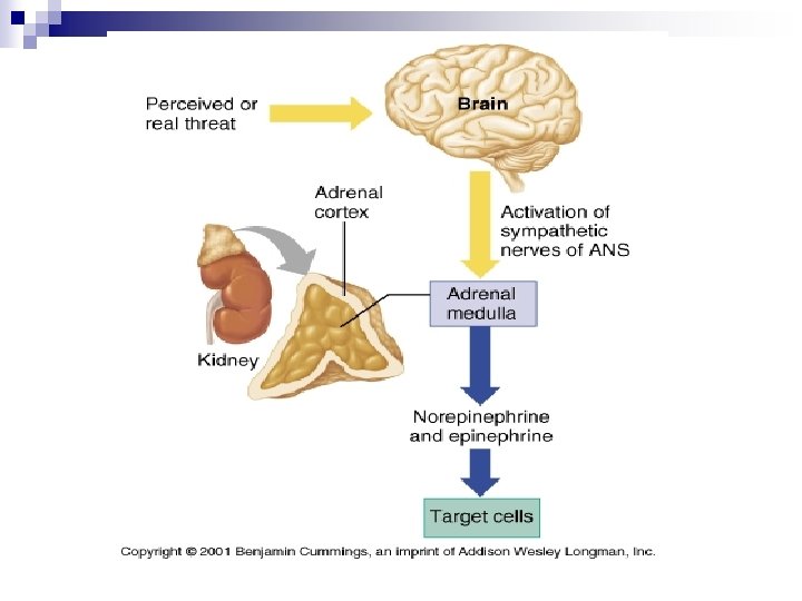
General Adaptation Syndrome
General Adaptation Syndrome n Hans Selye Alarm Phase ¨A stressor disturbs homeostasis ¨ Cerebral Cortex alerts Hypothalamus which alerts the Sympathetic Nervous System
General Adaptation Syndrome n Resistance Phase ¨ Body reacts to stressor ¨ Attempts to return to homeostasis ¨ Down and Up Regulation
General Adaptation Syndrome n Exhaustion Phase ¨ Physical and Psychological energy is sapped n Atypical depression ¨ n n n Mood disorder Dysphoria -generally characterized as an unpleasant or uncomfortable mood, such as sadness (depressed mood), anxiety, irritability, or restlessness Serious illness(es) may occur Hits person at weakest genetic point ¨ ¨ Autoimmune Disease(s) Endorphins Increase and inhibit the immune system response
General Adaptation Syndrome n Final Phase is Death
Dermatomes
Dermatomes
Bipolar Neuron n Two processes ¨ An axon and a dendrite n They extend in opposite directions ¨ Used for sensory organs n n Olfactory neurons Retina
Unipolar Neurons n n Presence of only a single axon, branching at the terminal end. True unipolar neurons not found in adult human; common in human embryos and invertebrates