VIRTUAL NEURONS LAB Lab 05 SCIENCE STANDARDS VIRTUAL

VIRTUAL NEURONS LAB Lab 05

SCIENCE STANDARDS: VIRTUAL NEURONS • Objectives—Students will • model working neural circuits using three types of neurons (motor, sensory, and interneurons). • draw and label a neural circuit using a motor neuron, sensory neuron, and interneuron, indicating the direction of neural communication. • identify and draw a motor neuron, a sensory neuron, and an interneuron. • calculate, predict, and test muscle twitch rate. • test and describe how neuronal communication in a circuit is affected by the neuron’s threshold, by the presence of excitatory and inhibitory neurons in the circuit, and by the pattern of connections within the circuit. • design a circuit that behaves according to a predetermined goal or pattern. • write rules for making circuits that work.

Vocabulary • action potential – an electrical signal that travels along the axon, away from the cell body, to the axon terminal where it triggers the release of neurotransmitters • excitatory neuron – a neuron whose neurotransmitter excites, stimulates, or depolarizes (cell membrane becomes less negative) another neuron, increasing the probability that the target neuron will fire an action potential • inhibitory neuron – a neuron whose neurotransmitter inhibits or hyperpolarizes (cell membrane becomes more negative) another neuron, decreasing the probability that the target neuron will fire an action potential • interneuron – any neuron that is not a sensory or motor neuron, interneurons carry information between neurons, for example, between sensory and motor neurons • motor neuron – a neuron that carries information away from the central nervous system to muscles or glands • neuromuscular junction – a specialized synapse onto a muscle • neuron – the principal cell of the nervous system, also called a nerve cell, specialized for the transmission of information and characterized by long fibrous projections called axons and short, branch-like projections called dendrites • pacemaker neuron – interneurons that fire spontaneously • sensory neuron – a neuron that carries information from the body’s sensory receptors in the skin, tongue, ear, nose, and eyes towards the central nervous system • threshold – the minimum amount of depolarization (becoming more positive) of the cell membrane potential needed to cause firing of a neuronal action potential

Materials • computer • Virtual Neurons software • Virtual Neurons Student Guide

Explore moving neurons, starting and stopping operation of the circuit. Getting Started The muscle twitches or contracts when it receives a signal from the motor neuron. What might cause changes in how fast the muscle twitches.

Simple Circuits Construct a simple circuit that makes the muscle twitch using only 2 neurons. Try it again with three neurons and note how the number of twitches changes over time.
")
• Try the simple circuit (one sensory, one inter, and one motor neuron) with different interneurons. • Some neurons are excitatory and some inhibitory. • How does the rate of muscle twitching change with different interneurons in the circuit? Simple Circuits • HINT: [An inhibitory interneuron will not excite the next neuron in the circuit and the muscle will not twitch. ]

• a motor neuron sends information away from the CNS toward an effector (muscle or gland). • an interneuron lies in the CNS and connects a sensory neuron to a motor neuron. • a sensory neuron sends information from a sensory receptor to the CNS. Types of Neurons

Excitatory neurons • Excitatory neurons receive information and transmit action potentials that release an excitatory neurotransmitter. • The excitatory neurotransmitter causes the post-synaptic neuron to be more likely to produce an action potential.

Inhibitory neurons • Inhibitory neurons receive information and transmit action potentials that release an inhibitory neurotransmitters. • Inhibitory neurotransmitters cause the postsynaptic neuron to be less likely to produce an action potential.

http: //brainu. org/lesson/virtual-neurons • Neurons, or nerve cells, are the basic functional units of the nervous system. • You will use the Virtual Neurons software to construct neural circuits and visualize how messages travel through the circuits.

Using the Software While you use the software, once you've set up a circuit, click on the "Zoom in. . . " button at the left. You'll see blue circles over the axon end of the neuron and gold circles over the dendrites. If you click on the area where the two circles overlap, you'll see a pop-up animation of the synapse. Click on Learn! to get more information.

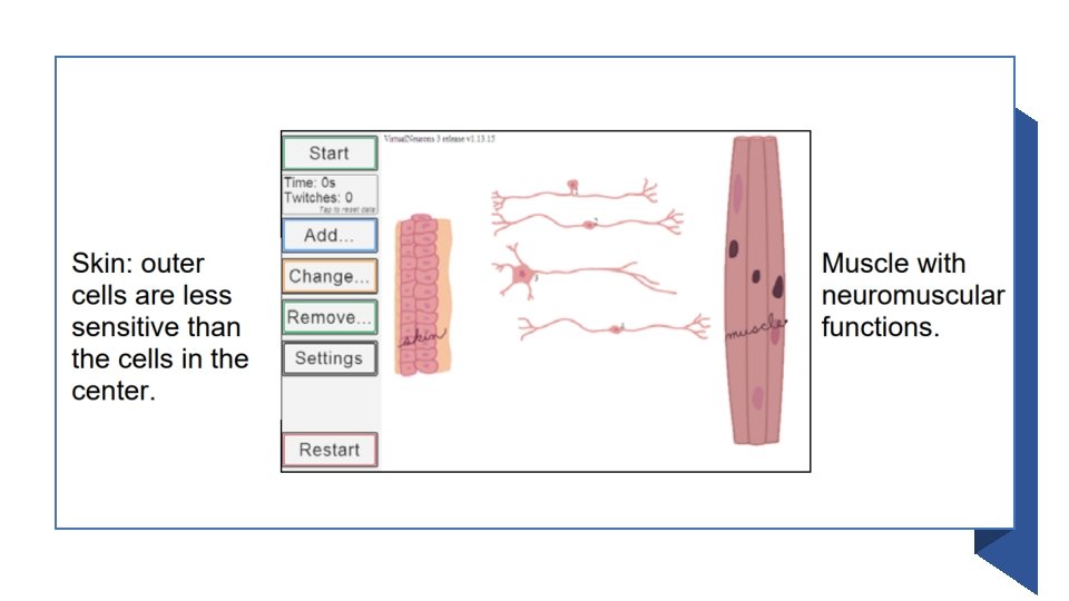
The initial window that opens up has a skin picture on the left side of the screen, a muscle picture on the right side of the screen, and four neurons in between -- one sensory neuron, one motor neuron, and two interneurons [one excitatory and one inhibitory].

• Your goal in this window is to get a message from the skin to the muscle via a neuronal circuit. Using the Software • The muscle, neurons, and skin can be moved around at any time by clicking on the image and dragging it to a new location. • Click on the cell body to move the neurons.

• To activate the circuit, click the Start/Resume button. Using the Software • To inactivate the circuit, press the Pause button. • When the circuit is active, the number of seconds the circuit is running and how many times the muscle twitches is recorded and displayed just below the Start button.

• The axon of the sensory neuron must come into contact with the skin cells. • The center skin cells are the most sensitive and will cause the sensory neuron to send the most signals in a set period of time. • The outer skin cells are less sensitive and will cause the sensory neuron to send fewer signals in a set period of time. • The axons of the motor neuron must come into contact with the neuromuscular junction. Using the Software

Using the Software The excitatory neurons emit green neurotransmitters. The inhibitory neurons emit red neurotransmitters. • You may add any type of neuron by clicking on the Add button and selecting from the panel which shows the 4 basic shapes (see image at right). • From top to bottom, the images are: • a sensory neuron • an excitatory interneuron • an inhibitory interneuron (with the red highlight) • a motor neuron

Optional: Enable Advanced Neuron Shapes If you’ve activated Enable Advanced Neuron Shapes in the Settings screen, you’ll see many neuron shapes when you click on the Change button and then click on the neuron that you want to swap out. NOTE: If you change the shape of an inhibitory neuron, the new shape will still be inhibitory.

Settings Features can be added at any time from the Settings screen. You may visit this screen by clicking the Settings button.

Check the boxes to enable the following features: • Virtual Neurons starts with a set of simple neuron shapes. Once you have enabled advanced neuron shapes, you may use the Change and Add buttons on the main screen to change the configuration of your neuronal network.

Check the boxes to enable the following features: • After activating this feature, action potential meters for each neuron will appear at the left of the screen below the Settings button. The small number in the upper left corner of the meter is the neuron number that appears by the neuron’s cell body.

Check the boxes to enable the following features: • The large number in the meter indicates the neuron’s threshold; in the image above, neuron #1 has a threshold of 1. • Since the meter is filled with green, that indicates that one action potential has fired, the threshold has been met, and neuron #1 will release neurotransmitter.

Check the boxes to enable the following features: • Neuron #2 has a threshold of 3 and, as one-third of the meter is filled with green, that indicates that 1 action potential has fired. • When 2 more action potentials fire, neuron #2 will release neurotransmitter.

Check the boxes to enable the following features: • At any time in the program, the threshold for each neuron can be increased or decreased by clicking inside the meter and changing the number (possible values 0 -5). • The circuit need not be inactive to change threshold.

Check the boxes to enable the following features: • Activate this feature and click Continue to return to the main screen. • At the bottom of the main screen you’ll see a graph. • The graph has a line for each neuron in your circuit. Shown are the neuron’s action potential and the muscle twitches vs. time while the circuit is running.

Check the boxes to enable the following features: • Each action potential and muscle twitch is recorded as a spike in the graph line. The graph will remain on the screen after the circuit is stopped. To get the graph to disappear, go to Settings and deselect Show the Graph.

Part 3 of the student guide. In this activity, students will work with the Settings screen where they may enable additional features in the program. • Action Potential Meters: After activating this feature, action potential meters for each neuron will appear at the left of the screen. • In addition, the neuron number will appear by the neuron’s cell body. • The action potential meter records the number of times a neuron receives a synaptic signal that moves it closer to threshold, indicated by the large number in the meter.

• When the green color fills the meter, the neuron fires an action potential. • When the action potential travels to the next nerve terminal, it releases neurotransmitter onto the dendrites of the next neuron or onto the muscle at the neuromuscular junction. • The filling green color represents neuronal membrane potential depolarizing, or going up, a little (+1) with each received excitatory synaptic signal and depolarizing fully with an action potential once membrane potential reached threshold.
 • What happens to neuronal membrane potential when an inhibitory")
Inhibitory Post-Synaptic Signaling (IPSP) • What happens to neuronal membrane potential when an inhibitory synaptic signal is reached? • [Membrane potential hyperpolarizes, or goes down, a little (-1) with each inhibitory synaptic signal received. ]
, the neuron fires")
Threshold • When the threshold is set to the minimum (0), the neuron fires spontaneously at a fixed rate. • You may change threshold for a neuron by clicking in its action potential meter.

• The number will increment +1 for each click on the meter. Action Potential Meter • Possible values are 0 -5. If you click in a meter set at maximum, the value will change to 0. 7. • Select Action Potential Meters in the Settings screen and click Continue. • At the program’s main screen, they should create a simple circuit, observe the changes in the action potential meters, and answer the questions in step 6 of Part 3. 8.

Understanding the Graph • How does the information on the graph correlate with the action potential meters?

Understanding the Graph • What does each vertical line on the graph represent? [The line represents the firing of an action potential in that neuron. ] • What would happen if I change threshold of neuron X? Why does the muscle twitch less frequently? [A higher threshold will result in less frequent firing. ]

Circuit Illustrating Summation of Excitation and Inhibition • In this circuit all neurons are excitatory except neuron 2 which is inhibitory. • Since the skin is depressed less farther away from the pin, neuron 1 fires at a slower rate than neuron 5 where the pin depresses the skin maximally. • This illustrates the big concept that the rate of firing in the sensory neuron is proportional to the strength of the sensory signal.

Negative Feedback • Negative Feedback works to decrease the event that caused it. • This is the more common type of feedback. • For example, as you pick up a box that is lighter than expected, negative feedback from your muscle receptors decreases the rate of motor neuron firing and muscle contraction since less effort is needed to lift a light box.
- Slides: 36