
Kimberle M Jacobs Ph D Sanger 11 046
Kimberle M. Jacobs, Ph. D Sanger 11 -046 827 -2135 kmjacobs@vcu. edu
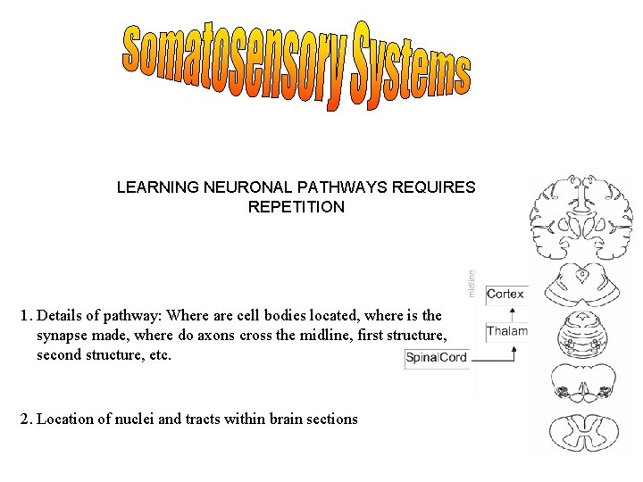
LEARNING NEURONAL PATHWAYS REQUIRES REPETITION 1. Details of pathway: Where are cell bodies located, where is the synapse made, where do axons cross the midline, first structure, second structure, etc. 2. Location of nuclei and tracts within brain sections
Somatosensory Systems – All senses other than Olfaction, Gustation, Audition, Vision, and Vestibular Sensation: Modalities Touch (and Pressure), Vibration Sense, Proprioception, Kinesthesia, Stereognosis, Pain & Temperature Proprioception - sense of static and dynamic position of limbs and body Kinesthesia - the ability to feel movements of the limbs and body Stereognosis – ability to recognize objects based on touch alone A. Pain and Temperature B. Fine Discrimination Touch
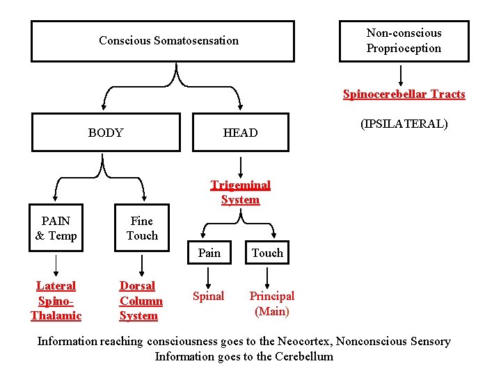
Non-conscious Proprioception Conscious Somatosensation Spinocerebellar Tracts BODY HEAD (IPSILATERAL) Trigeminal System PAIN & Temp Fine Touch Pain Lateral Spino. Thalamic Dorsal Column System Spinal Touch Principal (Main) Information reaching consciousness goes to the Neocortex, Nonconscious Sensory Information goes to the Cerebellum
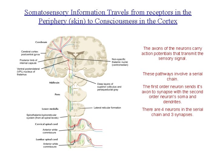
Somatosensory Information Travels from receptors in the Periphery (skin) to Consciousness in the Cortex The axons of the neurons carry action potentials that transmit the sensory signal. These pathways involve a serial chain. The first order neuron sends it’s axon to synapse with the second order neuron’s soma and dendrites. There are 4 neurons in the serial chain and 3 synapses.
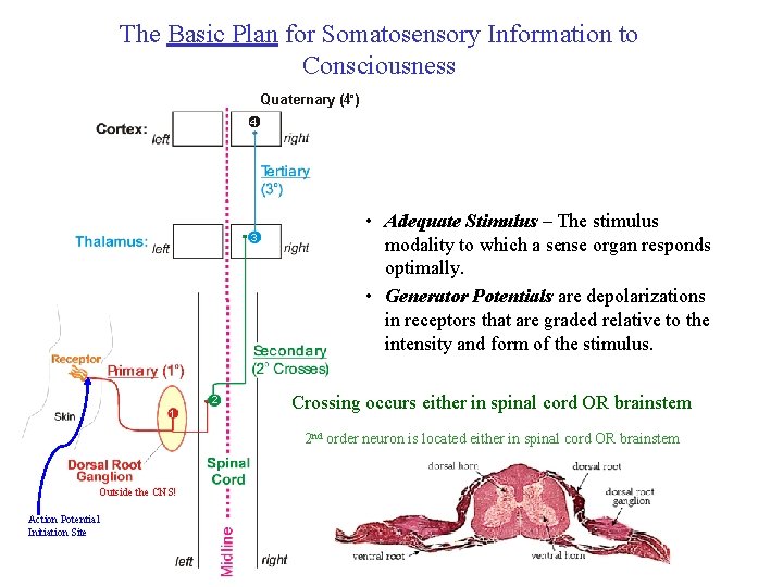
The Basic Plan for Somatosensory Information to Consciousness Quaternary (4 o ) 4 3 2 1 • Adequate Stimulus – The stimulus modality to which a sense organ responds optimally. • Generator Potentials are depolarizations in receptors that are graded relative to the intensity and form of the stimulus. Crossing occurs either in spinal cord OR brainstem 2 nd order neuron is located either in spinal cord OR brainstem Outside the CNS! Action Potential Initiation Site
Key Elements of the Basic Plan: SS to Consciousness § The basic plan for somatosensory information to consciousness involves 4 neurons and 3 synapses. § Cell bodies of the primary afferents are located outside of the central nervous system (in the dorsal root ganglion for body information and in the Trigeminal ganglion for head information). § Cell bodies of the second order neurons are located either in the spinal cord (anterolateral system) or in the brainstem (dorsal column system). § The axon of the second order neuron crosses the midline. § Cell bodies of the third order neurons are located in the thalamus (within VPL for body information and VPM for head information). § The fourth order neuron is located within the Primary Somatosensory cortex (Brodman’s areas 3, 1, 2). § If you lesion the pathway before it crosses you create IPSILATERAL deficits (loss of sensation on the same side of the body as the lesion). § If you lesion the pathway after it crosses you create CONTRALATERAL deficits (loss of sensation on the opposite side of the body as the lesion). § If you lesion the pathway AS it is crossing, you create BILATERAL deficits (both sides of the body affected).
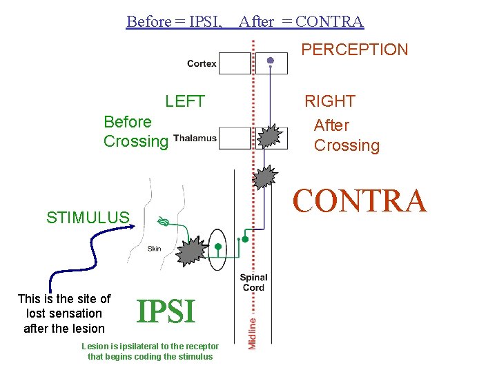
Before = IPSI, After = CONTRA PERCEPTION LEFT Before Crossing CONTRA STIMULUS This is the site of lost sensation after the lesion RIGHT After Crossing IPSI Lesion is ipsilateral to the receptor that begins coding the stimulus
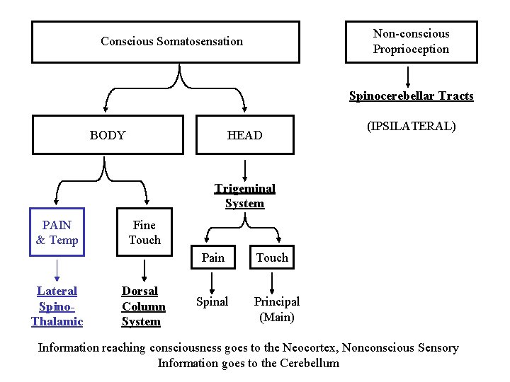
Non-conscious Proprioception Conscious Somatosensation Spinocerebellar Tracts BODY HEAD (IPSILATERAL) Trigeminal System PAIN & Temp Fine Touch Pain Lateral Spino. Thalamic Dorsal Column System Spinal Touch Principal (Main) Information reaching consciousness goes to the Neocortex, Nonconscious Sensory Information goes to the Cerebellum
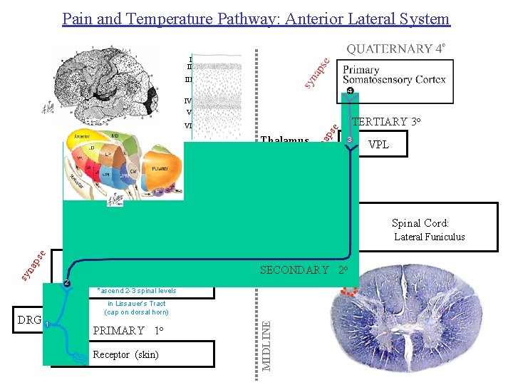
Pain and Temperature Pathway: Anterior Lateral System I II III 4 IV V TERTIARY 3 o na 3 sy Thalamus ps e VI VPL Lateral Spinothalamic Tract sy na ps e Crosses the midline in the Ventral White Commissure Of the Spinal Cord 2 Spinal Cord: Dorsal Horn SECONDARY 2 o *ascend 2 -3 spinal levels PRIMARY 1 o Receptor (skin) MIDLINE DRG 1 in Lissauer’s Tract (cap on dorsal horn) Spinal Cord: Lateral Funiculus

Dermatomes & Lissauer’s Tract Dermatome - the area of skin innervated by the sensory portion of a single cranial or spinal nerve. Primary afferents entering at T 4 will synapse mainly in T 2. Lesion at T 2 in spinal cord: Area affected will include T 4 since that is where the axons originally entered the spinal cord.
Key Elements - Lateral Spinothalamic Pathway § Transmits pain and temperature sensation from the body. § Located laterally in the spinal cord and remains relatively lateral through the brainstem and midbrain – (you will need to know the exact location – shown to you in video). § Crosses in the spinal cord. § Called the Lateral Spinothalamic Tract AFTER crossing in the spinal cord. § The tract travels from the spinal cord to the thalamus.
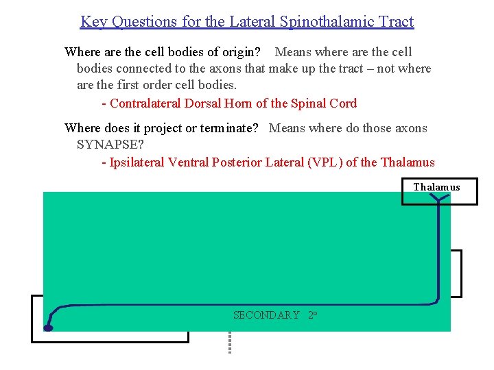
Key Questions for the Lateral Spinothalamic Tract Where are the cell bodies of origin? Means where are the cell bodies connected to the axons that make up the tract – not where are the first order cell bodies. - Contralateral Dorsal Horn of the Spinal Cord Where does it project or terminate? Means where do those axons SYNAPSE? - Ipsilateral Ventral Posterior Lateral (VPL) of the Thalamus MIDLINE Lateral Spinothalamic Tract Spinal Cord: Dorsal Horn SECONDARY 2 o Spinal Cord: Lateral Funiculus
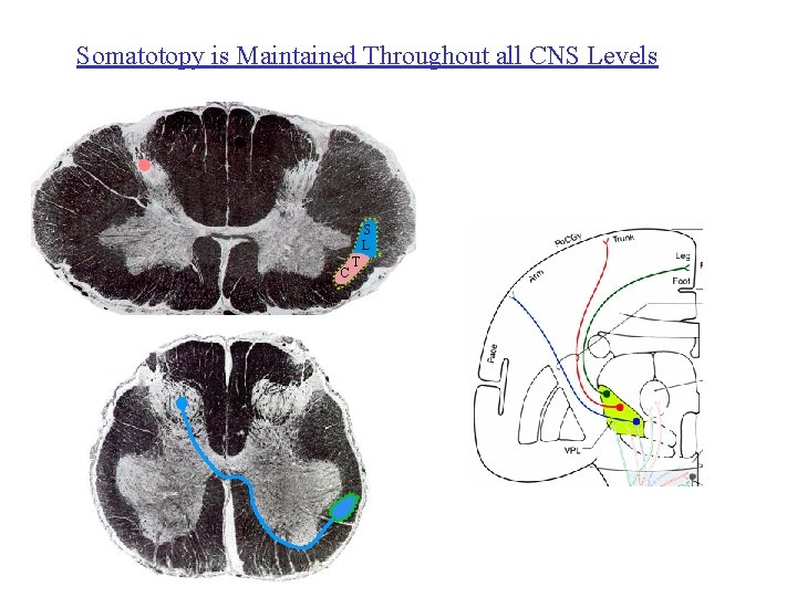
Somatotopy is Maintained Throughout all CNS Levels S L C T
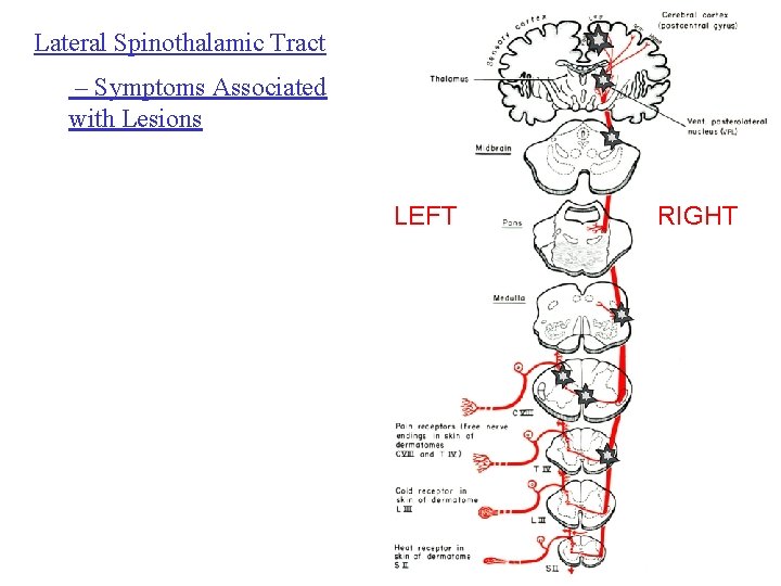
Lateral Spinothalamic Tract – Symptoms Associated with Lesions LEFT RIGHT
The Anterior Lateral System (ALS) The Lateral Spinothalamic Tract together with the Anterior Spinothalamic Tract is known as the ALS
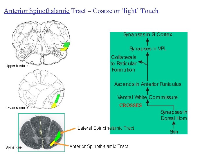
Anterior Spinothalamic Tract – Coarse or ‘light’ Touch Upper Medulla Lower Medulla CROSSES Lateral Spinothalamic Tract Spinal cord Anterior Spinothalamic Tract
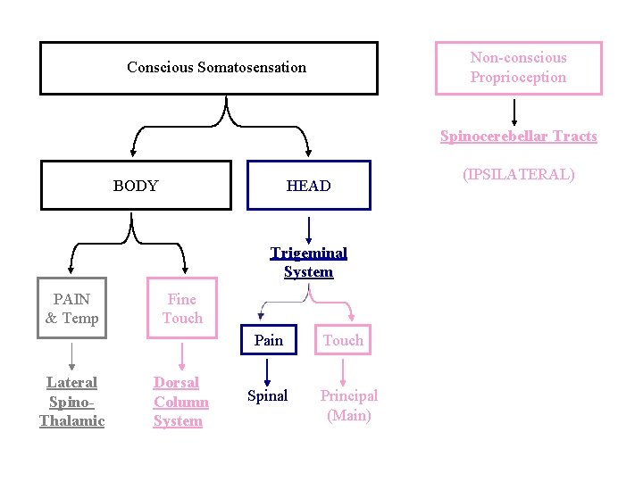
Non-conscious Proprioception Conscious Somatosensation Spinocerebellar Tracts BODY HEAD Trigeminal System PAIN & Temp Fine Touch Pain Lateral Spino. Thalamic Dorsal Column System Spinal Touch Principal (Main) (IPSILATERAL)
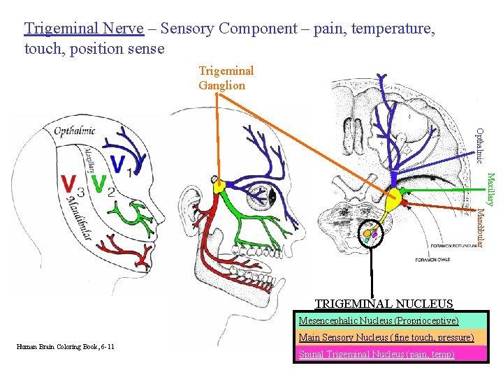
Trigeminal Nerve – Sensory Component – pain, temperature, touch, position sense Trigeminal Ganglion Opthalmic Maxillary Mandibular TRIGEMINAL NUCLEUS Mesencephalic Nucleus (Proprioceptive) Main Sensory Nucleus (fine touch, pressure) Human Brain Coloring Book, 6 -11 Spinal Trigeminal Nucleus (pain, temp)
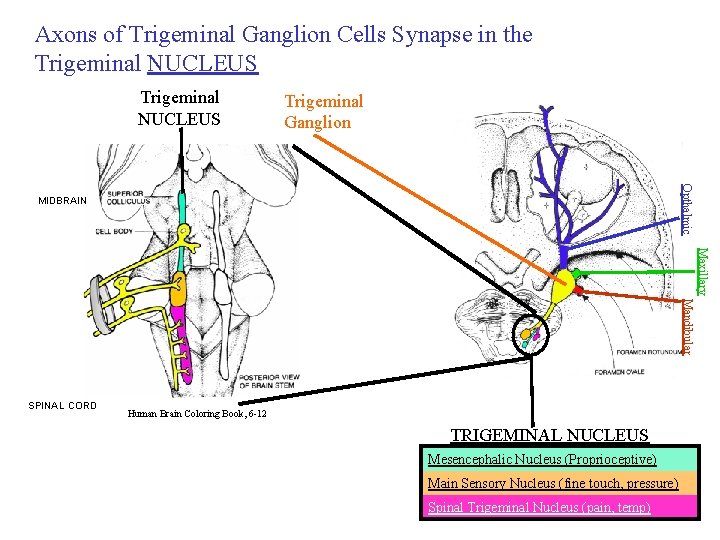
Axons of Trigeminal Ganglion Cells Synapse in the Trigeminal NUCLEUS Trigeminal Ganglion Opthalmic MIDBRAIN Maxillary Mandibular SPINAL CORD Human Brain Coloring Book, 6 -12 TRIGEMINAL NUCLEUS Mesencephalic Nucleus (Proprioceptive) Main Sensory Nucleus (fine touch, pressure) Spinal Trigeminal Nucleus (pain, temp)
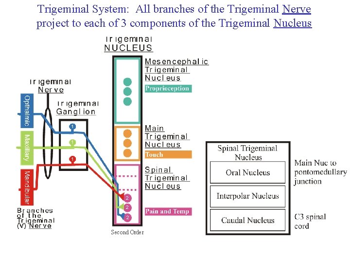
Trigeminal System: All branches of the Trigeminal Nerve project to each of 3 components of the Trigeminal Nucleus 1 1 1 2 2 2 Second Order
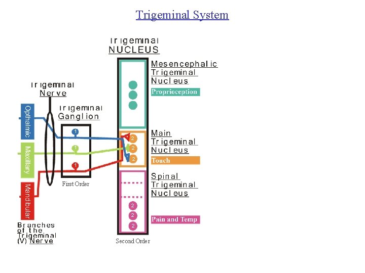
Trigeminal System 1 2 2 1 First Order 2 2 2 Second Order
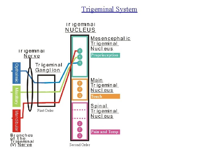
Trigeminal System 1 1 2 1 2 2 1 First Order 2 2 2 Second Order
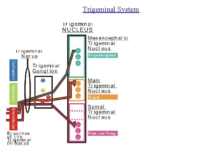
Trigeminal System 1 1 2 1 2 1 2 2 2
Similarities between Body and Face Pathways The trigeminal ganglion is functionally similar to the dorsal root ganglion. Both contain cell bodies of the PRIMARY AFFERENT pseudo-unipolar neurons. The Mesencephalic Nucleus of V is a special case because it is the only place within the CENTRAL nervous system that contains primary afferent cell bodies.
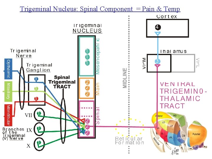
Trigeminal Nucleus: Spinal Component = Pain & Temp
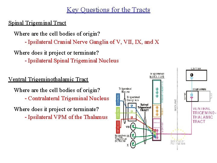
Key Questions for the Tracts Spinal Trigeminal Tract Where are the cell bodies of origin? - Ipsilateral Cranial Nerve Ganglia of V, VII, IX, and X Where does it project or terminate? - Ipsilateral Spinal Trigeminal Nucleus Ventral Trigeminothalamic Tract Where are the cell bodies of origin? - Contralateral Trigeminal Nucleus Where does it project or terminate? - Ipsilateral VPM of the Thalamus
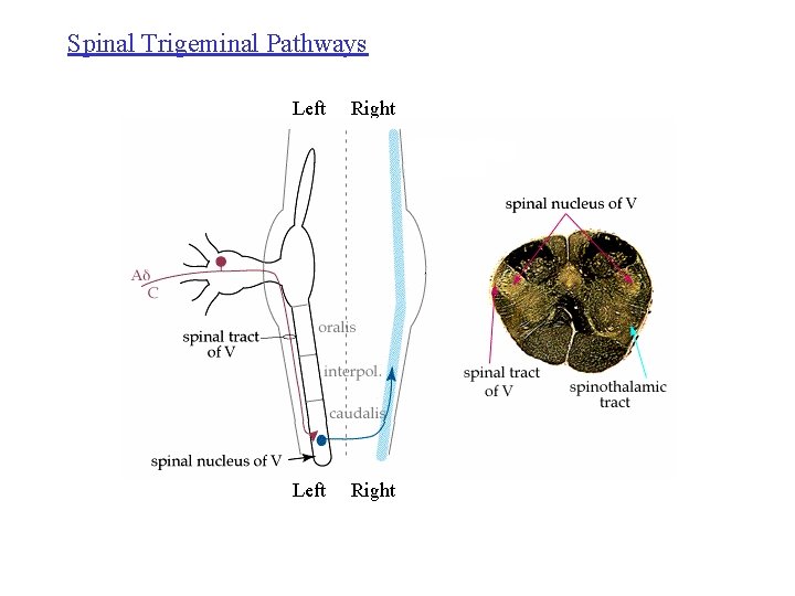
Spinal Trigeminal Pathways Left Right Ventral Trigeminothalamic tract
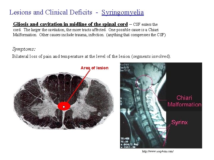
Lesions and Clinical Deficits - Syringomyelia Gliosis and cavitation in midline of the spinal cord – CSF enters the cord. The larger the cavitation, the more tracts affected. One possible cause is a Chiari Malformation. Other causes include trauma, infection. (anything that compresses the CSF) Symptoms: Bilateral loss of pain and temperature at the level of the lesion (segments involved). Area of lesion http: //www. asap 4 sm. com/
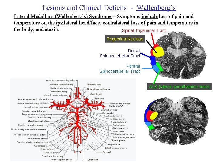
Lesions and Clinical Deficits - Wallenberg’s Lateral Medullary (Wallenberg’s) Syndrome – Symptoms include loss of pain and temperature on the ipsilateral head/face, contralateral loss of pain and temperature in the body, and ataxia. Spinal Trigeminal Tract Trigeminal Nucleus Dorsal Spinocerebellar Tract Ventral Spinocerebellar Tract ALS (lateral spinothalamic tract)
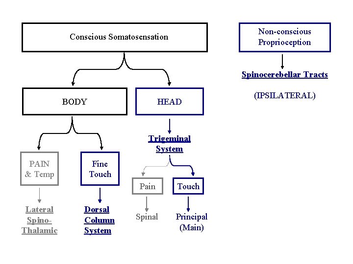
Non-conscious Proprioception Conscious Somatosensation Spinocerebellar Tracts BODY HEAD Trigeminal System PAIN & Temp Fine Touch Pain Lateral Spino. Thalamic Dorsal Column System Spinal Touch Principal (Main) (IPSILATERAL)
- Slides: 31